Introduction
Peripheral facial paralysis is an important disorder from psychosocial and functional perspectives. Although several related aetiological factors have been identified, the most common form is still idiopathic, which is also known as Bell's palsy.Reference Cirpaciu, Goanta and Cirpaciu1 Recurrent idiopathic peripheral facial paralysis is rare, and Bell's palsy recurs in approximately 12 per cent of patients, particularly within the first 2 years after diagnosis.Reference Cirpaciu, Goanta and Cirpaciu1,Reference Bibas, Jiang, Gleeson and Gleeson2
The aetiology of facial paralysis remains unclear. Ischemic neuropathy, microcirculatory perfusion failure of the vasa nervorum, and infectious, immunological and genetic causes have been suggested.Reference Bibas, Jiang, Gleeson and Gleeson2 The pathophysiology of the disease is attributed to facial nerve inflammatory oedema, which has been detected both by enhancement of the nerve in contrast-enhanced magnetic resonance imaging (MRI) and direct visual observation during decompression surgery.Reference Yetiser, Kazkayas, Altinok and Karadeniz3–Reference Tien, Dillon and Jackler7 Clinical studies that have demonstrated the effects of corticosteroids on the recovery and disease progression of peripheral facial paralysis support these observations.Reference Engström, Berg, Stjernquist-Desatnik, Axelsson, Pitkäranta and Hultcrantz8,Reference Sartoretti-Schefer, Brandle, Wichmann and Valavanis9
Recurrent idiopathic peripheral facial paralysis that recurs on the same side without a distinct pathology is extremely rare. We postulated that narrowness of the facial canal segments may predispose individuals to the development of recurrent idiopathic peripheral facial paralysis.Reference Hagino, Tsunoda, Tsunoda and Kishimoto10 The anatomy of the facial canal is best evaluated in high-resolution multidetector computed tomography (CT) images of the temporal bone, but the curvilinear pathway of the canal causes measurement difficulties. The measurement of the facial canal in axial and coronal reformatted images is restricted because the plane of the section may cross the facial canal obliquely. Curved planar reformation is an appropriate technique for illustrating the curvilinear anatomy of tubular tortious structures in a single image and allows more accurate measurements. To the best of our knowledge, the evaluation of facial canal segments using curved planar reformation in patients with facial paralysis has not been reported.
We evaluated the diameter of the facial canal segments in patients with ipsilateral recurrent idiopathic peripheral facial paralysis using curved planar reformation of temporal bone multidetector computed tomography images, and we investigated whether the diameter of the facial canal segments affected the recurrence of idiopathic facial paralysis by comparison with the healthy side in each patient.
Materials and methods
Patients
This prospective case–control study enrolled 568 consecutive patients suffering from peripheral facial paralysis between March 2012 and December 2015. Of the 568 patients, 34 patients (6 per cent) who had two or more attacks of facial paralysis on the same side were diagnosed with ipsilateral recurrent peripheral facial paralysis. From these 34 patients, the study included 20 patients diagnosed with ipsilateral recurrent idiopathic peripheral facial paralysis. The local ethics committee approved the study (2013, decision number: 13). After a detailed explanation of the study, informed consent was obtained from each patient.
All patients were examined by two otorhinolaryngologists and one neurologist. The neurologist performed electrophysiological tests (electromyography and nerve conduction studies).
Patients were excluded if they had undergone any head and neck surgery or had peripheral facial paralysis with determined causes, such as a tumour, metastasis, iatrogenic injury, polyneuropathy or Melkersson–Rosenthal syndrome (recurring facial paralysis, swelling of the face and lips, and the development of folds and furrows in the tongue).
The white blood cell count, C-reactive protein, erythrocyte sedimentation rate, rheumatoid factor, thyroid function tests, anti-nuclear antibody, blood biochemical analyses, viral herpes simplex virus, varicella-zoster virus, Epstein–Barr virus, human immunodeficiency virus and bacterial (Borrelia burgdorferi and Treponema pallidum) antibody levels were assessed. Pure tone audiometry and acoustic impedance were performed in routine audiological examinations. MRI of the brain and temporal bone with gadolinium was performed to exclude any temporal bone or intracranial pathologies (e.g. multiple sclerosis, vasculitis, sarcoidosis or tumour).
The patients were prescribed 1 mg/kg oral methylprednisolone during the acute phase of paralysis after assessing their blood pressure and glucose levels. Patients were followed for 6–36 months to avoid possible misdiagnosis.
Curved planar reformation technique
Curved planar reformation is a way to visualise the entire length of tortious tubular structures within a single image. The curved planar reformation technique is used to show vascular abnormalities (i.e. stenosis, occlusions and vessel wall calcifications)Reference Hagino, Tsunoda, Tsunoda and Kishimoto10 and to evaluate the entire course of the ureter and biliary stents.Reference Ochi, Shimizu, Yasuhara, Shigesawa, Mochizuki and Ikezoe11,Reference Dillman, Caoili, Cohan, Ellis, Francis and Schipper12
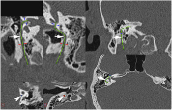
The procedure is as follows. First, an axial volumetric dataset is transferred to the workstation. Within contiguous reference images, the course of the facial canal is traced manually by a series of mouse clicks on the arbitrarily rotated imaging volume until the entire course of the facial canal within the volume of interest has been traced. Then, along the defined curved line, a single-voxel-thick plane, orthogonal to the reference plane, is extruded through the entire dataset. Finally, curved planar reformation of the facial canal is shown with a track. This track along the canal made it possible to determine the mid-level and the narrowest and widest parts of the facial canal segments (Figure 1).
Computed tomography image of the curved planar reformation technique showing the curved planar reformation tool selected at the workstation. The course of the facial canal is manually traced by series of mouse clicks on the arbitrarily rotated imaging volume until the entire course of the facial canal within the volume of interest has been traced. Curved planar reformation of the facial canal is displayed with a track (arrows). The track along the canal made it possible to determine the mid-level and also the narrowest and widest part of the facial canal segments with the measurements displayed at the axial images (arrowheads).

Computed tomography and image analyses
All CT examinations were performed using a 64-slice CT scanner (Aquilion 64, Toshiba Medical Systems, Tochigi, Japan). The scanning parameters were 120 kV, 200 mA, section thickness 0.5 mm and reconstruction interval of 0.5 mm with attenuation-based tube current modulation. All CT data were transferred to an Aquarius workstation (TeraRecon, San Mateo, USA), with multiplanar reformatting and curved planar reformation options. All measurements were made by a neuroradiologist blinded to the side of the facial paralysis. Both the affected and unaffected temporal bones were evaluated.
After obtaining the curved planar reformation images, the facial canal segments were measured. First, the meatal foramen (canalicular entrance to the labyrinthine segment) and mid-level of the labyrinthine segment were determined using the curved planar reformation technique. Then, in the mastoid and tympanic segments, both the narrowest (minimum) and widest (maximum) diameters of the canals were measured automatically from the trace of the curved planar reformation image.
Results
All data were analysed using SPSS® statistical software (version 20). The patients’ demographic characteristics were analysed using descriptive statistics. The data were normally distributed according to the Kolmogorov–Smirnov test. The diameters of each facial canal segment obtained from the ‘paralysed’ and ‘healthy’ sides were compared with paired sample t-tests. P-values less than 0.05 were accepted as indicating statistical significance.
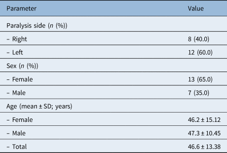
Table 1 presents the patients’ demographic characteristics. Same-side recurrent idiopathic peripheral facial paralysis was seen in 20 patients (13 females, 7 males; mean age 46.6 (range, 23–67) years). The patients described two to six attacks on the same side. Twelve patients suffered from left-side paralysis and eight from right-side paralysis. House–Brackmann classification scores ranged between two and five. The electrophysiological findings were compatible with acute or chronic axonal degeneration of the peripheral facial nerve.
Demographic characteristics of the patients

SD = standard deviation
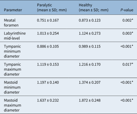
Comparing the diameters of the facial canal segments in each patient with the healthy side, we found that the diameters of labyrinthine segments measured at the meatal foramen and mid-levels were significantly smaller on the paralysed side than the healthy side (p = 0.002 and p = 0.003, respectively). The narrowest (minimum) and widest (maximum) diameters of the tympanic (p < 0.001 and p = 0.017, respectively) and mastoid (p < 0.001 and p < 0.001, respectively) segments were also significantly smaller on the paralysed side than on the healthy side (Table 2).
The measurements of facial canal segments on paralytic and healthy sides

*Statistical significance. SD = standard deviation
Discussion
The mechanism underlying idiopathic facial paralysis is suggested to be tissue oedema that leads to nerve compression.Reference May and Hardin13 However, the cause of the inflammation and the effects of segmental location of the compression remain controversial. Moreover, the effects of the size variability of the facial canal anatomy on nerve compression have not been documented. Because facial nerve palsy occurs via compression, it is reasonable to hypothesise that differences in the facial canal diameter may cause the development of paralysis. This study evaluated the diameters of the labyrinthine, mastoid and tympanic facial canal segments in patients with ipsilateral recurrent idiopathic peripheral facial paralysis using the curved planar reformation technique.
Ipsilateral recurrent idiopathic peripheral facial paralysis is a very rare entity with an unpredictable incidence. It is difficult to determine the recurrence rate and confirm its aetiology. The medical and drug treatment histories of all patients were reviewed from the hospital database to confirm the diagnosis of ipsilateral recurrent peripheral facial paralysis. A complete, careful assessment is performed for the differential diagnosis of peripheral facial paralysis, including blood analyses for neurotropic infections, electrophysiological studies of the facial nerve, and brain and temporal bone MRI with gadolinium enhancement. The patients were followed for at least six months to avoid possible misdiagnosis. To the best of our knowledge, this was the first prospective study to investigate the facial canal diameters in patients with ipsilateral recurrent idiopathic peripheral facial paralysis.
All causes (ischemia, infections, genetic, immunological and so forth) of Bell's palsy can be bilateral. However, there must be another explanation for why a patient has paralysis on one side rather than the other. For comparison, we chose the opposite canal as a benchmark to understand what the paralysis depends on.
Evaluating the dimensions of the facial canal segments using conventional axial, coronal and sagittal multiplanar reformatting images has some difficulties. These routine reformatted images may not be perpendicular to the longitudinal axis of the facial nerve canal. The curved planar reformation technique has been proven useful for displaying the entire length of tubular structures within a single image. It has been used to evaluate curved anatomical structures in clinical practice, such as vessels and ureter, airway, biliary, and carotid stents.Reference Hagino, Tsunoda, Tsunoda and Kishimoto10–Reference Dillman, Caoili, Cohan, Ellis, Francis and Schipper12,Reference Roos, Fleischmann, Koechl, Rakshe, Straka and Napoli14 The curved planar reformation image has a central track that is formed manually or automatically. This track enables more accurate measurements than with conventional multiplanar reformatting images. This curved planar reformation technique has not been used before for evaluating facial canal, and we postulated that it may be a feasible technique for measuring tiny differences in the facial canal segments.
Several studies have measured the diameter of facial canal segments in CT and compared the paralysed and healthy sides.Reference Watanabe, Sugai, Hosoya, Yamaguchi and Aoyagi15–Reference Murai, Kariya, Tamura, Doi, Kozakura and Okano19 Kefalidis et al. measured the canalicular entrance and middle part of the labyrinthine segment using CT for 25 patients with a history of Bell's palsy and found that the paralysed sides were significantly smaller than the healthy sides.Reference Kefalidis, Riga, Argyropoulou, Katotomichelakis, Gouveris and Prassopoulos16 Celik et al. examined the facial canal segments of 34 patients with a history of Bell's palsy and determined that narrowness of the labyrinthine segment may be an anatomic risk factor for facial paralysis.Reference Celik, Eskiizmir, Pabuscu, Ulkumen and Toker17 By contrast, Eksi et al. measured the labyrinthine, tympanic and mastoid segments of the facial canal and found no difference between the healthy and paralysed sides. All of these studies used conventional axial, coronal and sagittal multiplanar reformatting images for the measurements.Reference Eksi, Akbay, Bayarogullari, Cevik, Yengil and Ozler18
Murai et al. compared the cross-sectional area of the facial canal segments on the paralysed and healthy sides.Reference Murai, Kariya, Tamura, Doi, Kozakura and Okano19 The mean cross-sectional areas of the labyrinthine and tympanic segments of 16 patients with Bell's palsy were significantly smaller on the paralysed sides.
• Recurrent peripheral facial paralysis is uncommon in clinical practice
• Recurrent peripheral facial paralysis that recurs on the same side without a distinct pathology is extremely rare
• Anatomical narrowness in the facial canal segments may predispose patients to recurrences
• Curvilinear pathway of the facial canal contributes significant measurement difficulties
• The curved planar reformation is an appropriate technique for illustrating tortious structures
A post-mortem study of the facial nerve and facial canal demonstrated an anatomical difference in the diameter of the facial canal in the tympanic and mastoid but not in the labyrinthine segment in patients with Bell's palsy.Reference Vianna, Adams, Schachern, Lazarini, Paparella and Cureoglu20 Another study involving temporal bone examinations of cadavers found no significant difference in the diameter of the facial nerve or facial canal between the affected and unaffected sides in six patients with a history of unilateral Bell's palsy. However, on comparing results with a control group, the mean diameters of the labyrinthine, tympanic and mastoid segments were smaller in the Bell's palsy group.Reference Dawidowski, Branica, Batelja, Dawidowski, Kovac-Bilic and Simunic-Veselic21
There were several limitations to our study. First, in clinical practice the ipsilateral recurrence of recurrent idiopathic peripheral facial paralysis is rare; therefore, the study group was small. More patients with longer follow-up periods are needed to clarify our results. Second, the generation of the curved planar reformation images is not fully automated. The facial canal segments are marked manually, and it took a long time to measure the segments, which might cause minor errors.
Conclusion
The diameters at the meatal foramen, mid-level of the labyrinthine segment, and minimum and maximum diameters of the tympanic and mastoid segments were significantly smaller on the paralysed side than the healthy side according to curved planar reformation images of high-resolution CT of the temporal bone. This suggests that the narrowness of facial canal segments may be an important risk factor in the development of ipsilateral recurrent idiopathic peripheral facial paralysis.
These results need to be supported by further studies, including more case series. Facial nerve decompression may become an alternative treatment for patients unresponsive to corticosteroid therapy with high House–Brackman scores.
Competing interests
None declared