Introduction
In vitro culture of ovarian cortical tissue has become an important tool to elucidate the unknown mechanisms of early folliculogenesis (Jones and Shikanov, Reference Jones and Shikanov2019) and to support the development of the primordial follicle pool, to allow their future use in assisted reproductive techniques in humans and other mammalian species (Figueiredo et al., Reference Figueiredo, Cadenas, Lima and Santos2019). Moreover, experiments using in vitro ovarian cultures allow faster screening of the follicular response to different substances such as growth factors, hormones, antioxidants and/or reproductive toxicants (Stefansdottir et al., Reference Stefansdottir, Fowler, Powles-Glover, Anderson and Spears2014; Guerreiro et al., Reference Guerreiro, Lima, Rodrigues, Carvalho Ade, Castro, Campello, Pessoa Cdo, Gadelha, Figueiredo, Bordignon and Rodrigues2016, Reference Guerreiro, Mbemya, Bruno, Faustino, de Figueiredo and Rodrigues2019; Santos et al., Reference Santos, Lins, Barberino, Menezes, Gouveia and Matos2019). Improvements in in vitro follicle culture will therefore enhance current fertility preservation strategies (Bus et al., Reference Bus, Langbeen, Martin, Leroy and Bols2019). In this context, considerable research efforts have been undertaken to optimize the culture medium by supplying additional substances in order to reduce follicle apoptosis and to ensure optimum follicle growth. One potential candidate for medium supplementation is the hormone leptin.
Leptin, a 16 kDa adipokine primarily secreted by adipose tissue, was initially identified as a signal that regulates food intake and energy expenditure (Tsai et al., Reference Tsai, Asakawa, Amitani and Inui2012). In recent years, it has also been implicated in a wider range of physiological functions, including reproduction (Bilbao et al., Reference Bilbao, Di Yorio, Galarza, Varone and Faletti2015; Taskin et al., Reference Taskin, Kocabay, Ebrahimi, Karahuseyinoglu, Sahin, Ozcimen, Ruacan and Onder2019; Zieba et al., Reference Zieba, Biernat and Barć2020). In mammals, in vitro studies have demonstrated that leptin enhances the development of isolated preantral (ovine 10 ng/ml leptin: Kamalamma et al., Reference Kamalamma, Kona, Praveen Chakravarthi, Siva Kumar, Punyakumari and Rao2016; Kumar et al., Reference Kumar, Sivakumar, Pathipati, Chakravarthi, Brahmaiah and Rao2019; and ovine 25 ng/ml: Macedo et al., Reference Macedo, Santos, Bezerra, Menezes, Gouveia, Barbosa, Lins, Monte, Barberino, Batista, Barros, Wischral, Queiroz, Araújo and Matos2019) and early antral follicles (ovine 2 ng/ml: Menezes et al., Reference Menezes, Monte, Gouveia, Lins, Donfack, Macedo, Barberino, Santos, Matos, Batista and Wischral2019), improves the developmental capacity of the oocytes and increases blastocyst rates (bovine 1 and 10 ng/ml: Boelhauve et al., Reference Boelhauve, Sinowatz, Wolf and Fabíola2005; bubaline 10 and 50 ng/ml: Sheykhani et al., Reference Shafiei Sheykhani, Batavani and Najafi2016; 10 ng/ml: Panda et al., Reference Panda, Pandey, Somal, Parmar, Bhat, Baiju, Bharti, Sai Kumar, Chandra and Sharma2017; and swine 10 and 100 ng/ml: Craig et al., Reference Craig, Zhu, Dyce, Petrik and Li2004).
Some effects of leptin are mediated by its interaction with its receptor (LEPR) (Fruhbeck, Reference Fruhbeck2006). Both leptin and LEPR proteins have already been demonstrated in follicular cells in caprine (Batista et al., Reference Batista, Silva, Rêgo, Silva, Silva, Beltrão, Gomes Filho, Wischral and Guerra2013) and ovine (Macedo et al., Reference Macedo, Santos, Bezerra, Menezes, Gouveia, Barbosa, Lins, Monte, Barberino, Batista, Barros, Wischral, Queiroz, Araújo and Matos2019) species. The interaction between leptin and LEPR can activate the phosphatidylinositol-3-kinase/protein kinase B (PI3K/Akt) pathway (Aragonès et al., Reference Aragonès, Ardid-Ruiz, Ibars, Suárez and Bladé2016; Seoane-Collazo et al., Reference Seoane-Collazo, Martínez-Sánchez, Milbank and Contreras2020), which appears to be the main signalling pathway that regulates the growth and survival of primordial follicles (mice: Zhao et al., Reference Zhao, Ma, Sun, Ye, Zhang, Sun, Xu, Wang and Li2014; swine: Moniruzzaman et al., Reference Moniruzzaman, Lee, Zengyo and Miyano2010; ovine: Santos et al., Reference Santos, Lins, Barberino, Menezes, Gouveia and Matos2019). In a human ovarian carcinoma cell line (OVCAR-3), leptin stimulates cell growth through the PI3K pathway (Chen et al., Reference Chen, Chang, Lan and Breslin2013).
Despite the satisfactory results of leptin on isolated follicles, there has been no report of the effects of leptin on the in vitro culture of ovarian tissue in large mammals. Moreover, to optimize current culture systems for ovarian cortex, a better understanding of the signalling processes involved in follicle survival and activation is needed (Devos et al., Reference Devos, Grosbois and Demeestere2020). However, it is not known whether the PI3K/Akt pathway is involved in leptin effects on ovarian tissue. Therefore, the aims of this study were to analyse the effects of leptin on the survival, activation and in vitro growth of sheep primordial follicles cultured in ovarian cortical fragments and to verify if the PI3K/Akt signalling pathway is involved in leptin action.
Materials and methods
Unless noted otherwise, all chemicals used in this study were purchased from Sigma Aldrich Chemical Co. (St. Louis, MO, USA).
Source of ovarian tissue
Ovaries (n = 20) were collected at a local abattoir from 10 adult (1–3 years old) mixed-breed sheep for in vitro culture with different concentrations of leptin (n = 10 ovaries) or with the PI3K inhibitor (n = 10 ovaries). Immediately postmortem, the ovaries were washed once in 70% alcohol (Dinâmica, São Paulo, Brazil) followed by two washes in minimum essential medium buffered with HEPES (MEM–HEPES) and supplemented with antibiotics (100 μg/ml penicillin and 100 μg/ml streptomycin) and transported to the laboratory in MEM–HEPES at 4ºC (Chaves et al., Reference Chaves, Martins, Saraiva, Celestino, Lopes, Correia, Verde, Matos, Báo, Name, Campello, Silva and Figueiredo2008).
In vitro culture of ovarian tissue
In vitro culture was performed according to Santos et al. (Reference Santos, Lins, Barberino, Menezes, Gouveia and Matos2019). In the laboratory, ovarian cortex samples from each ovarian pair (n = 10 ovaries from five sheep) were cut into slices approximately 3 mm × 3 mm (1-mm thick) in size using a needle and scalpel under sterile conditions. For each animal, one tissue slice was randomly selected and fixed for histological analysis (fresh control). The remaining slices of the ovarian cortex were cultured individually in 1 ml of culture medium in 24-well culture dishes for 7 days at 39°C in humidified air containing 5% CO2. The base culture medium (control) consisted of α-MEM (pH 7.2–7.4) supplemented with 10 ng/ml insulin, 5.5 μg/ml transferrin, 5 ng/ml selenium, 2 mM glutamine, 2 mM hypoxanthine, 50 µg/ml ascorbic acid and 1.25 mg/ml bovine serum albumin (BSA), which is referred to as α-MEM+. To verify the influence of leptin on the in vitro culture of sheep ovarian cortex, fragments were cultured in α-MEM+ (control) or in α-MEM+ supplemented with different concentrations of human recombinant leptin (1, 5, 10, 25 or 50 ng/ml), which were chosen based on previous studies (Kammalama et al., Reference Kamalamma, Kona, Praveen Chakravarthi, Siva Kumar, Punyakumari and Rao2016; Macedo et al., Reference Macedo, Santos, Bezerra, Menezes, Gouveia, Barbosa, Lins, Monte, Barberino, Batista, Barros, Wischral, Queiroz, Araújo and Matos2019). The culture medium was replenished every 2 days. Each treatment was repeated five times, therefore using the ovaries of five different animals.
Morphological analysis and evaluation of in vitro follicular activation and growth
For morphological examination, tissues from the fresh control and from all cultured treatments were fixed in 10% buffered paraformaldehyde (Dinâmica) for 18 h, dehydrated using graded ethanol (Dinâmica), clarified in xylene (Dinâmica), and embedded in paraffin (Dinâmica). Haematoxylin and eosin (Vetec, São Paulo, Brazil) was used to stain 5-μm thick serial sections for routine histological examination under a light microscope (Nikon, Tokyo, Japan; ×400 magnification). The preantral follicles were classified as histologically normal when an intact oocyte was present and surrounded by granulosa cells that were well organized in one or more layers and had no condensed nuclear chromatin. Atretic follicles were defined as those with a shrunken or vacuolated oocyte, condensed chromatin, disorganized granulosa cells detached from the basement membrane, and/or cell swelling (Santos et al., Reference Santos, Lins, Barberino, Menezes, Gouveia and Matos2019). Blind follicle counting was performed on every fifth section of the ovaries by one experienced investigator.
For assessment of the follicular activation and growth (follicle and oocyte diameters), only histologically normal follicles with visible oocyte nucleus were recorded. The evaluation of follicular activation was performed by quantifying the follicles at different stages of development (Silva et al., Reference Silva, van den Hurk, Costa, Andrade, Nunes, Ferreira, Lôbo and Figueiredo2004), i.e. primordial (one layer of flattened granulosa cells around the oocyte) or growing follicles (intermediate: one layer of flattened to cuboidal granulosa cells; primary: one layer of cuboidal granulosa cells or secondary: two or more layers of cuboidal granulosa cells around the oocyte). The proportion of primordial and growing follicles was calculated at day 0 (fresh control) and after 7 days of culture. In addition, from the basement membrane, the major and minor axes of morphologically normal follicles and oocytes were measured using the Image-Pro Plus software (Media Cybernetics Inc.). The average of these two measurements was used to determine the diameters of the follicle and the oocyte, respectively.
Assessment of apoptosis by immunohistochemistry to active-caspase-3
After histological analysis, for a more in-depth evaluation of follicular quality, immunohistochemical analysis was performed in the fresh control and in treatments that showed the best results regarding follicular morphology. Immunohistochemistry was performed as described previously with some modifications. Briefly, sections (3-μm thick) from each block were cut using a microtome (EasyPath, São Paulo, Brazil) and mounted in Starfrost glass slides (Knittel, Braunschweig, Germany). The slides were incubated in citrate buffer (Dinâmica) at 95ºC in a decloaking chamber (Biocare, Concord, USA) for 40 min to retrieve antigenicity; endogenous peroxidase activity was prevented by incubation with 3% H2O2 (Easypath) for 10 min. Nonspecific binding sites were blocked using 1% normal goat serum (Easypath). Subsequently, the sections were incubated in a dark humidified chamber for 50 min at room temperature with rabbit polyclonal anti-activated caspase-3 antibody (1:100; Santa Cruz Biotechnology, Santa Cruz, CA, USA). Thereafter, the sections were incubated for 20 min in EasyLink One polymer (Easypath). Protein localization was demonstrated with diaminobenzidine (DAB; Easypath), and the sections were counterstained with haematoxylin (Vetec) for 1 min. Negative controls (reaction control) were performed in the absence of the primary antibody. Follicles were analyzed using the above-mentioned light microscope connected to a computer equipped with Image-Pro Plus® software (Media Cybernetics), and if the oocyte and/or 70% of the surrounding granulosa cells were positively stained (brown staining), the follicle was considered apoptotic (adaptation of Langbeen et al., Reference Langbeen, Ginneken, Fransen, Bosmans, Leroy and Bols2016). The percentage apoptosis was calculated as the number of apoptotic follicles out of the total number of follicles (×100).
Pharmacologic inhibition of the PI3K pathway
The aim was to test the hypothesis that pharmacological inhibition of the PI3K pathway would inhibit the actions of leptin on primordial follicle survival and activation in vitro. For this in vitro culture, additional pairs of sheep ovaries (n = 10 ovaries from five animals) were collected, transported to the laboratory, and fragmented as described above. Ovarian fragments were cultured for 7 days in α-MEM+ supplemented with 1 ng/ml leptin (concentration that showed the best results in the previous culture) in the absence or the presence of the PI3K inhibitor. For PI3K inhibition, the manufacturer recommended treating the cultured cells with LY294002 for 1 h before and for the duration of the stimulation. Therefore, 50 µM of the PI3K inhibitor LY294002 (Cell Signalling Technologies, Danvers, MA) was added to the α-MEM+ for 1 h at 39°C under 5% CO2. Next, the medium was supplemented with leptin. Total replacement of the medium was performed every 2 days for the culture in the absence or the presence of the PI3K inhibitor (the inhibitor was also replaced). The LY294002 concentration (50 μM) was chosen according to Adhikari et al. (Reference Adhikari, Risal, Liu and Shen2013). Each treatment was repeated five times. After this culture, the ovarian tissue was fixed, processed and used for histological (morphology and activation) and immunohistochemical (apoptosis) analyses as described above.
Evaluation of p-AKT immunostaining
To check the effectiveness of LY294002 in inhibiting the PI3K pathway and considering that Akt is one of the main downstream targets of PI3K (Cecconi et al., Reference Cecconi, Mauro, Cellini and Patacchiola2012), Akt phosphorylation was evaluated after 7 days of culture with 1 ng/ml leptin in the absence or presence of LY294002. Immunohistochemistry was carried out as described above, but the slides were incubated with a rabbit polyclonal anti-p-Akt antibody (1:40; Santa Cruz Biotechnology) instead of the anti-activated caspase-3 antibody. Immunostaining was subjectively classified as absent, weak, moderate or strong using a microscope (Nikon) under ×400 magnification (Santos et al., Reference Santos, Lins, Barberino, Menezes, Gouveia and Matos2019).
Statistical analysis
The percentages of morphologically normal, apoptotic, primordial and growing follicles were compared by chi-squared test. Data of follicle and oocyte diameters were submitted to Shapiro–Wilk test to verify normal distribution of residues and homogeneity of variances. Thereafter, data were submitted to the Kruskal–Wallis non-parametric test and the Student–Newman–Keuls test for comparisons among treatments. The results were expressed as the mean ± standard error of mean (SEM). Differences were considered to be statistically significant when P < 0.05. Data were analyzed using the BioEstat 5.0 procedure.
Results
Follicular morphology and development after in vitro culture
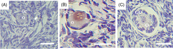
Overall, normal (Fig. 1 A) and atretic (Fig. 1 B) follicles were observed in all treatments. After 7 days of culture, the percentage of morphologically normal follicles decreased significantly in all treatments compared with the fresh control (82%; Fig. 1 C). However, the control medium (56%) and the medium containing 1 ng/ml leptin (57%) had similar (P > 0.05) percentages of normal follicles, which were significantly higher than those observed for other treatments (39, 38, 39 and 36% for 5, 10, 25 and 50 ng/ml, respectively).
Histological sections of ovine ovarian fragments. (A, B) Normal follicle after in vitro culture in medium containing 1 ng/ ml leptin (A) and atretic follicle after culture in 50 ng/ ml leptin (B). GC, granulosa cells; O, oocyte; Scale bars: 30 μm (×400). (C) Percentages of ovine morphologically normal follicles in the fresh control and after 7 days of in vitro culture in α-MEM+ or in different concentrations of leptin. *Differs significantly from the fresh control (P < 0.05). A,BDifferent letters denote significant differences among the treatments (P < 0.05).

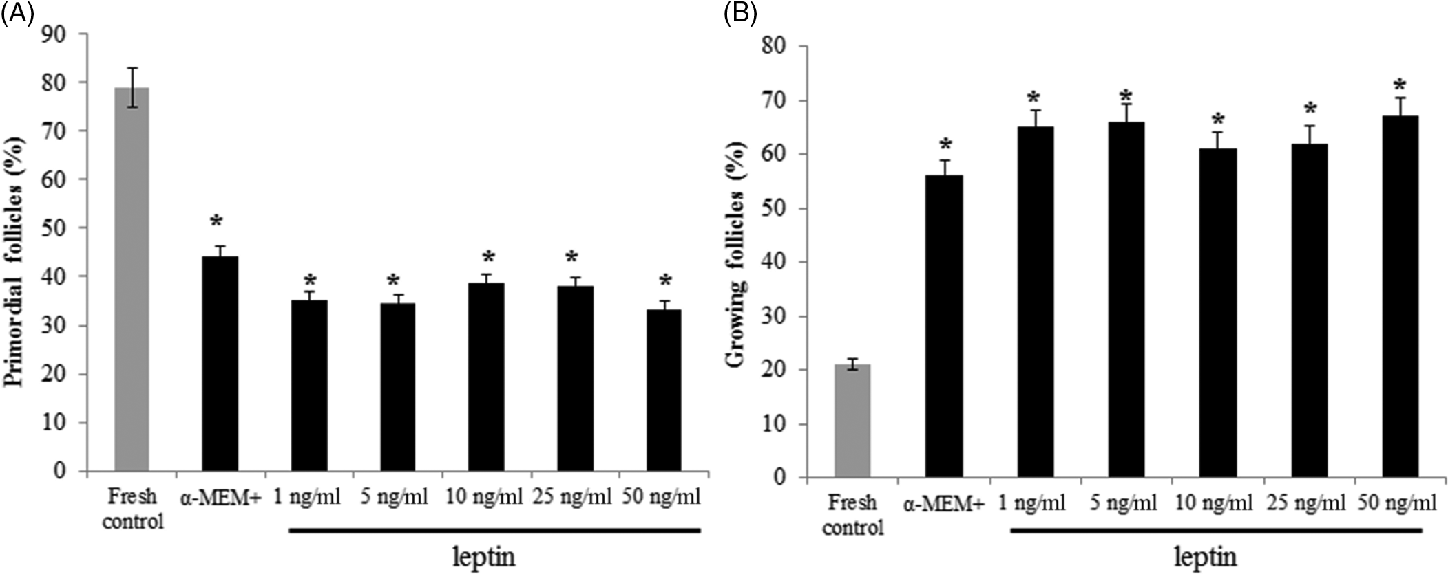
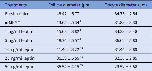
After in vitro culture, there was a significant reduction in the percentage of primordial follicles (Fig. 2 A) and an increase in the percentage of growing follicles (Fig. 2 B) in all treatments compared with the fresh control. However, no difference (P > 0.05) in the follicular activation was observed between the control medium (α-MEM+) and leptin-treated groups. As shown in Table 1, a significant reduction in the follicle diameter was observed when the ovarian tissues were cultured in medium containing 10, 25 or 50 ng/ml leptin compared with the fresh control and other treatments. Additionally, there was no significant influence of leptin on the oocyte diameter.
Percentages of primordial (A) and growing (B) follicles in the fresh control and after 7 days of in vitro culture in α-MEM+ or with different concentrations of leptin. *Differs from fresh control (P < 0.05).

Follicle and oocyte diameters (mean ± SEM) in the fresh control and after in vitro culture of ovine ovarian tissue in different concentrations of leptin

* Differs significantly from fresh control (P < 0.05).
A,B Different letters denote significant differences among treatments (P < 0.05).
Follicular apoptosis
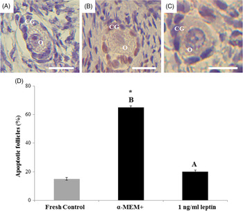
Immunohistochemical analysis for apoptosis was performed in the fresh control (Fig. 3 A) and in treatments that showed the best results for the percentage of morphological normal follicles, i.e. α-MEM+ (Fig. 3 B) and medium containing 1 ng/ml leptin (Fig. 3 C). The percentage of apoptotic follicles was similar (P > 0.05) between 1 ng/ml leptin (20%) and the fresh control (15%), and both groups had less apoptosis (P < 0.05) than that observed in the control medium (65%; Fig. 3 D).
Immunohistochemical analysis for the expression of activated caspase-3 in ovine preantral follicles. (A–C) Normal follicle in the fresh control (A), apoptotic follicle (brown staining granulosa cells) cultured in α-MEM+ (B), and normal follicle cultured in medium containing 1 ng/ml leptin. GC: granulosa cells; O: oocyte. Scale bars: 25 μm (×400). Percentages of apoptotic follicles in the fresh control and after 7 days of culture in α-MEM+ or in 1 ng/ml of leptin. *Differs significantly from the fresh control (P < 0.05). A,BDifferent letters denote significant differences among the treatments (P < 0.05).

Follicular morphology, activation and apoptosis after inhibition of the PI3K pathway
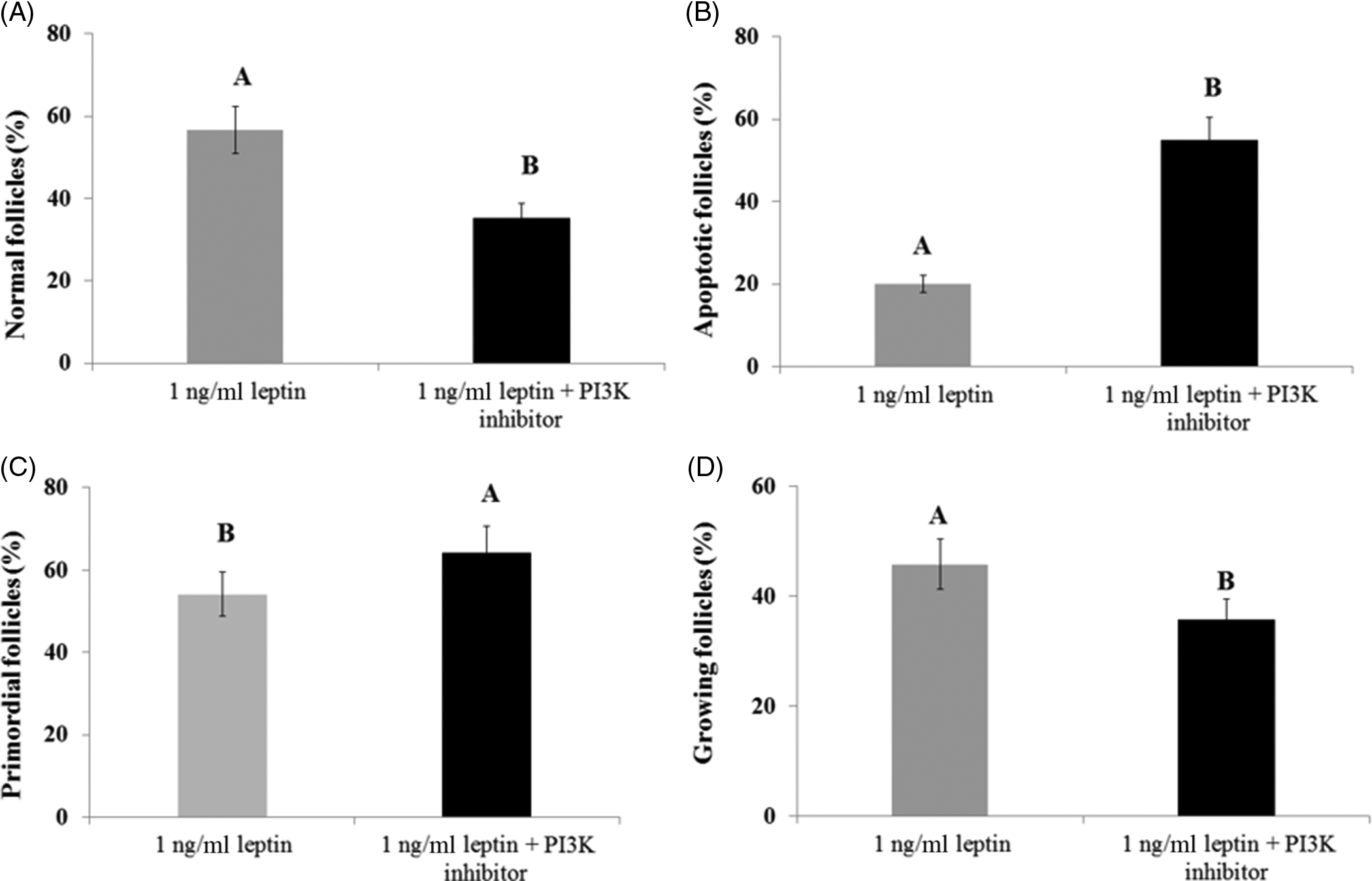
The PI3K pathway inhibition was performed in follicles cultured with 1 ng/ml leptin because this treatment had less apoptosis than control medium (α-MEM+). In vitro culture of ovine ovarian tissue in 1 ng/ml leptin with LY294002 decreased significantly the percentage of normal follicles (35.33%; Fig. 4 A) and increased significantly the percentage of apoptosis (55%; Fig. 4 B) compared with the medium containing 1 ng/ml leptin without PI3K inhibitor (56.67% and 20% for normal and apoptotic follicles, respectively). Moreover, the culture of ovine ovarian tissue with 1 ng/ml leptin and LY294002 significantly inhibited the in vitro activation of primordial follicles compared with medium without the inhibitor (Fig. 4 C, D).
Percentages of morphologically normal (A), apoptotic (B), primordial (C) and growing follicles (D) after in vitro culture in 1 ng/ml leptin in the absence or presence of LY294002 (PI3K inhibitor). A,BDifferent letters denote significant differences among the treatments (P < 0.05).

Immunohistochemistry for Akt phosphorylation (p-Akt) after PI3K inhibition
The negative control did not show staining for phosphorylated Akt (Fig. 5 A). After in vitro culture with 1 ng/ml leptin without LY294002, a moderate immunostaining for p-Akt was observed in the oocytes (Fig. 5 B). Nevertheless, after PI3K inhibition, p-Akt immunostaining was absent (Fig. 5 C).
Immunohistochemical expression of p-AKT in the sheep ovary: follicles in 1 ng/ml leptin in the absence of LY294002 (A) or in the presence of LY294002 (PI3K inhibitor; B); Negative control (C). GC: granulosa cells; O: oocyte. Scale bars: 25 μm (×400).

Discussion
In recent years, some in vitro studies have demonstrated the beneficial effects of leptin on isolated ovarian follicle development, oocyte maturation and embryo production (Jin et al., Reference Jin, Cui, Han and Kim2009; Kamalamma et al., Reference Kamalamma, Kona, Praveen Chakravarthi, Siva Kumar, Punyakumari and Rao2016; Keshrawani et al., Reference Keshrawani, Aruna Kumari and Reddy2016; Macedo et al., Reference Macedo, Santos, Bezerra, Menezes, Gouveia, Barbosa, Lins, Monte, Barberino, Batista, Barros, Wischral, Queiroz, Araújo and Matos2019; Menezes et al., Reference Menezes, Monte, Gouveia, Lins, Donfack, Macedo, Barberino, Santos, Matos, Batista and Wischral2019). It is worth noting that in vitro culture of ovarian cortical tissue is an important tool to elucidate the regulatory mechanism and the signalling pathways involved in the activation of primordial follicles, which are the most predominant type of follicle in the ovary (Shea et al., Reference Shea, Woodruff and Shikanov2014; Telfer, Reference Telfer2019). Our study showed for the first time the effects of leptin on the follicles cultured within ovarian tissue slices.
In this study, ovarian tissue cultured in the control medium (α-MEM+) or 1 ng/ml leptin resulted in greater percentages of histologically normal follicles than those found in other leptin concentrations. Although histological analysis remains the most classic method to determine the follicular atresia, the detection of early apoptotic changes may be beyond the scope of histological assessment (Bedaiwy and Hussein, Reference Bedaiwy and Hussein2004). Therefore, the use of a more sensitive method to evaluate cell quality is recommended. Using immunohistochemical analysis, we demonstrated that 1 ng/ml leptin was effective in preventing follicular apoptosis (only 20%, which was similar to that of the fresh control) compared with α-MEM+ (65%). Previous studies have shown the anti-apoptotic activity of leptin on bubaline cumulus–oocyte complexes (Panda et al., Reference Panda, Pandey, Somal, Parmar, Bhat, Baiju, Bharti, Sai Kumar, Chandra and Sharma2017), on bovine blastocysts from in vitro-fertilized oocytes (Boelhauve et al., Reference Boelhauve, Sinowatz, Wolf and Fabíola2005) and human trophoblast cells triggered by high temperature (Pérez-Pérez et al., Reference Pérez-Pérez, Toro, Vilarino-Garcia, Guadix, Maymó, Dueñas, Varone and Sánchez-Margalet2016). It is likely that leptin is exerting its anti-apoptotic effects by interacting with its receptor (LEPR), which is expressed in sheep preantral follicles (Macedo et al., Reference Macedo, Santos, Bezerra, Menezes, Gouveia, Barbosa, Lins, Monte, Barberino, Batista, Barros, Wischral, Queiroz, Araújo and Matos2019) and/or by enhancing the intracellular levels of antioxidant enzymes and decreasing the levels of inflammatory factors (Zwirska-Korczala et al. Reference Zwirska-Korczala, Adamczyk-Sowa, Sowa, Pilc, Suchanek, Pierzchala, Namyslowski, Misiolek, Sodowski, Kato, Kuwahara and Zabielski2007; Bilbao et al., Reference Bilbao, Di Yorio, Galarza, Varone and Faletti2015). In our study, as the ovaries from the fresh control had more morphologically normal follicles than those cultured in 1 ng/ml leptin and similar percentages of apoptotic follicles, it could be speculated that follicles cultured in medium containing 1 ng/ml leptin are likely to undergo both necrosis and apoptosis, and that leptin is protecting the follicles from cell death by apoptosis. Considering that leptin is produced by oocytes from ovine primordial and primary follicles (Macedo et al., Reference Macedo, Santos, Bezerra, Menezes, Gouveia, Barbosa, Lins, Monte, Barberino, Batista, Barros, Wischral, Queiroz, Araújo and Matos2019), we believe that the addition of a lower concentration of leptin (1 ng/ml) to the medium is sufficient to maintain satisfactory follicle survival and to prevent apoptosis in small follicles.
Interestingly, the decrease in the percentage of normal follicles after in vitro culture in 10, 25 or 50 ng/ml leptin was associated with a reduction in follicle diameter, which is indicative of follicle degeneration (Lunardi et al., Reference Lunardi, de Aguiar, Apolloni, Duarte, de Sá, Leal, Sales, Lobo, Campello, Smitz, Apgar, de Figueiredo and Rodrigues2017). Another study has shown that exposure to high leptin concentrations inhibited human granulosa cell proliferation and increased caspase-3 mRNA levels (Lin et al., Reference Lin, Wang, Wu, Ullah, Yu, Ur Rahman and Huang2017). Moreover, in vitro studies showed that rat ovarian tissue exposed to high concentrations of leptin (100 ng/ml) inhibited superoxide dismutase (SOD) activity compared with the control tissues (Bilbao et al., Reference Bilbao, Di Yorio, Galarza, Varone and Faletti2015). Therefore, we suggest that leptin may have a concentration-dependent activity in follicular survival and development. At high concentrations, leptin seems to have a pro-oxidant effect and to be involved in cell death.
In the current study, all treatments promoted the activation of primordial follicles compared with the fresh control. The initiation of follicle growth starts with the differentiation of flattened granulosa cells into cuboidal, followed by their proliferation. Subsequently, the oocyte grows in size (Picton, Reference Picton2001; Braw-Tal, Reference Braw-Tal2002; Zhang et al., Reference Zhang, Risal, Gorre, Busayavalasa, Li, Shen, Bosbach, Brännström and Liu2014). Although our findings showed that leptin did not influence the percentage of growing follicles or the oocyte diameter compared with the control medium (α-MEM+), we did not evaluate if leptin had a positive effect on the number of granulosa cells after in vitro culture, and this should be performed in future studies. It could be speculated that the period of culture of sheep ovarian tissue or the concentration of leptin used may not be adequate to promote an apparent increase in follicle activation and oocyte growth.
Considering that leptin maintains survival and promotes follicle growth by the PI3K/Akt pathway (Cirillo et al., Reference Cirillo, Rachiglio, la Montagna, Giordano and Normanno2008; Ghasemi et al., Reference Ghasemi, Saeidi, Azimi-Nejad and Hashemy2019), we hypothesized that the PI3K/Akt pathway would be involved in ovine follicular survival and activation in response to leptin. In addition, as Akt is one of the main targets of PI3K (Cecconi et al., Reference Cecconi, Mauro, Cellini and Patacchiola2012), it is important to note the direct effect of LY294002 on Akt phosphorylation (Gharbi et al., Reference Gharbi, Zvelebil, Shuttleworth, Hancox, Saghir, Timms and Waterfield2007). Our results showed that the expected inhibition of Akt phosphorylation after culture in 1 ng/ml leptin + LY294002 was associated with an increase in follicular apoptosis and inhibition of primordial follicle activation. Akt modulates granulosa cell apoptosis (Zhou et al., Reference Zhou, Peng and Mei2019) and is a marker of primordial follicle activation (Reddy et al., Reference Reddy, Adhikari, Zheng, Liang, Hämäläinen, Tohonen, Ogawa, Noda, Volarevic, Huhtaniemi and Liu2009; Santos et al., Reference Santos, Lins, Barberino, Menezes, Gouveia and Matos2019). One in vitro study has suggested that the PI3K/Akt pathway may be involved in the proliferative and anti-apoptotic effects of leptin on goose granulosa cells (Wen et al., Reference Wen, Hu, Xiao, Han, Gan, Gou, Liu, Li, Xu, He and Wang2015). Therefore, although other pathways downstream may also come into play, we suggest that leptin reduces follicular apoptosis and stimulates in vitro activation of primordial follicles in sheep possibly through the activation of the PI3K/AKT pathway.
In conclusion, a 1 ng/ml leptin concentration reduced follicular apoptosis after in vitro culture of sheep ovarian cortical tissue. In addition, the PI3K/AKT pathway is possibly involved in the effects of leptin on survival and activation of sheep primordial follicles.
Declaration of interest
None of the authors have any conflict of interest to declare.
Financial support
This work was supported by FACEPE (Process APQ-0895–5.05/14). T.J.S. Macedo holds a scholarship from the FACEPE. A. Wischral, M.A.A Queiroz, G.G.L. Araújo and M.H.T. Matos are supported by a grant from CNPq. A.M. Batista is supported by a grant from CAPES.
Ethical standards
Not applicable.