Case report
A male fetus was diagnosed at 21 weeks gestation with critical aortic stenosis by fetal echocardiography. The criterions for diagnosis included a dilated and dysfunctional left ventricle, with retrograde flow around the aortic arch, and reversed shunting from left to right across the patent oval foramen. Antegrade flow across the dysplastic aortic valve was virtually undetectable, with the ascending aorta noted to be markedly hypoplastic in the absence of any poststenotic aortic dilation. Despite normal morphology of the mitral valve, which showed moderate to severe regurgitation, shunting across the oval foramen was unequivocally from left to right. After obtaining informed consent, and under a protocol approved by our Institutional Review Board, we performed aortic valvotomy at 23 weeks gestation under transabdominal echocardiographic guidance. The fetus weighed 430 grams at the time of the intervention. The mother received sedation and local anesthesia, while the fetal position was manipulated so that the thorax faced forward. Once an ideal position was attained, a trochar of 14 gauge was inserted through the uterine wall. The fetus received a combination of fetanyl at 2 micrograms per kilogram, vecuronium at 1 microgram per kilogram, and atropine at 20 micrograms per kilogram through a 25-gauge needle passed intramuscularly through the trochar. An 18-gauge needle with a blunt tip was then inserted through the trochar and across the fetal thorax and the left ventricular apex, aiming towards the aortic valve using echocardiographic guidance. As emphasized already, we had not previously detected any antegrade flow across the aortic valve, either by colour Doppler or pulse wave interrogation. Thus, we could not determine with precision whether there was complete aortic atresia as opposed to severe aortic stenosis. Due to this, we decided to use fibreoptic technology to visualize the abnormal ventricular outflow tract. A fibreoptic cardioscope of 0.8 millimetres dimensions was inserted through the needle into the left ventricle. Through the cardioscope, we noted whitish endocardial fibroelastosis in the left ventricular endocardium, as well as a dysplastic aortic valve with a pinhole opening. The normal mitral valvar apparatus was also visualized (Fig. 1). The diameter of the hypoplastic aortic valve was measured at 2.7 millimetres. After having confirmed the patency of the aortic valve, we entered the valvar orifice using a 014 guidewire, following this with a coronary angioplasty balloon measuring 3.7 millimetres by 1 centimetre. The balloon was positioned across the valvar mechanism, and inflated twice, retaining fetal echocardiographic guidance. The balloon was chosen to be 135% of the measured size of the diameter of the aortic valve. After withdrawal of the balloon, we noted immediate antegrade flow across the aortic valve, as well as a moderate degree of aortic regurgitation. Although minimal, there was an immediate improvement in the left ventricular systolic function and a decrease in the severity of mitral regurgitation. Over the ensuing 3 weeks, the aortic regurgitation progressively decreased, disappearing completely by the fourth week. The aortic stenosis, however, became worse as aortic valvar regurgitation was resolving. There was no further regression in the hypoplasia of the left ventricle, but the echogenicity of the fibroelastotic endocardial surface became more evident. The fetus was delivered at term, having a dysplastic and critically stenotic aortic valve, with severe left ventricular dysfunction. He underwent postnatal aortic balloon valvotomy, with mild residual aortic stenosis and regurgitation. Although the systolic function had completely normalized, and prostaglandins were discontinued successfully after the valvotomy, the aortic stenosis became recurrent, requiring two subsequent balloon valvotomies. Each valvotomy resulted in increasing aortic regurgitation, and thus increased left ventricular end-diastolic volume (Fig. 2). The aortic regurgitation, along with the persisting fibroelastosis, caused significant left ventricular diastolic dysfunction. A pseudoaneurysm also developed in the left ventricular outflow tract following his last balloon valvotomy. Due to these complications, the patient underwent replacement of the aortic root combined with a Konno procedure, siting the Konno incision at the location of the pre-existing pseudoaneurysm in the left ventricular outflow tract. A concurrent endocardial resection of the abnormal fibrotic tissues in the left ventricle was also performed (Fig. 3). Postoperatively, there was significant improvement in the left ventricular diastolic function, as reflected by an immediate decrease in the left atrial pressure. The postoperative echocardiogram demonstrated normal biventricular function and physiology (Fig. 4). Despite achieving normal biventricular physiology, with both normal systolic and diastolic ventricular function, the patient eventually succumbed to sepsis, and died at 4 months of age.

Figure 1. A pinhole opening in the aortic valve is shown as seen through the cardioscope inserted via the left ventricle in this fetus of 23 weeks gestation.

Figure 2. The left ventricular end-diastolic volume is compared for balloon aortic valvotomy performed before and after birth. The increase in volume resulted from allowing antegrade flow across the aortic valve, and from aortic regurgitation.

Figure 3. In Panel (a), we show surgically resected fragments of left ventricular endocardium, with Panel (b) showing the endocardial fibroelastosis revealed subsequent to staining with hematoxylin and eosin.

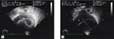
Figure 4. Echocardiographic four chamber views are shown comparing the size of the left ventricle immediately after birth (a) with the situation seen subsequent to the endocardial resection (b).
Discussion
The object of prenatal aortic valvotomy is to preserve the potential for left ventricular growth, hopefully preventing the development of hypoplastic left heart syndrome. It is argued that antegrade flow across the left ventricular outflow tract during fetal development, in face of normal flow across the mitral valve, permits the left ventricle to develop and grow more normally during the second and the third trimesters. There are some accounts of biventricular physiology being achieved postnatally in patients subsequent to fetal aortic valvotomy. All reported cases, however, to the best of our knowledge, have required postnatal catheter or surgical interventions, such as repeat balloon valvotomy, repair of coarctation, or even conversion of the physiology of the hypoplastic left heart syndrome into a biventricular physiology by taking down the first stage of Norwood palliation or reconstruction of a bidirectional Glenn or hemi-Fontan procedure.11 Despite having an adequate left ventricular end-diastolic volume and normal systolic function, biventricular physiology cannot be attained without preserving adequate diastolic function.
There are two components of diastolic function: an active phase of relaxation and a passive phase of compliance. In the setting of diastolic dysfunction, the level of sarcoplasmic endoreticulum calcium activase activity is decreased in the active phase. This, in turn, decreases the removal of calcium from the cytosol. Decreased enzymic activity is found in patients with ventricular hypertrophy, which may be due to the presence of systemic hypertension or an obstructive lesion, such as aortic stenosis or coarctation of the aorta. The second phase is the passive phase of compliance. In this phase, diastolic dysfunction can result from increased stiffness of the ventricle, such as produced by endocardial fibroelastosis, which replaces the normal endocardial or myocardial tissues with fibrotic collagen.12, 13 Surgical resection of the accompanying endocardial fibroelastosis, therefore, may play an important role in alleviating the passive phase of dysfunction.
Our experience in collaboration with the group from Boston supports the notion that fetal intervention can prevent the development of hypoplasia of the left heart in increasing numbers of patients as more technically successes are achieved.12, 13 Resection of fibroelastosis in afflicted patients may also be a determining factor in attaining normal diastolic function. This then can result in maintaining the desired biventricular physiology. Although fetal intervention is increasingly successful from the technical stance, the goal of achieving biventricular physiology remains dependent on two key issues, namely, the appropriate selection of patients and the timing of the intervention. As yet, we can only speculate that we are changing the natural history of hypoplastic left heart syndrome, and that genetic predisposition is being challenged.
Acknowledgment
Dr James Huhta was supported by the Daicoff-Andrews Chair in Perinatal Cardiology, University of South Florida, and All Children's Foundations.