INTRODUCTION
Leishmania, an intracellular parasite of macrophages, is responsible for over 12 million leishmaniasis cases in more than 98 countries (de Vries et al. Reference de Vries, Reedijk and Schallig2015). Depending on the species, the protozoan can promote different clinical manifestations that are associated with cutaneous and visceral pathologies (Adade et al. Reference Adade, Carvalho, Tomaz, Costa, Godinho, Melo, Lima, Rodrigues, Zingali and Souto-Padrón2014). Leishmania (L.) amazonensis is an important agent that induces cutaneous leishmaniasis and is responsible for the inhibition of the immune system, which promotes a high rate of parasite proliferation (Pereira and Alves, Reference Pereira and Alves2008).
Macrophage activation was formerly termed as simply ‘activated’, but is currently divided into two major profiles: classically activated, or M1, and alternatively activated macrophages, or M2. As regards their characteristics, M1 macrophages are effector cells during the type 1 T helper (Th1)-type immune response and are differentiated into this profile by activation stimuli, such as the presence of interferon (INF)-γ and lipopolysaccharide (LPS) (Mosser, Reference Mosser2003). These M1 cells have markers that distinguish them from other profiles, such as the high production of cytokines interleukin (IL)-12, IL-23, IL-6, tumour necrosis factor (TNF)-α and microbicidal substances such as nitric oxide (NO) and reactive oxygen species (ROS), besides morphological alterations in cell spreading and formation of cytoplasmic projections, providing the ability to react against intracellular parasites and tumour cells (Cassado et al. Reference Cassado, de Albuquerque, Sardinha, Buzzo, Faustino, Nascimento, Ghosn, Lima, Alvarez and Bortoluci2011; Xuan et al. Reference Xuan, Qu, Zheng, Xiong and Fan2015).
In contrast, M2 macrophages are activated during the Th2 lymphocyte response, being stimulated by IL-4, aiming to repair and heal tissue (Cassado et al. Reference Cassado, de Albuquerque, Sardinha, Buzzo, Faustino, Nascimento, Ghosn, Lima, Alvarez and Bortoluci2011). These macrophages are generally characterized by the high production of IL-10 and decreased productions of IL-12 and IL-23. M2 macrophages are characterized by producing low or no NO and ROS, characterizing a reduced microbicidal capacity (Liu et al. Reference Liu, Zou, Chai and Yao2014; Bashir et al. Reference Bashir, Sharma, Elahi and Khan2016).
The drugs that are currently indicated for the treatment of leishmaniasis are of high costs and present high toxicity, and have considerable side-effects. The first line of treatment uses pentavalent antimonials, and when this approach fails in a patient, second-line drugs are employed, such as amphotericin B or pentamidine. These therapies induce adverse reactions, with the most commonly reported adverse reactions being nephrotoxicity and heart failure, respectively (McGwire and Satoskar, Reference McGwire and Satoskar2014).
Given the limited efficacy of the current drugs available to treat Leishmania, the development of alternative treatments is critical. Natural products have been identified as critical substances during drug discovery processes, and they are the basis for most first drugs used (Butler, Reference Butler2008; Schepetkin et al. Reference Schepetkin, Khlebnikov, Kirpotina and Quinn2015).
Animal venoms and their purified compounds are known to exhibit a variety of pharmacological activities, including activities against pathogens in vitro (Quintana et al. Reference Quintana, Chacón, Vargas, Segura, Gutiérrez and Alarcón2012). In addition, snake venom proteins have been widely investigated in different areas of the life sciences. The interactions of these compounds with different cells can involve a variety of mechanisms that can result in various cellular responses, leading to the activation or blocking of physiological functions in the cell (Marcinkiewicz, Reference Marcinkiewicz2013). The venom of the South American rattlesnake Crotalus durissus terrificus (Cdt) is reported to have various biological effects, ranging from anti-inflammatory to antitumoural activities (Sampaio et al. Reference Sampaio, Brigatte, Sousa-e-Silva, dos-Santos, Rangel-Santos, Curi and Cury2003; Costa et al. Reference Costa, Faiad, Landgraf, Ferreira, Brigatte, Curi, Cury and Sampaio2013). Cdt crude venom is composed of gyroxin, crotamine, convulxin and crotoxin (CTX), which have proteolytic, analgesic, coagulative and neurotoxic effects, respectively (Passero et al. Reference Passero, Tomokane, Corbett, Laurenti and Toyama2007). CTX is the major toxic component of this venom and is composed of a non-covalent association between crotapotin, an acidic subunit, with phospholipase A2 (PLA2), a basic subunit.
Several studies have demonstrated the antileishmanial activity of PLA2 derived from the venom of diverse snake species (Torres et al. Reference Torres, Dantas, Toyama, Filho, Zara, Rodrigues de Queiroz, Pinto Nogueira, Rosa de Oliveira, de Oliveira Toyama, Monteiro and Martins2010; Nunes et al. Reference Nunes, Figueira, Lopes, De Souza, Izidoro, Ferro, Souza, Rodrigues, Rodrigues and Yoneyama2013; de Moura et al. Reference de Moura, Kayano, Oliveira, Setúbal, Ribeiro, Barros, Nicolete, Moura, Fuly, Nomizo, da Silva, Fernandes, Zuliani, Stábeli, Soares and Calderon2014; Barros et al. Reference Barros, Pereira, Barros, L., Calvi, Santos, Barraviera and Ferreira2015). Additional studies have used venoms derived from Bothrops mattogrossensis, Bothrops pauloensis, Bothrops lutzi, Philodryas patagoniensis and Cdt, which have also been reported to have antileishmanial activity (Passero et al. Reference Passero, Tomokane, Corbett, Laurenti and Toyama2007, Reference Passero, Laurenti, Tomokane, Corbett and Toyama2008; Peichoto et al. Reference Peichoto, Tavares, DeKrey and Mackessy2011; Nunes et al. Reference Nunes, Figueira, Lopes, De Souza, Izidoro, Ferro, Souza, Rodrigues, Rodrigues and Yoneyama2013; Adade et al. Reference Adade, Carvalho, Tomaz, Costa, Godinho, Melo, Lima, Rodrigues, Zingali and Souto-Padrón2014). However, few studies have studied the effects of the whole CTX molecule against Leishmania parasites, and most of these have demonstrated the effects of CTX that are related to the growth of the promastigote forms (Passero et al. Reference Passero, Tomokane, Corbett, Laurenti and Toyama2007). There is currently no evidence of the activity of CTX, derived from Cdt, during macrophage–L. amazonensis interactions. Moreover, following on from findings showing that CTX obtained from Cdt can selectively modulate secretory activity in macrophages, by reinforcing the immunomodulatory actions of this toxin (Costa et al. Reference Costa, Faiad, Landgraf, Ferreira, Brigatte, Curi, Cury and Sampaio2013), we demonstrate in this study, for the first time, the immunomodulatory activity of CTX during macrophage–L. amazonensis interactions.
MATERIALS AND METHODS
CTX isolation
CTX (CTX-complex) was obtained from the lyophilized venom of Cdt, which was supplied by the Laboratory of Herpetology, Butantan Institute, São Paulo, Brazil, and maintained at −20 °C. A crude venom solution was subjected to anion-exchange chromatography as previously described in Rangel-Santos et al. (Reference Rangel-Santos, Dos-Santos, Lopes-Ferreira, Lima, Cardoso and Mota2004) using a Mono-Q HR 5/5 column in an fast protein liquid chromatography system (Pharmacia, Uppsala, Sweden). The fractions (1 mL min−1) were eluted using a linear gradient of NaCl (0–1 mol L−1 in 50 mmol L−1 Tris-HCl, pH 7·0). Three peaks (p1, p2 and p3) were obtained: p2 corresponded to the pure CTX fraction (approximately 60% of the crude venom), whereas peaks 1 and 3 included the other Crotalus durissus terrificus venom (CdtV) toxins. Prior to pooling, the fractions containing CTX were tested for homogeneity using non-reducing sodium dodecyl sulphate-polyacrylamide gel electrophoresis (12·5%) (Laemmli, Reference Laemmli1970), and PLA2 activity was assessed using a colorimetric assay with a synthetic chromogenic substrate (Lôbo de Araújo and Radvanyi, Reference Lôbo de Araújo and Radvanyi1987).
Murine macrophage isolation
Macrophages were harvested from the peritoneal cavities of male BALB/c mice into Dulbecco's modified Eagle's medium (DMEM, pH 7·2) and incubated at 37 °C in an atmosphere containing 5% CO2 for 1 h. Non-adherent cells were washed away with phosphate-buffered saline (PBS, pH 7·2), and the remaining macrophages were then maintained in DMEM supplemented with 10% fetal bovine serum (FBS – Gibco® Thermo Fisher Scientific) at 37 °C in 5% CO2 for 24 h.
Murine macrophage cytotoxicity assays
Macrophages (106 macrophages in 1 mL of medium per well) were seeded in 24-well tissue culture plates during the adhesion step. The cells were then treated with CTX at concentrations ranging from 0·3 to 9·6 µg mL−1 at 37 °C in 5% CO2 for 48 h. Subsequently, cell supernatants were collected, and the cells were washed with PBS (pH 7·2). These concentrations of CTX are the same as those used in previous studies (Sampaio et al. Reference Sampaio, Brigatte, Sousa-e-Silva, dos-Santos, Rangel-Santos, Curi and Cury2003, Reference Sampaio, Santos, Costa, Rangel-Santos, Carneiro, Curi and Cury2006a , Reference Sampaio, Alba-Loureiro, Brigatte, Landgraf, dos Santos, Curi and Cury b ; Costa et al. Reference Costa, Faiad, Landgraf, Ferreira, Brigatte, Curi, Cury and Sampaio2013). The cytotoxicity of CTX was evaluated using MTT [3-(4,5-dimethylthiazol-2-yl)-2,5-diphenyl tetrazolium bromide] assays in murine macrophage cultures. The cells were washed and incubated with MTT for 3 h, and the formazan crystals were then diluted in dimethyl sulfoxide. The absorbance of the resulting solution was recorded at an optical density of 570 nm, as described (Fotakis and Timbrell, Reference Fotakis and Timbrell2006). As a negative control, cells were killed with methanol.
Evaluation of ROS in murine macrophages in response to CTX treatment
After treatment with CTX for 24 or 48 h, the macrophages were washed with PBS (pH 7·2) and then incubated with CellRox Green® (Life Technologies™) at a concentration of 5 µ m (diluted in DMEM culture medium) for 30 min at 37 °C and 5% CO2. After incubation, the cells were washed with PBS and mechanically harvested with cell scrapers, for flow cytometry (BD FACSCanto II) analysis. The results were evaluated using WinMDI (NIH) software to determine fluorescence intensity.
Parasites
Leishmania amazonensis promastigotes were isolated by the inoculation of lesion tissue fragments obtained from patients with localized cutaneous leishmaniasis (MHOM/BR/26361) into axenic Novy-MacNeal-Nicolle medium. Regular infections of hamsters (Mesocricetus auratus) were performed to maintain parasite infectivity. The parasites were maintained in Roswell Park Memorial Institute (RPMI) 1640 medium supplemented with 10% FBS at 27 °C.
In vitro antileishmanial assays
Antipromastigote assays
Promastigote forms were harvested at the logarithmic phase of growth, washed twice with PBS by centrifugation, counted in a Neubauer haemocytometer and seeded at a concentration adjusted to 106 promastigotes per well in a final volume of 1 mL in 24-well plates with RPMI medium, supplemented with 10% FBS. CTX was added to the culture at concentrations of 2·4 and 4·8 µg mL−1 (from a stock concentration of 1·28 mg mL−1), according to the more responsive concentrations that were observed in the assays using murine macrophages. The number of parasites was counted daily for up to 96 h using a Neubauer chamber, and IC50 values were calculated using GraphPad Prism 6·0. Amphotericin B® (0·8 µg mL−1) was used as a reference drug in analyses of parasite promastigotes.
Antiamastigote assay
Murine macrophages (5 × 10⁵ mL−1) that had been allowed to adhere to coverslips were challenged with L. amazonensis promastigotes at a proportion of 1:10 for 3 h at 37 °C in 5% CO2. After incubation, the cultures were washed with PBS to remove non-adherent parasites and then treated with 2·4 and 4·8 µg mL−1 CTX for 24 and 48 h at 37 °C in 5% CO2. Cells were then fixed with 4% paraformaldehyde, stained with Giemsa and mounted with Entellan® (Merck). For each sample, at least 200 cells were randomly counted using an Olympus BX41 microscope. The results are shown as phagocytic indices, which were calculated as follows: [percentage of macrophages with ingested parasites] × [mean number of intracellular parasites per macrophage].
NO production
Adherent cells were incubated with promastigotes and treated as previously described. After 24 h of treatment, the supernatants were collected, and NO production was evaluated indirectly (nitrite production) using Griess reagent, according to the methods described in Ding et al. (Reference Ding, Nathan and Stuehr1988). At the end of the culture period, 50 µL of the supernatant was removed and incubated with an equal volume of Griess reagent (1% sulfanilamide, 0·1% naphthalene diamine dihydrochloride, 2·5% H3PO4) at room temperature for 10 min. Absorbance was determined using a microplate spectrophotometer apparatus (BIO-RAD Model 450 Microplate reader) at 570 nm. The nitrite concentration was determined using sodium nitrite as the standard, and the results were expressed as μ m. Control cells were activated using LPS and INF-γ at a concentration of 2 µg mL−1 and 0·01 ng mL−1, respectively.
Morphometric analysis
We next evaluated the spreading area of macrophages that were infected with L. amazonensis and treated with CTX (2·4 and 4·8 µg mL−1) for 24 h in an attempt to evaluate the phagocytic capacity of these cells after CTX treatment. Images of stained cells were acquired using a Carl Zeiss Axiophot A1. The area measurements and analyses were performed using ImageJ (NIH) as described (Sokol et al. Reference Sokol, Hudson, James, Frost and Wales1987). Briefly, cell areas were measured from five random images obtained from each sample, and the mean areas were recorded.
Cytokine quantification
Cytokines present in the supernatants of Leishmania–macrophage interaction solutions, both treated and untreated, were quantified using cytometric bead array (CBA Th1/Th2/Th17 kit) technology, according to the manufacturer's instructions. Data were collected using a FACSCanto II flow cytometer with FACSDiva (BD Biosciences, San Jose, CA, USA), and the results were analysed using a FCAP Array 3·0 and expressed in pg mL−1, in comparison to the standard curve.
Statistical analysis
Statistical analyses were performed using GraphPad Prism 6·0. All experiments were performed with three independent experiments in triplicate. The results were tested using one-way analysis of variance with Tukey's post hoc tests and unpaired Student's t-tests. P < 0·05 was considered significant.
Ethics statement
The study was carried out in strict accordance with the Brazilian animal protection law (Lei Arouca number 11·794/08) of the National Council for the Control of Animal Experimentation (CONCEA, Brazil). The protocol was approved by the Committee on the Ethics of Animal Experiments of the Federal University of Para (BIO053-12 CEPAE/ICB/UFPA).
The promastigotes of L. amazonensis used in this study were conceived by Evandro Chagas Institute, Pará, and its use in scientific research was approved by the institution. All samples were identified by strain number, ensuring the patients’ anonymity.
RESULTS
Effect of CTX on macrophage viability and ROS production
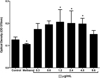
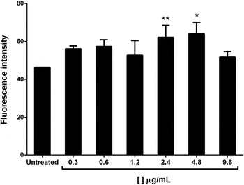
Cells treated with CTX (0·3–9·6 µg mL−1) for 48 h did not show signs of cytotoxicity, but at 1·2, 2·4 and 4·8 µg mL−1, the cells demonstrated a significant increase in mitochondrial activity (Fig. 1). To evaluate ROS production, the CellRox® Green assay was performed and analysed by flow cytometry. Results demonstrated that macrophages treated with 2·4 and 4·8 µg mL−1 CTX showed higher concentrations of ROS than the control group (1·34±0·13-fold and 1·38±0·13-fold, respectively; Fig. 2).
Murine macrophage viability was determined using MTT assays after treatment with different concentrations of CTX for 48 h. Cells treated with 2·4 and 4·8 µg mL−1 CTX demonstrated a significant increase in mitochondrial activity compared with the control group. Methanol was used as the negative control. *P < 0·05, values are expressed as means ± s.d. and compared with the control group.

A CellRox Green assay was used to analyse ROS production after macrophages were treated with different concentrations of CTX.*P < 0·05; ** P < 0·01, values are expressed as means ± s.d. compared with untreated group.

Evaluation of in vitro antileishmanial activity
Antipromastigote activity
The direct effect of CTX on L. amazonensis promastigotes was monitored for up to 96 h. Promastigotes treated with 4·8 µg mL−1 CTX exhibited a significant decrease in their growth rate (24%, ±2·7), while those treated with 2·4 µg mL−1 of CTX exhibited a 14·6% ±12·5 decrease in the growth of promastigotes. After 96 h of treatment, CTX was determined to have an IC50 = 22·86 µg mL−1 (Fig. 3).
Promastigote growth curve for Leishmania amazonensis that were treated with different concentrations of CTX for 96 h. AMP-B, amphotericin B. *P < 0·05, values are compared with the untreated group.

Antiamastigote activity
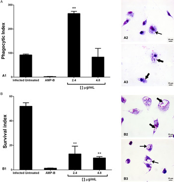
To analyse whether CTX stimulates L. amazonensis phagocytosis by macrophages, phagocytic indices were calculated using cultured macrophages that were infected with L. amazonensis and treated with CTX (2·4 and 4·8 µg mL−1) for 24 h. CTX administration resulted in an increased capacity to phagocytose promastigotes (2·85 ± 0·01-fold), compared with the untreated control group (Fig. 4A1–A3). However, to determine whether continuous treatment with CTX would eliminate the internalized parasites, treatment was performed for 48 h. Macrophages treated with CTX eliminated amastigotes once the survival index of the treated cells was reduced significantly in comparison with the untreated infected host cells (4·0 ± 2·47-fold and 5·33 ± 0·29-fold, respectively; Fig. 4B1–B3).
Macrophages were infected with Leishmania amazonensis and treated with CTX for 24 h (A) and 48 h (B). (A1) The phagocytic index of host cells that were treated with CTX for 24 h. Note the significant increase in phagocytosis after treatment with 2·4 µg mL−1 CTX. (A2) and (A3) Giemsa staining of infected macrophages untreated (CTL) or treated with 2·4 µg mL−1 of CTX, respectively. Note the greater amount of amastigotes inside the macrophages (large arrows). (B1) The survival index of host cells that were treated with CTX for 48 h. (B2) and (B3) Giemsa staining of macrophages that were infected (CTL) or infected and treated with 2·4 µg mL−1 of CTX, respectively. **P < 0·01 indicates statistical significance compared with the control group; values are expressed as means ± s.d.

Morphometric analysis of macrophages infected with L. amazonensis and treated with CTX
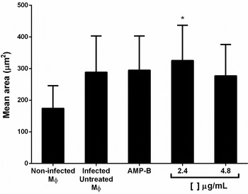
After macrophages were infected with L. amazonensis and incubated with CTX (2·4 µg mL−1) for 24 h, we observed a significant increase in the mean area, compared with the untreated infected group (1·21 ± 0·91-fold; Fig. 5).
Morphometric analysis demonstrating the significant increase in macrophage (Mɸ) mean areas after infected macrophages were treated with 2·4 µg mL−1 of CTX. *P < 0·05, values are expressed as means ± s.d. and compared with the untreated group.

CTX induces NO production in infected macrophages
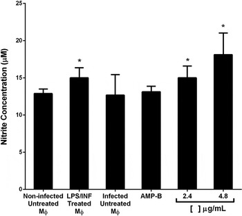
NO production was also analysed in the infected macrophages because L. amazonensis is known to have an inhibitory effect against NO production by macrophages. In the positive control group, macrophages were incubated with LPS (1 µg mL−1, Zhou et al. Reference Zhou, Zhang, Wang, Wei, Chen, Guo, Zhang and Wang2015). CTX reversed the inhibitory effect of infection after treatment with 2·4 and 4·8 µg mL−1 CTX for 24 h and increased NO production in these cells, compared with the control (1·17 ± 0·12-fold and 1·41 ± 0·23-fold, respectively; Fig. 6).
Nitrite concentrations in macrophages that were infected with Leishmania and treated with CTX. NO production was observed indirectly by analysing nitrite concentrations and was found to be higher in the infected macrophages that were treated with CTX. Control cells were activated with LPS and INF-γ at concentrations of 2 µg mL−1 and 0·01 ng mL−1, respectively. *P < 0·05, values are expressed as means ± s.d. and compared with the untreated group.

Cytokine production in infected macrophages treated with CTX
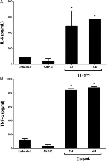
Cytokine secretion by treated macrophages that were infected with L. amazonensis was assessed using culture supernatants. The results obtained using flow cytometry demonstrated increased IL-6 (Fig. 7A) and TNF-α (Fig. 7B) levels in the macrophages treated with 2·4 µg mL−1 CTX (5·5-fold and 6·9-fold, respectively, compared with the control) and 4·8 µg mL−1 CTX (6·4-fold and 7·1-fold, respectively, compared with the control).
Cytokine concentrations in macrophages that were infected with Leishmania and treated with CTX. Treated and infected macrophages produced a higher concentration of IL-6 (A) and TNF-α (B) compared with the untreated controls. *P < 0·05, values are expressed as means ± s.d. and compared with the untreated group.

DISCUSSION
Leishmaniasis is a neglected disease, and available therapies present many disadvantages, including high toxicity and emerging drug resistance in the parasite, indicating that there is a need to develop novel compounds to treat these infections (Adade et al. Reference Adade, Carvalho, Tomaz, Costa, Godinho, Melo, Lima, Rodrigues, Zingali and Souto-Padrón2014). Macrophages are crucial to the establishment and persistence of this parasitic infection (Podinovskaia and Descoteaux, Reference Podinovskaia and Descoteaux2015). Therefore, investigating new substances that show potential to improve the microbicidal and immune capacity of macrophages can be of substantial assistance in controlling the survival of the parasite. Few reports in the literature have described the use of CTX against Leishmania parasites or during interactions between the parasites and the host cell.
This study demonstrates that CTX did not display cytotoxicity in host cells, at the concentrations studied. Interestingly, macrophages seemed to exhibit an increase in mitochondrial activity following treatment. Based on this activity, we evaluated how CTX can modulate mitochondrial metabolism. Thus, ROS quantification was performed in host cells, and we found that CTX increased ROS production in macrophages after treatment. ROS increase oxidative stress and act as microbicidal molecules. In fact, CTX stimulated the maximal activity of hexokinase, glucose-6-phosphate dehydrogenase, citrate synthase and 14CO2 production from [U-14C]-glucose and [U-14C]-glutamine in macrophages (Faiad, Reference Faiad2012), which led to an increase in NO or H2O2 production in these cells (Faiad, Reference Faiad2012; Costa et al. Reference Costa, Faiad, Landgraf, Ferreira, Brigatte, Curi, Cury and Sampaio2013). The importance of the activation of macrophage metabolism by other venoms during Leishmania infection was demonstrated in Bhattacharya et al. (Reference Bhattacharya, Ghosh, De, Gomes, Gomes and Dungdung2013), who showed that treating splenocytes with Bungarus caeruleus venom during a Leishmania donovani infection stimulated these cells to produce ROS, enabling the destruction of intracellular parasites.
One of the new findings of the present study was that CTX derived from Cdt displayed activity against Leishmania promastigotes. A significant decrease in the parasite growth rate was observed even when a low concentration of CTX was applied (IC50 = 22·86 µg mL−1). Barros et al. (Reference Barros, Pereira, Barros, L., Calvi, Santos, Barraviera and Ferreira2015) demonstrated an IC50 of 52·07 µg mL−1 using PLA2-derived Cdt against Leishmania infantum chagasi, and Torres et al. (Reference Torres, Dantas, Toyama, Filho, Zara, Rodrigues de Queiroz, Pinto Nogueira, Rosa de Oliveira, de Oliveira Toyama, Monteiro and Martins2010) observed an IC50 of 86·56 µg mL−1 for crude venom derived from Brothrops marajoensis, while Muhammad et al. (Reference Muhammad, Dunbar, Khan, Tekwani, Bedir, Takamatsu, Ferreira and Walker2003) reported an IC50 of 0·17 µg mL−1 for amphotericin B. Several studies have demonstrated leishmanicidal effects by toxins derived from snakes, such as B. caeruleus toxin, which was capable of reducing the population by up to 60% in vitro, and succeeded in eliminating the parasite in mice with visceral leishmaniasis (Bhattacharya et al. Reference Bhattacharya, Ghosh, De, Gomes, Gomes and Dungdung2013). Recently, Nunes et al. (Reference Nunes, Figueira, Lopes, De Souza, Izidoro, Ferro, Souza, Rodrigues, Rodrigues and Yoneyama2013) used a fraction of B. pauloensis venom and observed a reduction in the growth of L. amazonensis promastigotes of 60–70%. These important findings provide good support for further studies of those toxins, since their activities are selective against parasites and may maintain host-cell viability.
As CTX was not toxic to host cells, and because it presented anti-promastigote activity, it was important to evaluate the activation profile that was induced by CTX during macrophage–Leishmania interactions. Our study demonstrated that macrophages treated with CTX were able to engulf a higher number of parasites after 24 h of treatment; moreover, CTX eliminated intracellular amastigotes after 48 h of treatment.
This stimulatory effect of CTX on phagocytic capacity differs from the inhibitory effect observed before (Sampaio et al. Reference Sampaio, Brigatte, Sousa-e-Silva, dos-Santos, Rangel-Santos, Curi and Cury2003, Reference Sampaio, Santos, Costa, Rangel-Santos, Carneiro, Curi and Cury2006a , Reference Sampaio, Alba-Loureiro, Brigatte, Landgraf, dos Santos, Curi and Cury b ). Therefore, it is possible that incubating macrophages with parasites causes distinct signalling pathways to induce phagocytosis and the microbicidal activity that eliminates intracellular parasites. In fact, the initial interaction of the promastigote with the cells may trigger the release of intracellular survival factors by the parasite, which may subsequently modulate the phagocytic capacity of macrophages (Rotureau et al. Reference Rotureau, Morales, Bastin and Späth2009). In addition, it has been demonstrated that the inhibitory effect of CTX on macrophage spreading and phagocytic activities is caused by the PLA2 subunit and that this inhibitory effect is not dependent on the type of receptor involved in the phagocytic process (Sampaio et al. Reference Sampaio, Rangel-Santos, Peres, Curi and Cury2005). In contrast, the present study demonstrated that, when added to the culture of macrophages infected with Leishmania, this toxin stimulates the phagocytic capacity of macrophages at 24 h. Costa et al. (Reference Costa, Faiad, Landgraf, Ferreira, Brigatte, Curi, Cury and Sampaio2013) demonstrated that monoculture of macrophages treated with CTX present a decreased secretion of proinflammatory mediators, compatible with the anti-inflammatory profile described for this toxin. On the other hand, the secretion of proinflammatory mediators is enhanced by pretreated macrophages with CTX, when co-cultured with tumour cells, reinforcing the immunomodulatory effect described for this CTX. Therefore, we believe that the action of CTX on the phagocytic capacity of macrophages may be more associated with the immunomodulatory activity of the CTX complex than the enzymatic activity of PLA2. Different receptors are involved in the process of phagocytozing the parasite, including complement receptors, mannose receptors, fibronectin receptors and FCγ receptors. These receptors may be modulated according to inflammatory conditions, which may lead to the inhibition or activation of NADPH oxidase in the newly formed phagosome, which promotes promastigote clearance (Ueno and Wilson, Reference Ueno and Wilson2012; Podinovskaia and Descoteaux, Reference Podinovskaia and Descoteaux2015). Thus, macrophage phagocytic receptors may involve a potentially distinct CTX-mediated modulatory action during their interactions with the parasite.
Studies using crude venom and toxins isolated from Bothrops alternatus have shown them to increase the phagocytic capacity of treated macrophages (Setubal et al. Reference Setubal, Pontes, Furtado, Kayano, Stábeli and Zuliani2011). Interestingly, Furtado et al. (Reference Furtado, Oliveira, Pontes, Setúbal, Xavier, Lacouth-Silva, Lima, Zaqueo, Kayano, Calderon, Stábeli, Soares and Zuliani2014) obtained similar results when they treated J774A.1 macrophages with a Bothrops atrox-derived toxin. However, Macedo et al. (Reference Macedo, de Barros, Ferreira, Moreira-Dill, Calderon, Soares and Nicolete2015) studied the effect of crotamine, another toxin derived from Cdt venom, and did not observe any antileishmanial activity after treating infected peritoneal macrophages with up to 100 µg mL−1 of the toxin. Thus, the higher phagocytic capacity induced by CTX in macrophages that were challenged with Leishmania parasites after 24 h appears to induce the destruction of the intracellular amastigotes after 48 h of treatment, and this is directly related to the development of an effective immune response. Interestingly, Costa et al. (Reference Costa, Faiad, Landgraf, Ferreira, Brigatte, Curi, Cury and Sampaio2013) demonstrated that macrophages, after 24 h in medium containing CTX, demonstrated a significant increase in the secretion of lipoxin A4 (LXA4) and its stable analogue, 15-epi-LXA4. However, the anti-inflammatory activity of these lipid mediators (LXs) did not compromise the ability of INF-γ-stimulated macrophages to kill intracellular parasites or to upregulate inducible nitric oxide synthase or NO synthesis, which are key antimicrobial mechanisms of macrophages. These results suggest that the LXs and their analogues are immunomodulatory rather than immunosuppressive agents (Aliberti et al. Reference Aliberti, Serhan and Sher2002a , Reference Aliberti, Hieny, Reis e Sousa, Serhan and Sher b ).
In fact, the antileishmanial activity induced by host cell activation is extremely important because there has been an increase in the number of parasites that are resistant to the drugs currently available to treat leishmaniasis (Fernández et al. Reference Fernández, Diaz-Toro, Ovalle, Valderrama, Muvdi, Rodríguez, Gomez and Saravia2014). Moreover, it is well documented that L. amazonensis can inactivate cellular immune responses, which is an important peculiarity of this protozoa infection and can cause anergic immune responses (Silveira et al. Reference Silveira, Lainson, Gomes, Laurenti and Corbett2009). In the current study, during infection, we observed that treated macrophages presented larger cell areas, indicating that they have undergone morphological changes suggestive of cell activation. Furthermore, to understand how murine macrophages might eliminate Leishmania amastigotes, we evaluated NO production and cytokine profiles. CTX induced an increase in the production of NO, even when macrophages were challenged with L. amazonensis, and was able to induce macrophages to switch to a M1 profile during Leishmania infection, presenting higher levels of IL-6 and TNF-α. The M1 profile is known to be involved in cellular responsiveness, as characterized by analyses of NO and proinflammatory cytokine production, such as IL-6 and TNF-α. The M2 profile represents the anti-inflammatory response, in which there is an increase in the production of a different set of cytokines, such as IL-10 and IL-4. This polarization profile is very beneficial to the host, and it helps to combat Leishmania infection and eliminate the parasite. Therefore, the data obtained in this study indicate the selective action of CTX on macrophage metabolism. Another study using crotamine isolated from Cdt venom also demonstrated that a higher concentration of TNF-α was observed in Leishmania-infected macrophages compared with the untreated and infected controls (Macedo et al. Reference Macedo, de Barros, Ferreira, Moreira-Dill, Calderon, Soares and Nicolete2015). These data indicate that the Cdt venom or its isolates exhibit immunomodulatory activity.
The modulation of the macrophage response against Leishmania, caused by CTX, indicates a classical M1 activation profile, which is characterized by the increased production of NO and ROS, as well as proinflammatory cytokines such as IL-6 and TNF-α, which were observed in this study (Liu et al. Reference Liu, Zou, Chai and Yao2014; Rath et al. Reference Rath, Müller, Kropf, Closs and Munder2014; Venturin et al. Reference Venturin, Chiku, Silva, de Almeida and de Lima2016). In addition to these biochemical changes, the morphological changes, characteristic of the M1 profile of activation, such as cell spreading and an increase in cytoplasmic projections become evident in these cells (Cassado et al. Reference Cassado, de Albuquerque, Sardinha, Buzzo, Faustino, Nascimento, Ghosn, Lima, Alvarez and Bortoluci2011).
To our knowledge, this report shows, for the first time, that CTX isolated from Cdt venom induces leishmanicidal activity against intracellular parasites and substantial activation of infected macrophages. Thus, CTX is a promising and potent antileishmanial agent and a potentially potent immunomodulatory compound that is able to induce macrophages to polarize to the M1 type during Leishmania infection, leading to the elimination of intracellular parasites.
ACKNOWLEDGEMENTS
We are grateful to Instituto Evandro Chagas and Instituto Butantan.
FINANCIAL SUPPORT
This work was supported by Conselho Nacional de Desenvolvimento Científico e Tecnológico (CNPq), Coordenação de Aperfeiçoamento de Pessoal de Nível Superior (CAPES), FAPESPA (ICAAF No. 161/2014), FADESP, PROPESP-UFPA and Instituto Nacional de Biologia Estrutural e Bioimagem-INBEB (CNPq, grant number 573767/2008-4).