


The phenotypic spectrum in individuals with variants in the COL4A1 gene includes autosomal dominant familial porencephaly type I (OMIM 175780), autosomal dominant brain small vessel disease with or without ocular anomalies (OMIM 175780), hereditary angiopathy with nephropathy, aneurysms and muscle cramps (HANAC) syndrome (OMIM 611773), isolated retinal artery tortuosity (OMIM 180000) and/or congenital cataract, pontine autosomal dominant microangiopathy and leukoencephalopathy (PADMAL) (OMIM 618564). Reference Plaisier, Ronco, Adam, Bick, Mirzaa, Pagon, Wallace and Amemiya1,Reference Tambala, Vassar and Snow2 Here, we wish to describe a previously unacknowledged association between COL4A1 and neuroblastoma and discuss the possible mechanisms underlying this co-occurrence.
A 17-year-old right-handed male presented with a history of recurrent stroke from 6 months of age. He was born at term by vaginal delivery to non-consanguineous parents of Han Chinese ancestry. Prenatal ultrasound revealed ventricular dilatation from 34 weeks of gestation. He had a global delay in the attainment of milestones. He developed a palpable abdominal mass around 11 months of age and was evaluated with a CT abdomen. The metaiodobenzylguanidine scan was positive in the abdominal mass alone, and the bone marrow examination was negative. Biopsy confirmed the diagnosis of a poorly differentiated neuroblastoma. Favorable prognostic factors, including absence of NYMC amplification, favorable Shimada histology, absence of deletion in the 1p chromosome and DNA index greater than 1, were detected. Left retroperitoneal neuroblastoma stage III was managed with chemotherapy followed by surgical resection. He began experiencing left focal motor seizures from 2.5 years of age that were managed with carbamazepine. Parents discontinued antiseizure medication after 3 years. He had an acute onset of left-sided UMN facial weakness at 15 years of age, and a brain MRI (Figure 1A–H) revealed an acute infarct in the right corona radiata, hyperintense signal changes in cerebellar and cerebral white matter and bilateral basal ganglia. His family history was non-contributory. Genetic testing identified a de novo heterozygous pathogenic variant c.2494G>A (p. Gly832Arg) in exon 32 of the COL4A1 gene, previously reported without oncological association. Reference Hatano, Daida and Hoshino3
(A–H): Brain MRI at 15 years of age. T2 FLAIR axial images (A–D) show signal changes in right > left cerebellar white matter, right occipital lobe, bilateral basal ganglia and bilateral cerebral white matter. DWI (E) and ADC (F) images show foci of diffusion restriction in the right corona radiata; SWI (G–H) show multiple foci of abnormal susceptibility in cerebral white matter bilaterally.

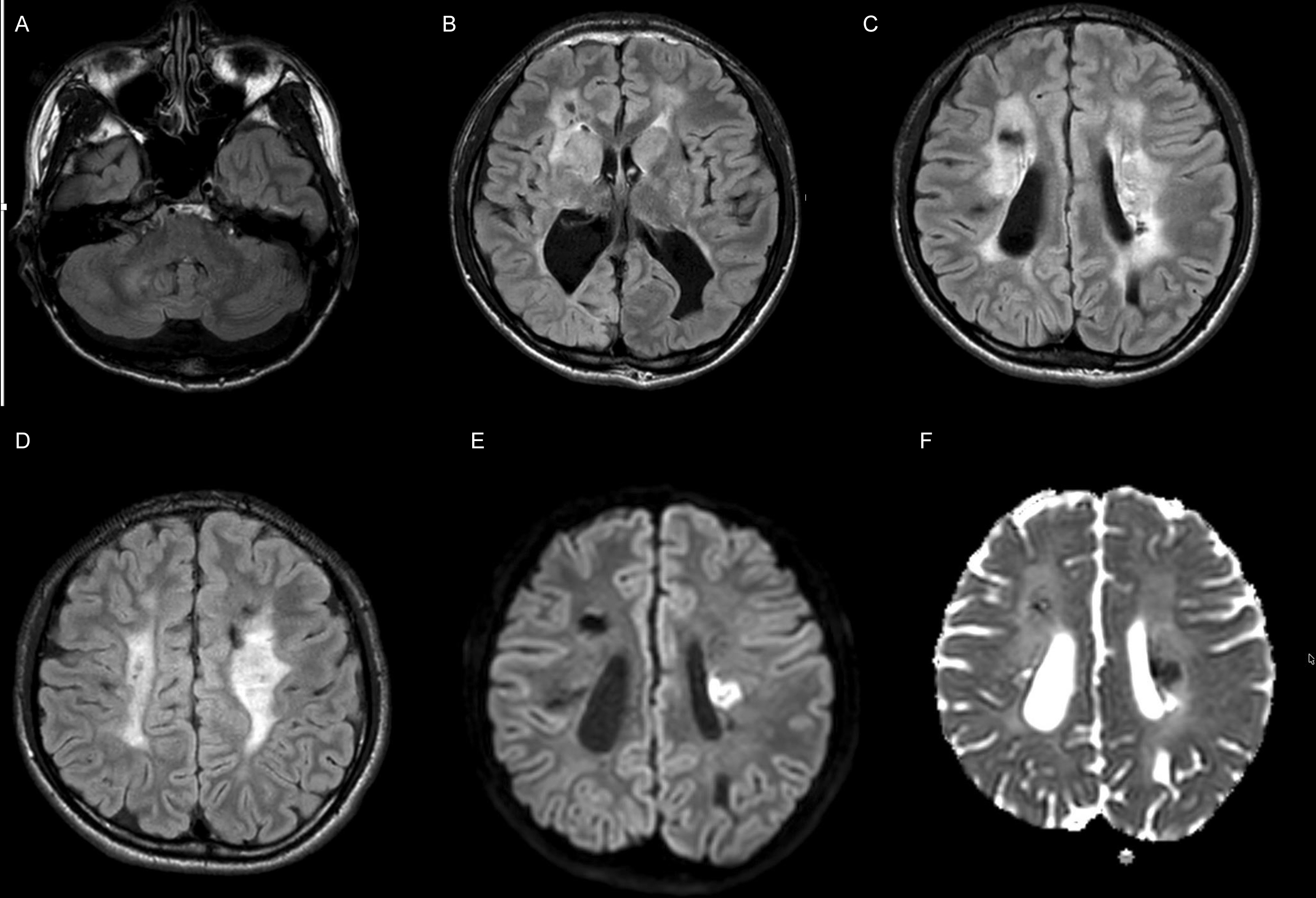
At 17 years of age, he experienced an acute onset of right-sided numbness and weakness with dysarthria. Brain MRI (Figure 2A–F) revealed acute ischemic stroke in the left corona radiata and multifocal chronic infarcts. Ten days after admission for the acute stroke management, he had rapid deterioration in the level of consciousness with recurrent episodes of gaze deviation. Follow-up brain MRI (Supplemental Figure 1A–F) revealed extensive hemorrhagic transformation with innumerable new small hemorrhages in the left temporal and occipital regions, left striatum and bilateral frontal subcortical white matter. Continuous electroencephalography monitoring captured multiple subclinical seizures with onset from the right posterior quadrant that were managed with levetiracetam. He lost all motor and cognitive skills and became bedbound and nonverbal. He developed frequent episodes of generalized dystonic posturing of variable duration and progressive spasticity of all four limbs. Escalating pharmacotherapy included baclofen, gabapentin, clonidine and benzodiazepines. He was transitioned to the adult movement disorders clinic. Neurological examination in an awake state revealed limited eye contact with no comprehensible speech. Bilateral pupils were equal in size and reactive to light. There was spastic quadriparesis next to dystonia. Further pharmacological optimization, including botulinum toxin injections to selective muscle groups, provided minimal additional benefit.
(A–F): Brain MRI at 17 years of age. T2 FLAIR axial images (A–D) show signal changes in cerebellar white matter, right occipital lobe, bilateral basal ganglia and bilateral cerebral white matter; expected interval evolution of multifocal chronic infarcts and hemorrhages in bilateral cerebral hemispheres. DWI (E) and ADC (F) images show an acute infarct in the left corona radiata.
Additional investigations, such as blood pressure recordings and transthoracic echocardiography with bubble study, were normal. Ophthalmology assessment revealed tortuous retinal vessels. Persistent microscopic hematuria was detected. A 24-hour urinary protein excretion was normal. Ultrasound of the abdomen and pelvis revealed increased echogenicity in bilateral renal pyramids.
It was striking that an individual, harboring a pathogenic variant in the COL4A1 gene, had also been diagnosed with neuroblastoma, an unexpected co-occurrence that prompted us to explore an underappreciated link.
The basement membrane of the epithelial and endothelial cells in humans is composed of type IV procollagen and laminin. Type IV procollagen consists of α1 and α2 chains with diversity in structure and tissue distribution as compared to collagen I–III and V, but similarity in the presence of a central collagenous domain and noncollagenous propeptides. COL4A1 and COL4A2 encoding proα1 (IV) and proα2 (IV) chains assemble to form a heterotrimeric helix (2 alpha1 and 1 alpha2 chains). Variants in the COL4A1 and COL4A2 genes cause pleiotropic symptoms with multisystem involvement and are increasingly recognized as having oncogenic properties. Reference Kuo, Labelle-Dumais and Gould4,Reference Wang, Liu and Li5
Abnormal expression of COL4A1, a novel oncogene, has been linked with the occurrence, development and outcome of malignancies, including glioma, glioblastoma, meningioma, breast malignancy and gastrointestinal tract malignancy, by promoting angiogenesis and tumor progression. Reference Wang, Liu and Li5–Reference Jin, Shen and Zhang7 Interestingly, neuroblastoma has been reported in only one other individual with a COL4A1-related disorder, early intracerebral hemorrhage and porencephaly, who was diagnosed at six months of age. Reference Meuwissen, Halley and Smit6 This patient harbored a different de novo heterozygous missense variant, c.2008G>A, p. Gly670Arg, in the COL4A1 gene, and a causal association with neuroblastoma was not established. Reference Meuwissen, Halley and Smit6 A broader literature search identified no additional cases of neuroblastoma associated with pathogenic COL4A1/2 variants. Previous mouse model experiments with a missense variant have shown mechanistic heterogeneity of COLA4A1 in a tissue-specific manner. Given the diverse role of COL4A1, the same variant may exhibit dichotomous behavior with loss of function in one tissue and gain of function in another tissue. Reference Labelle-Dumais, Schuitema and Hayashi8 This can possibly explain the clinical presentations in our proband, with loss-of-function effects resulting in vasculopathy in different organs and gain-of-function effects resulting in the risk of oncogenesis. It is unclear whether environmental or epigenetic modifiers may also influence gene expression selectivity, thereby enhancing focal adhesion and altering JAK-STAT and Toll‑like receptor signalling pathways. Reference Wang, Liu and Li5 Tumor tissue molecular analysis might provide more information if the haploinsufficiency or dominant negative variant affects the selectivity of expression or function of COL4A1.
We propose that neuroblastoma may represent a novel association, expanding the phenotypic spectrum in individuals with COL4A1 variants. This association warrants systematic evaluation in larger cohorts, and functional studies should be considered to elucidate the pathomechanisms of oncogenesis in individuals with missense variants. With emerging evidence suggesting an increased risk of oncogenesis in individuals harboring COL4A1 variants, it may be necessary to develop guidelines for tumor surveillance and counsel patients and their families about this association.
Supplementary material
The supplementary material for this article can be found at https://doi.org/10.1017/cjn.2026.10698.
Author contributions
Conceptualization: AM, SY, CGanos. Literature search: SY, AM. Preparation of manuscript: SY, AM. Critical review and revision of manuscript: CGanos, CG.
Funding statement
The authors received no specific funding for this work.
Competing interests
The authors declare that they have no competing interests for the work reported in this manuscript.
Adriana Mititelu: No disclosures to report. Sangeetha Yoganathan: No disclosures to report. Carolina Gorodetski: Grants or contracts from the Dystonia Medical Research Foundation, Center for Brain and Mental Health, consulting fees and payment for honoraria from Medtronic Inc. and Acadia. Leadership or fiduciary role in the Movement Disorder society (Education Committee). Christos Ganos: Wolf Chair in Neurodevelopmental Psychiatry and Research Support from the Stewart Family. Educational honoraria from the Movement Disorder Society and Merz Pharmaceuticals. Medicolegal work related to litigation cases.



