Introduction
Nasopharyngeal carcinoma (NPC) is unique among head and neck cancers because of its epidemiology. It is relatively rare worldwide and occurs predominantly in males.Reference Parkin, Pisani and Ferlay1 The highest age-adjusted incidence rates for nasopharyngeal cancer have been reported in Southern China, ranging from 25 to 50 per 100 000 population.Reference Su and Wang2 Additionally, NPC occurs in a younger population, grows rapidly in local and regional areas, and is highly metastatic to lung and bone.
Although NPC is markedly radiosensitive, there is a high rate of treatment failure because of its invasiveness and metastatic behaviour. It is estimated that five-year survival rates are available for 90 per cent of the patients with stage I NPC. The efficacy of radiotherapy is not so prominent in stage III patients, and the therapeutic efficacy in stage II patients remains controversial owing to a limited improvement in survival. The resistance of NPC tumour cells to radiotherapeutic drugs represents a major problem in clinical oncology.
During carcinogenesis, some cancer cell genes (e.g. tumour suppressor genes) lose some aspect of their normal functionality, while others gain a function or are activated despite normally being dormant (e.g. oncogenes). Furthermore, cancer cells require the continuous presence of certain stimuli (e.g. growth factors) in order to maintain their malignant phenotype. Activated oncogenes and growth factor loops represent potential targets for ribonucleic acid (RNA) interference strategies.Reference Tong, Zhang and Nemunaitis3
Insulin-like growth factor I receptors and epidermal growth factor receptors are required for carcinogenesis and tumour progression in many human malignancies. The concept of targeting specific receptors has been validated by the successful clinical applications of multiple new drugs, such as trastuzumab and gefitinib. The genetic blockade of insulin-like growth factor I receptors or epidermal growth factor receptors has been accomplished using the following: antisense inhibition; dominant negative inhibition; small interference RNA (SiRNA); and triplex formation mediated by oligonucleotide, plasmid vector transfection or viral transduction.
SiRNA against insulin-like growth factor I receptors or epidermal growth factor receptors has already been described in the literature. Bohula et al. Reference Bohula, Salisbury, Sohail, Playford, Riedemann and Southern4 described a SiRNA sequence which could effectively block insulin-like growth factor I receptor messenger (m) RNA. Using a scanning oligonucleotide array technique, they selected an antisense oligodeoxynucleotide that selectively blocked insulin-like growth factor I receptors but not insulin receptors. Furthermore, these authors also demonstrated that SiRNA homologous to accessible insulin-like growth factor I receptor regional targets (determined by examining the secondary structure of the insulin-like growth factor I receptor) induced strong, sequence-specific insulin-like growth factor I receptor gene silencing. Xiangbai et al. Reference Xiangbai, Yongzhi, Guobin and Kaixiong5 described combining SiRNA at two different sites in the epidermal growth factor receptor in order to suppress its expression, induce apoptosis and enhance the 5 fluorouracil (5-FU) sensitivity of colon cancer cells. However, when short hairpin RNA to epidermal growth factor receptor was combined with short hairpin RNA to insulin-like growth factor I receptor, the effects of this, and the resulting chemosensitivity of nasopharyngeal cell lines to chemotherapeutic drugs, were not well understood.
In this study, we constructed short hairpin RNA sequences which would transcribe short hairpin RNA to insulin-like growth factor I receptor and epidermal growth factor receptor. We then combined U6-insulin-like growth factor I receptor short hairpin RNA with U6-epidermal growth factor receptor short hairpin RNA, in order to study their combined effects on NPC cell lines, regarding: expression; apoptosis; phosphorylation of phospho-ser/Thr protein kinase (pAkt) and phospho-extracellular signal-regulated kinase (pErk) (major components of the receptors' signalling pathways); and chemosensitivity to chemotherapeutic drugs.
Materials and methods
Selection of sequences and construction of short hairpin RNA segments
Potential target sequences for Homo sapiens insulin-like growth factor I receptor mRNA (NM 000875) and epidermal growth factor receptor mRNA (NM_201283) were screened, from sequences provided by Gen-Bank (Wuhan, China). From the potential SiRNA sequences, we selected two sites for each receptor (380 and 3430 for insulin-like growth factor I receptor, and 349 and 643 for epidermal growth factor receptor) after one of plasmids used by filtering (Figure 1). Four sequences were designed according to the short hairpin RNA design principle,Reference Tuschl6 and were proven to be effective.Reference Zhang, Zhang, Bai, Song, Chen and Gao7, Reference Lee, Imsumran, Park, Kwon, Yoon and Lee8 It was ensured that the selected SiRNA sequence had at least four basyls that were different from other genes in the human genome.Reference Jackson, Bartz, Schelter, Kobayashi, Burchard and Mao9 The EST (name of database) database was searched by using basic local alignment and search tool (BLAST) (see http://www.ncbi.nlm.nih.gov/BLAST), and no other isogenous genes were found. The negative control gene sequence was not isogenous with any human genes (Figure 1).
Sequences of oligonucleotides for the U6-short hairpin epidermal growth factor receptor and the U6-small hairpin (si) insulin-like growth factor I receptor. Each sequence consists of sense (19 bp), loop and antisense (19 bp) for either insulin-like growth factor I receptor or epidermal growth factor receptor. Sequences of SiRNA were started at the 380th and 3430th IGF-1R for insulin-like growth factor I receptor, and at the 349th and 643th EGFR for epidermal growth factor receptor. These deoxyribonucleic acid (DNA) strands were ligated with corresponding complimentary DNA strands.

In this study, in order to overcome the disadvantages of SiRNA regarding short acting time and lack of regenerating ability,Reference Brummelkamp, Bernards and Agami10 the encoding short hairpin RNA plasmid expressive vectors were used with the U6 promoter and neor gene. The hairpin loop sequence of the short hairpin RNA comprised TTCAAGACG (where T is thymines, C is cytosine, A is adenine and G is guanine) and the transcription terminator was TTTTT. The plasmid Pgenesil-1 was digested with BamHI and HindIII, and ligated with annealed segments of target gene template deoxyribonucleic acid (DNA). Competent bacteria (DH5a) were then transformed. After kanamycin selection and identification of the plasmids by digestion with PstI and SalI, the bacteria contained in the resulting solution were tested for the sequence.
Once segments of selected gene template DNA was correctly inserted, SalI produced a 400 bp DNA segment and it was correctly identified. Four sequences were identified, namely: pU6-epidermal growth factor receptor short hairpin RNA 1 (349); pU6-epidermal growth factor receptor short hairpin RNA 2 (643); U6-insulin-like growth factor 1 receptor short hairpin RNA 1 (380); and pU6-insulin-like growth factor 1 receptor short hairpin RNA 2 (3430).
The T4 DNA ligase, BamHI, HindIII, PstI and SalI were obtained from NEB (London, UK). The plasmid Pgenesil-1 was obtained from Wuhan Genesil Biotechnology (Wuhan, China). After annealing these oligonucleotides, double-stranded DNA with sense–loop–antisense was cloned into pGenesil-1 containing a U6 promoter and an enhanced green fluorescence protein gene.
Cell culture and transfection
The human nasopharyngeal carcinoma (NPC) cell lines CNE2 and TW03 were preserved by Wuhan University. These CNE2 and TW03 cells were then cultured in RPMI 1640 medium supplemented with 10 per cent calf bovine serum, and humidified with 5 per cent CO2 at 37°C. Twelve hours before transfection, cells were seeded onto 24-well plates at a density of 5 × 10⁴ cells per well. The confluence reached approximately 70–80 per cent at the time of transfection. Cells were washed twice using OPTI-MEMI (Gibco, Wuhan, China) and then a further 2 ml of OPTI-MEMI was added to each well. Two hundred and fifty microlitres of RPMI was used to dilute 2–2.5 µg plasmid vector and 10 µl Lipofectamine 2000 (Invitrogen, San Diego, California, USA). These were then left at room temperature for 20 minutes. The compound was added to each well and mixed thoroughly.
The CNE2 and TW03 cells were seeded onto 24-well culture plates at a density of 5 × 10⁴ cells per well and cultured in RPMI 1640 medium with 10% FCS, until the cells reached the 70–80% confluence. According to the experiment design, the cultures were arranged in 7 groups and transfected by adding a mixture of 2ug shRNAs and 10 uL Lipofectamine 2000, respectively, in 250 uL OPTI-MEM I to each well. After incubation for 24 h, the Lipofectamine 2000 and the shRNAs-containing medium were removed and the transfected cells selected by the G418 (800–1200 µg/ml G418 in medium for first 15 days, followed by 500–600 µg/ml G418 for next 15 days, and then 200 µg/ml G418 as a constant).
Seven treatment groups were used. Group one comprised the plasmid vector transfected with a mixture of half U6-insulin-like growth factor I receptor short hairpin RNA 2 (3430) and half U6-epidermal growth factor receptor short hairpin RNA 2 (643). Group two comprised the plasmid vector transfected with a mixture of half U6-insulin-like growth factor I receptor short hairpin RNA 1 (380) and half U6-epidermal growth factor receptor short hairpin RNA 1 (349). Group three comprised the plasmid vector transfected with pU6-epidermal growth factor I receptor short hairpin RNA 2 (643). Group four comprised the plasmid vector transfected with pU6-insulin-like growth factor I receptor short hairpin RNA 2 (3430). Group five comprised the plasmid vector transfected with pU6-epidermal growth factor receptor short hairpin RNA 1 (349). Group six comprised the plasmid vector transfected with pU6-insulin-like growth factor I receptor short hairpin RNA 1 (380). Group seven comprised negative control plasmid vector HK.
Western blot to detect receptor expression
Twenty-four hours after transfection, G418 was used for four-week selection. Proteins were extracted and Western blotting was performed with antibodies to insulin-like growth factor I receptor β chain and to epidermal growth factor receptor (C20; biotechnology, Santa Cruz, California, USA). The experiment was repeated three times.
Flow cytometry to detect apoptosis
One million CNE2 cells were collected, washed twice at 4°C with precooled phosphate-buffered solution, and then palin-suspended in 5 ml 70 per cent precooled alcohol. Each tube was sealed with 5 µl liquid paraffin, mixed well and incubated at −20°C for 12 hours. The cells were then recollected, washed again with phosphate-buffered solution, and RNAse A was added (200 µl of 1 mg/l solution, 30 minutes at 37°C). The solution was then gently mixed with propidium lodide (800 µl of 100 mg/l solution, with 1 per cent Triton X-100) and left at 4°C, away from light, for 30–60 minutes. Flow cytometry was then used to detect apoptosis (through channels two and three).
Effects of a mixture of half U6-insulin-like growth factor I receptor short hairpin RNA 2 (3430) and half U6-epidermal growth factor receptor short hairpin RNA 2 (643) on the insulin-like growth factor I receptor and epidermal growth factor receptor downstream pathways
The CNE2 human NPC cells were transfected with either U6-insulin-like growth factor 1 receptor short hairpin RNA 2 (3430), U6-epidermal growth factor receptor short hairpin RNA 2 (643), or a half-and-half mixture of these two sequences, and screened. We then investigated the effects of the various U6 short hairpin RNA segments on the downstream pathways of insulin-like growth factor I receptor and epidermal growth factor receptor. Cells containing the various segments were incubated in serum-free conditions for 16 hours and then stimulated with insulin-like growth factor I (10 ng/ml) or epidermal growth factor (10 ng/ml). The resulting proteins were then extracted and Western blotting was performed. BandScan 5.0 software was used for grey scale scanning in order to evaluate the relative values of protein expression.
Effect of a mixture of half U6-insulin-like growth factor I receptor short hairpin RNA 2 (3430) and half U6-epidermal growth factor receptor short hairpin RNA 2 (643) on caspase activation and apoptosis induced by chemotherapeutic agents
The CNE2 and TWO3 human NPC cells were transfected with either U6-insulin-like growth factor I receptor short hairpin RNA 2 (3430), U6-epidermal growth factor receptor short hairpin RNA 2 (643), or a half-and-half mixture of these two sequences, and screened. The CNE2 cells were then treated with 5-FU (20 µM) for 48 hours, and the TWO3 cells with cisplatin (50 µM) for 24 hours. Proteins were extracted and immunoblotted with cleaved caspase 3 and caspase 3 antibodies (Cell Signaling Technology, Inc., Maryland, USA).
Under the same conditions, apoptotic cells were detected using the Tunel assay and the Pod in situ cell death detection kit (Roche, Mannheim, Germany).
Effects of a mixture of half U6-insulin-like growth factor I receptor short hairpin RNA 2 (3430) and half U6-epidermal growth factor receptor short hairpin RNA 2 (643) on nasopharyngeal cancer cell chemosensitivity to 5-fluorouracil and cis-diamnodichloroplatinum (CDOP)
The TWO3 human NPC cell line was transfected with either U6-insulin-like growth factor I receptor short hairpin RNA 2 (3430), U6-epidermal growth factor receptor short hairpin RNA 2 (643), or a half-and-half mixture of these two sequences, and screened. The cells were then seeded into 96-well plates (5 × 10³ per well). After 24-hour incubation, adriamycin and cisplatin of appropriate concentrations were added. After 72 hours, viable cell counts were determined by 3-(4,5)-dimethylthiahiazo (-z-y1)-3,5-di- phenytetrazoliumrotmide (MTT) assay and compared with those for negative control gene sequence-transfected cells.
Statistical analysis
Results are presented as means ± standard error of the mean, for each sample. The statistical significance of differences was determined by one-way analysis of variance (ANOVA) or two-factor factorial ANOVA. A p value of less than 0.05 was considered to indicate statistical significance.
Results
Successful construction of short hairpin RNA plasmid expression vector
The plasmid Pgenesil-1 has PstI digest sites. After insertion of segments of target gene template DNA, these digest sites were replaced. In the inserted target gene template DNA, a SalI digest site was designed between BamHI and HindIII in the Pgenesil-1 plasmid. Once correctly inserted, the SalI produced a 400-base pair DNA segment, which was correctly identified. The transformation bacteria solution was tested by the Shanghai Invitrogen company, with results meeting the design.
Successful transfection of short hairpin RNA expression into target cells
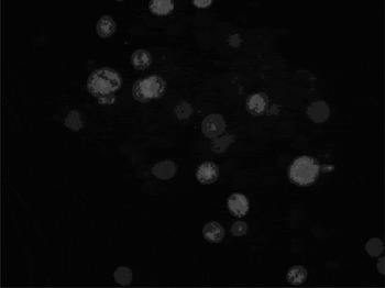
After the plasmid vector (containing a fluorescent green protein) was transfected into the cells, it was visible on light microscopy as a fluorescent green wave. Twenty-four hours after transfection, confocal microscopy demonstrated with green fluorescent appearance, as shown in Figure 2, indicating success.
Photomicrographs showing cells transfected with a plasmid vector containing fluorescent protein, 24 hours after transfection (EGFP ×40).

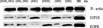
Western blot assay for antibodies to insulin-like growth factor I receptor β chain and antibody to epidermal growth factor receptor revealed the expressions of insulin-like growth factor I receptor and/or epidermal growth factor receptor in seven groups. After transfection of the U6-short hairpin RNA sequences, we confirmed that RNA interference, using the RNA sequences for insulin-like growth factor I receptor and/or epidermal growth factor receptor, suppressed expression of insulin-like growth factor I receptor and/or epidermal growth factor receptor (Figure 3).
Western blot assay for insulin-like growth factor I receptor (IGF1R) and epidermal growth factor receptor (EGFR), confirming reduced expression of both proteins, compared with a negative control sample. The CNE2 nasopharyngeal cancer cells were transfected with the following U6-short hairpin ribonucleic acid segments: 3430 and 643; 380 and 349; 643; 3430; 349; 380; and negative control gene sequence (see text for details).

Apoptosis detected by flow cytometry
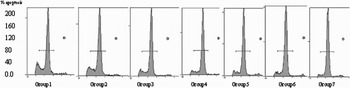
The proportions of cellular apoptosis in treatment groups one, two, three, four, five, six and seven (as described above) were respectively: 10.96 ± 0.08 per cent; 10.92 ± 0.21 per cent; 6.35 ± 0.61 per cent; 5.34 ± 0.35 per cent; 4.33 ± 0.51 per cent; 4.20 ± 0.71 per cent and 2.42 ± 0.39 per cent. There were significant differences beween apoptosis rates of group seven and apoptosis rates of groups one, two, three, four, five and six (Figure 4).
Flow cytometry results for CNE2 nasopharyngeal cancer cells transfected with U6-short hairpin ribonucleic acid segments. Flow cytometry showed an apoptotic peak or hypodiplod peak before the cells are resting or non-cycling (Go)/presynthesis period during synthesis of a protein initiatior of chromosome replication (G1) peak. Combinations of half U6-epidermal growth factor receptor (EGFR) 2 and half U6-insulin-like growth factor I receptor (IGF1R) 2, or of half U6-epidermal growth factor receptor 1 and half U6-insulin-like growth factor I receptor 1, showed the strongest apoptotic peaks and highest apoptosis percentages, compared with other preparations. HK = negative control gene sequence

Inhibition of ligand-induced Ser/Thr protein kinase (Akt) and extracellular signal-regulated kinese (Erk) phosphorylation in human nasopharyngeal cancer cell lines by pretreatment combined U6-short hairpin RNA sequences
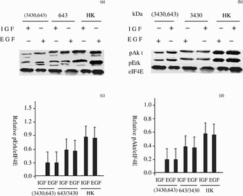
Phosphorylation of Akt and Erk, induced by insulin-like growth factor I and epidermal growth factor, is known to be a major component of the downstream signalling pathways of insulin-like growth factor I receptor and epidermal growth factor receptor, respectively. However, transfection with a mixture of half U6-insulin-like growth factor I receptor short hairpin RNA 2 (3430) and half U6 epidermal growth factor receptor short hairpin RNA 2 (643) inhibited this phosphorylation more effectively than did either insulin-like growth factor I receptor or epidermal growth factor receptor. This observation suggests that transfection with short hairpin RNA targeting both insulin-like growth factor I receptor and epidermal growth factor receptor more effectively blocks the downstream signalling pathways of these two receptors, compared with either insulin-like growth factor I receptor or epidermal growth factor receptor (Figure 5a and b). Densitometric analysis of signal changes is presented in Figure 5(c) and (d).
Results for inhibition of ligand-induced phospho-Ser/Thrprotein kinase (pAkt) and phospho-extracellular signal - regulated kinase (pErk) activation by a combination of half U6-insulin-like growth factor (IGF) 1 receptor short hairpin ribonucleic acid (RNA) 2 (3430) and half U6-epidermal growth factor (EGF) receptor short hairpin RNA 2 (643). The RNA interference, mediated by sequence-specific genes for these two receptors, blocked phosphorylation of Akt and Erk in nasopharyngeal cancer cell lines, indicating that these two major anti-apoptotic pathways were inhibited. (a) is expression of EGFR and IGF-1R protein in groups one, three and seven and (b) in groups one, four and seven. Densitometric analysis shows the differences in activation for (c) Erk and (d) Akt. HK = negative control gene sequence; eIF4E = eukaryotic translation initiation factor 4E (used as internal control)

Enhancement of nasopharyngeal cancer cell line chemosensitivity, activation of caspase 3 and apoptosis induction by combining a mixture of half U6-insulin-like growth factor I receptor short hairpin RNA 2 (3430) and half U6 epidermal growth factor receptor short hairpin RNA 2 (643) with chemotherapeutic agents
In CNE2 nasopharyngeal carcinoma cells, a mixture of half U6-insulin-like growth factor I receptor short hairpin RNA 2 (3430) and half U6-epidermal growth factor receptor short hairpin RNA 2 (643) enhanced the apoptotic effect (Figure 6a and b) of caspase 3 (following activation by 5-FU). These results were confirmed by Tunel assay (Figure 6c). The proportion of cells showing apoptosis after chemotherapy (5-FU and cisplatin) was markedly increased by prior treatment with a mixture of half U6-insulin-like growth factor I receptor short hairpin RNA 2 (3430) and half U6-epidermal growth factor receptor short hairpin RNA 2 (643). These findings suggest the possibility of combination therapy using chemotherapeutic agents plus such a mixture of short hairpin RNA sequences.
Results showing that a combination of half U6-insulin-like growth factor I receptor short hairpin ribonucleic acid (RNA) 2 (3430) and half U6-epidermal growth factor receptor short hairpin RNA 2 (643) increased the chemosensitivity of nasopharyngeal cancer cell lines. (a and b) Western blotting for caspase-3 showed that chemotherapeutic drugs reduced pro-caspase-3 (32 kDa) cleavage and induced active form (17 and 12 kDa); transfection with short hairpin RNA segments to insulin-like growth factor I receptor and to epidermal growth factor receptor enhanced the caspase cleavage in both (a) CNE2 and (b) TWO3 cell lines. (c) Tunel assay revealed that a combination of short hairpin RNA to insulin-like growth factor I receptor and to epidermal growth factor receptor improved induction of apoptosis by 5-fluorouracil (5-FU) in CNE2 cells. Blockage of insulin-like growth factor I receptor and epidermal growth factor receptor, by a combination of short hairpin RNA to insulin-like growth factor I receptor and to epidermal growth factor receptor, enhanced induction of apoptosis by cisplatin in TW03 cells more than in CNE2 cells. CDDP = cis-diamnodichloroplatinum ** = p < 0.05, compared with no-adding drug treatment (–) and dual silencing of EGF and IGF 1 receptors compared with either silencing EGF or IGF-1 receptor

Discussion
Although nasopharyngeal carcinoma (NPC) is markedly radiosensitive, there is a high rate of treatment failure because of its invasiveness and metastatic behaviour. It is estimated that five-year survival rates will be available for 90 per cent of patients with stage I NPC. The efficacy of radiotherapy is not so prominent in stage III patients, and its therapeutic efficacy in stage II patients remains controversial owing to a limited improvement in survival. The resistance of tumour cells to radiotherapy and chemotherapeutic drugs represents a major problem in clinical oncology.
We investigated the effect on NPC cells of combining chemotherapeutic drugs with a mixture of short hairpin RNA sequences to epidermal growth factor receptor and to insulin-like growth factor I receptor. Short hairpin RNA to insulin-like growth factor I receptor and to epidermal growth factor receptor was effectively expressed. A combination of short hairpin RNA both to insulin-like growth factor I receptor and to epidermal growth factor receptor resulted in effective expression of short hairpin RNA to both insulin-like growth factor I receptor and epidermal growth factor receptor. By comparison, null, mismatched short hairpin RNA plasmid vectors showed almost no inhibition.
Our flow cytometry results also indicate that the null, mismatched short hairpin RNA plasmid vectors had no effect on apoptosis. In contrast, short hairpin RNA to epidermal growth factor receptor and to insulin-like growth factor I receptor effectively induced apoptosis. Furthermore, a combination of short hairpin RNA sequences to epidermal growth factor receptor and to insulin-like growth factor I receptor induced cell apoptosis more effectively than did short hairpin RNA segments to either receptor alone. Both insulin-like growth factor I and epidermal growth factor induced phosphorylation of Akt and Erk, known major components of the downstream signalling pathways of both the insulin-like growth factor I receptor and the epidermal growth factor receptor. However, combining the short hairpin RNA sequences to both receptors (i.e. transfection with a mixture of half U6-insulin-like growth factor I receptor short hairpin RNA 2 (3430) and half U6-epidermal growth factor I receptor short hairpin RNA 2 (643)) inhibited this phosphorylation more effectively than did short hairpin RNA sequences to either receptor alone.
In CNE2 and TWO3 human NPC cell lines, a combination of short hairpin RNA sequences to insulin-like growth factor I receptor and to epidermal growth factor receptor enhanced the apoptosis induced by both 5-FU and cisplatin. Caspase 3 was activated by treatment with both drugs in CNE2 cells. It is known that both agents can activate caspase 3 in TWO3 cells. These results were confirmed by Tunel assay. These findings suggest the possibility of combination therapy using chemotherapeutic agents plus a combination of short hairpin RNA sequences to insulin-like growth factor I receptor and to epidermal growth factor receptor.
It is assumed that insulin-like growth factor I receptor down-regulates apoptosis due to the effect of insulin-like growth factor, which down-regulates antiapoptosis protein (Bcl-xl) expression via the mitogen-activated protein kinase pathway.Reference Oldak and Malejczyk11 A decrease in the rate of apoptosis is the main characteristic of malignant tumour cells. An acceleration in the rate of tumour cell apoptosis, seen in response to a combination of short hairpin RNA sequences to insulin-like growth factor I receptor and to epidermal growth factor receptor, may be one of the mechanisms by which repression of epidermal growth factor receptor and insulin-like growth factor I receptor induces an anti-tumour effect. It has been demonstrated that therapy which suppresses insulin-like growth factor I receptor and epidermal growth factor receptor together, with the aim of promoting apoptosis, is effective in tumour therapy.Reference Harari and Huang12
• During carcinogenesis, genes in cancer cells lose some aspect of their normal functionality, gain a function, or are activated (whereas they would normally lie dormant) (in the case of oncogenes)
• Insulin-like growth factor I receptor and epidermal growth factor receptor are required for carcinogenesis and tumour progression in many human malignancies
• Although nasopharyngeal carcinoma is markedly radiosensitive, there is a high rate of treatment failure because of its invasiveness and metastatic behaviour
• Combining short hairpin ribonucleic acid (RNA) segments to insulin-like growth factor I receptor and to epidermal growth factor receptor, via plasmid or virus transfection, can more effectively inhibit nasopharyngeal cancer cell growth, induce apoptosis and increase chemotherapy sensitivity
Five fluorouracil are widely used in the treatment of malignant intestinal tumours, but some type II and III NPC tumours are not sensitive to these agents. Although considerable progress has been made in the application of small molecule tyrosine kinase inhibitors and oncoprotein-targeted antibodies, none of these agents has been found to be curative.Reference Shawver, Slamon and Ullrich13
In our study, cell growth was inhibited and expression of insulin-like growth factor I receptor and of epidermal growth factor receptor was decreased, by administering a combination of short hairpin RNA sequences to epidermal growth factor receptor and to insulin-like growth factor I receptor. In NPC cells, combining 5-FU treatment with a mixture of short hairpin RNA sequences to epidermal growth factor receptor and to insulin-like growth factor I receptor significantly inhibited cancer cell growth, compared with the effect of short hairpin RNA to insulin-like growth factor I receptor or to epidermal growth factor receptor alone. We suggest that such a combination of agents has a synergistic effect. Improved protein reduction was achieved by combining chemotherapy with a mixture of short hairpin RNA sequences to epidermal growth factor receptor and to insulin-like growth factor I receptor, compared with the effect of either chemotherapy or short hairpin RNA treatment alone. This was because a greater reduction in insulin-like growth factor I receptor and epidermal growth factor receptor expression conferred increased sensitivity to 5-FU. The decrease in the expression of insulin-like growth factor I receptor and epidermal growth factor receptor, and the increase in sensitivity to 5-FU were linear-dependent.
We suggest that molecular targeting therapy represents the future of tumour therapy research in the twenty-first century. The use of a combination of short hairpin RNA sequences to insulin-like growth factor I receptor and to epidermal growth factor receptor, via plasmids or viruses, can more effectively inhibit NPC cell growth, induce apoptosis, and increase sensitivity to chemotherapeutic agents such as 5-FU (the latter especially when these agents are used concurrently with RNA treatment). Our study results lay a solid foundation for the use of a combination of multiple short hairpin RNA sequence gene therapy and chemotherapy.
In future research, we will expand our investigation of the effects of a mixture of half U6-insulin-like growth factor I receptor short hairpin RNA and half U6-epidermal growth factor receptor short hairpin RNA, using different cancer models. Furthermore, we suggest that a strategy of multi-targeted, sequence-specific, SiRNA-mediated gene therapy could be extended to other growth factors and activated oncogenes.