Introduction
Facial weakness is a troublesome feature that negatively impacts on facial aesthetics and can have a severe psychological impact on the patients. When compounded with as dreadful a disease as mucormycosis, the morbidity is devastating for the patients. Several clinicians from many parts of the Indian subcontinent, working during the coronavirus disease 2019 (Covid-19) associated mucormycosis epidemic, have reported dealing with numerous cases of facial neuritis. This condition may have been observed clinically in the past but was not categorically labelled as a ‘clinical sign’. Despite many theories being put forward, the exact pathogenesis remains obscure.
On the basis of our experience managing a large number of Covid-19-associated mucormycosis cases, we describe a pattern of facial neuritis manifesting as facial palsy in Covid-19-associated mucormycosis and outline the clinico-radiological characteristics of such patients. Although facial paresis has been previously described as occurring independently in Covid-19 and mucormycosis cases, to our knowledge, this is the first study to elucidate the mechanism of facial neuritis on a background of Covid-19-associated mucormycosis.
Materials and methods
A retrospective review of cases of Covid-19-associated mucormycosis was conducted at a tertiary care institute in India from January 2021 to July 2021. The study was approved by the institute's ethics committee (reference number: IEC-362/04.06.2021, RP-13/2021). The diagnosis of mucormycosis was confirmed by visualisation of aseptate or pauci-septate fungal hyphae, with right-angled branching, on tissue biopsy with a direct potassium hydroxide (KOH) mount or histopathological examination.
Those patients with pre-operative facial palsy were included in the study. The clinical and demographical details were noted. Head and neck radiological imaging studies comprised sequential contrast-enhanced computed tomography (CT) and contrast-enhanced magnetic resonance imaging (MRI) of the brain, orbits, nose, paranasal sinuses and temporal bones. The radiology findings were reviewed by radiologists in consensus, to determine the potential route of disease spread leading to facial paralysis.
Results
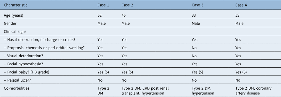
A total of 103 patients with Covid-19-associated mucormycosis were treated in the institute during the study period. Four patients were found to have facial paralysis, with a mean age of 46 years. The clinical and demographical details are summarised in Table 1.
Clinico-demographical details of Covid-19-associated mucormycosis patients with facial palsy

Covid-19 = coronavirus disease 2019; HB grade = House–Brackmann facial nerve palsy grade; DM = diabetes mellitus; CKD = chronic kidney disease
All patients underwent surgical debridement, with post-operative medical management comprising liposomal amphotericin B and oral posaconazole. The facial nerve was not explored in any of the four cases.
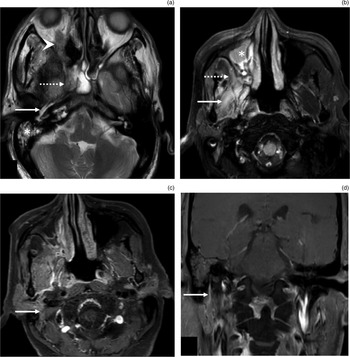
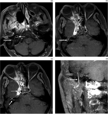
The radiological details are summarised in Figures 1 and 2. Radiological evaluation revealed diffuse facial nerve thickening extending proximally from the stylomastoid foramen to the internal auditory canal (Figures 2a–d). Facial nerve enhancement was seen on the contrast-enhanced MRI, with the CT scan demonstrating an intact facial canal. In all four cases, there was radiological evidence of inflammation in the infratemporal fossa, which extended laterally as fibro-fasciitis along the lateral pterygoid muscle (Figure 1c). A similar inflammatory pattern was evident in the deep lobe of the parotid (Figure 1c) and at the exit zone of the facial nerve in the stylomastoid foramen. Ascending neuritis, with enhancement of the facial nerve to the internal auditory canal, could be demonstrated in all cases. Middle-ear effusion was observed in three-quarters of cases (Figure 1a). The disease abutting the facial nerve tends to produce ascending facial neuritis, causing facial palsy.
Case one of facial neuritis in coronavirus disease 2019 associated mucormycosis. (a) Axial, T2-weighted magnetic resonance imaging (MRI) scan shows mastoiditis (asterisk) and T2 hyperintensity in the Eustachian tube (arrow). The T2 hypointense soft tissue is also seen in the right orbit (arrowhead) and right sphenoid sinus (dotted arrow). (b) Axial, fluid-attenuated inversion recovery (‘FLAIR’) MRI scan demonstrates extensive inflammation in the right maxillary sinus (asterisk), retro-antral space (dotted arrow) and masseteric space (arrow). (c) Axial, post-contrast, fat-suppressed, T1-weighted MRI scan shows abnormal enhancement extending from the right maxillary sinus, through the infratemporal fossa to the region of stylomastoid foramen (arrow). (d) Coronal, post-contrast, fat-suppressed, T1-weighted MRI scan demonstrates abnormal enhancement along the mastoid segment of the right facial nerve (arrow).

Case two of facial neuritis in coronavirus disease 2019 associated mucormycosis. (a–c) Axial, post-contrast, fat-suppressed, T1-weighted magnetic resonance imaging (MRI) scans, from a cranial to caudal direction, reveal abnormal, asymmetric enhancement of the facial nerve from the stylomastoid foramen to the internal auditory canal. There is enhancement of the mastoid segment (arrow in part (a)), the tympanic segment (arrow in part (b)), and the labyrinthine segment and first genu (arrow in part (c)). The internal carotid artery wall is also enhanced (dotted arrow in part (a)). Active inflammation is noted in the right ethmoid sinus, orbit, orbital apex and cavernous sinus (dotted arrow in part (c)). (d) Coronal, maximum intensity projection MRI image shows the abnormally enhanced mastoid segment of the facial nerve (arrow).

All patients underwent surgical debridement of the nose, paranasal sinuses and infratemporal fossa via an external approach. The mean duration of hospital stay was 63 days (range, 22–84 days). The detailed treatment protocols are still under investigation. We have employed a combination of liposomal amphotericin B and oral posaconazole. The patients received a cumulative mean dose of 5065 mg of liposomal amphotericin B (range, 3250–6450 mg) and 14 200 mg of oral posaconazole during admission.
All patients were discharged on oral posaconazole 300 mg once daily, which was continued for a mean duration of 52 days. The absence of pain and the presence of healthy mucosa, along with the absence of any necrotic tissue on clinical evaluation conducted via endoscopy, and radiological evidence showing no active disease on contrast-enhanced MRI, determined treatment completion. Three of the four patients completed treatment, while one (case one) died as a result of septic shock. The mean follow-up time was four months (range, 3–5 months). So far, there has been no improvement in facial nerve function in the three surviving patients.
Discussion
Acute invasive fungal sinusitis caused by Mucorales, termed as sinonasal mucormycosis, is a rapidly progressive devastating infection of the nose and paranasal sinuses, with a tendency for contiguous spread to the orbits, infratemporal fossa, brain and skin. Optimal treatment involves surgical debridement of necrotic tissues followed by medical management with liposomal amphotericin B, regarded as the first-line medical management in such patients.Reference Cornely, Alastruey-Izquierdo, Arenz, Chen, Dannaoui and Hochhegger1
Coronavirus disease 2019 associated mucormycosis has taken the form of an epidemic in the Indian subcontinent. Along with the common presentation of nasal blockage, facial hypoesthesia, proptosis, chemosis, dental pain and palatal ulcer in mucormycosis, several clinicians have noticed the presence of facial paralysis in Covid-19-associated mucormycosis patients. The available literature has reports of facial paralysis in rhino-cerebral mucormycosis; however, the exact pathophysiology of facial nerve involvement has not been explored. We evaluated the potential route of facial nerve involvement in Covid-19-associated mucormycosis through clinico-radiological correlation.
Facial paralysis has been reported in few isolated cases of mucormycosis. Mane et al. reported facial nerve paralysis in four patients with mucormycosis.Reference Mane, Patil, Mohite and Mohanty2 All patients were diabetic, and underwent surgical debridement followed by liposomal amphotericin B treatment. Two patients expired during ongoing treatment, while the other two showed improvement in facial nerve function following the completion of treatment. The authors speculated the pterygopalatine fossa to be the reservoir of infection in mucormycosis, with the spread of infection proceeding to the inferior orbital fissure, orbital apex and infratemporal fossa, as was described by Hosseini and Borghei.Reference Hosseini and Borghei3
In our study, involving four patients, disease in the sinonasal cavity travelled through the pterygopalatine fossa towards the infratemporal fossa via the pterygomaxillary fissure, and led to fibro-fasciitis of the lateral pterygoid muscle. Lateral extension along the lateral pterygoid muscle, resulting in inflammatory spread to the deep lobe of the parotid and the facial nerve at the exit from the mastoid (stylomastoid foramen), seems the most plausible explanation of facial palsy.
In a similar case scenario, Shekar et al. reported sinonasal orbital mucormycosis in a 75-year-old man with lower motor neuron facial paralysis and no alteration or loss of taste sensation.Reference Shekar, Sikander, Rangdhol and Naidu4 Facial nerve palsy has been reported in cases with isolated involvement of the parotid gland by mucormycosis, leading to Mucor parotitis.Reference Chandu, MacIsaac, MacGregor, Campbell, Wilson and Bach5,Reference Gochhait, Alexender, Dehuri, Rangarajan, Jacob and Siddaraju6
Invasion of the peripheral nerves in mucormycosis has also been reported in the literature, and the organism may travel along the peripheral nerves.Reference Margo, Linden, Strickland-Marmol, Denietolis, McCaffrey and Kirk7–Reference McLean, Ginsberg and Stanton9 The authors of these papers identified ascending neuritis of the trigeminal nerve on radiological evaluation, which was confirmed on histopathological assessment. Facial nerve involvement at the stylomastoid foramen through similar ascending neuritis is a strong possibility.
Some authors have hypothesised similar involvement of the Vidian nerve by the organism and ascending involvement of the geniculate ganglion.Reference Mane, Patil, Mohite and Mohanty2,Reference Aloosi10 In our case series of four patients, this explanation seems unlikely. There was no radiological enhancement along the Vidian nerve. In order to further validate our hypothesis, we performed Schirmer's test in two patients and both had normal lacrimation.
Another possible explanation for facial palsy is the reactivation of latent ‘viral’ infection, given the considerable immune compromise in such cases. Facial nerve involvement in diabetic patients has been described previously in Bell's palsy. It is hypothesised to be due to the pathology of resistance arteries, leading to facial nerve oedema and ischaemia.Reference Mane, Patil, Mohite and Mohanty2 The clear demonstration of intense inflammation on MRI and the uniform involvement of extratemporal facial nerve areas in all cases makes direct inflammation of the nerve by fungal elements a more likely explanation. Although middle-ear effusion was present in three of our patients, the involvement of the facial nerve in the intra-temporal segment by the effusion does not seem plausible, as the facial nerve is a robust nerve with good protection within a bony fallopian canal. In addition, middle-ear effusion is a common disease and does not usually cause facial palsy.
• Facial weakness has been observed in patients with coronavirus disease 2019 (Covid-19) associated mucormycosis
• Complete facial palsy is a rare complication of Covid-19-associated mucormycosis
• This study examined the clinico-radiological details of four Covid-19-associated mucormycosis patients presenting with facial palsy
• Serial radiological evaluation revealed disease extension from the paranasal sinuses along the fascial planes, involving the infratemporal fossa and reaching the stylomastoid foramen
• Ascending neuritis with enhancement of the facial nerve from the stylomastoid foramen to the internal auditory canal was demonstrated in all cases
• The prognosis of facial nerve function recovery was poor, despite medical and surgical treatment of Covid-19-associated mucormycosis
To the best of our knowledge, ours is the first study to elucidate the route of facial nerve involvement in Covid-19-associated mucormycosis. Familiarity with this route may better enhance our understanding of the pathogenesis of this manifestation.
Conclusion
We have described the clinical sign of facial palsy associated with Covid-19-associated mucormycosis. The possible explanation, backed by radiology, is disease or gangrene abutting the facial nerve leading to ascending neuritis of the facial nerve. The reactivation of Bell's palsy or middle-ear pathology as the cause of nerve damage seems unfounded because of the said description.
Competing interests
None declared