Introduction
Middle aortic syndrome is a rare clinical condition marked by segmental or diffuse narrowing of the abdominal or distal descending thoracic aorta, which may involve the renal arteries, coeliac artery, superior mesenteric artery (SMA), and other associated branches. Middle aortic syndrome accounts for only 0.5–2%, of all cases involving aortic narrowing. Reference Lazea1 The aetiology of middle aortic syndrome is incompletely understood. Congenital middle aortic syndrome is thought to result from aberrations in the fusion of the dorsal aorta during development, while acquired cases have been associated with genetically related vasculopathies. Reference Lazea1 These encompass a range of hereditary conditions including Williams syndrome, non-syndromic elastin arteriopathy, neurofibromatosis type-1, as well as autoimmune disorders such as Takayasu’s arteritis and giant cell arteritis. Reference Patel2–Reference Opoka-Winiarska5
Middle aortic syndrome typically presents in children or adolescents, with severe hypertension being the most common symptom. Other manifestations may include claudication, leg weakness, dyspnoea, headache, abdominal pain, nausea, vomiting, and eating intolerance. Inflammatory conditions can cause systemic symptoms such as fever, fatigue, or joint pain. Renovascular hypertension is the most prevalent complication of middle aortic syndrome. Reference Radford and Pohlner4 In severe cases, middle aortic syndrome can lead to left ventricular hypertrophy, diastolic heart failure, mesenteric ischaemia, renal failure, and limb ischaemia, contributing to significant morbidity and mortality. Reference Patel2,Reference Porras6 When medical management is insufficient in treating middle aortic syndrome, patients may necessitate interventions such as open surgical fixation or endovascular procedures. Reference Forman, Sinskey and Shalabi7
Williams syndrome is a congenital condition characterised by a microdeletion on chromosome 7, which involves the ELN gene responsible for encoding elastin. Reference Pober Barbara8 This deficiency in elastin results in cardiovascular anomalies, particularly arterial stenoses, which are prevalent in approximately 80–90% of Williams syndrome cases and typically emerge early in life. Reference Collins, Kaplan, Somes and Rome9,Reference Collins, Kaplan, Somes and Rome10 Among these anomalies, supravalvar aortic stenosis affects up to 65% of cases, while peripheral pulmonary artery stenosis is observed in approximately 61% of individuals. Reference Collins11 Additionally, various other less frequent lesions may manifest as part of this syndrome. The incidence of middle aortic syndrome remains largely unknown. In this case report, we detail the discovery of middle aortic syndrome in a 16-year-old female patient diagnosed with Williams syndrome admitted for evaluation of abdominal pain and hematochezia.
Case presentation
The patient is a 16-year-old female with a medical history notable for Williams syndrome, iron deficiency anaemia, and constipation-predominant irritable bowel syndrome. She presented to our institution for admission and evaluation with esophagogastroduodenoscopy and colonoscopy due to a three-month history of anaemia with hematochezia, in the context of chronic abdominal pain of unknown aetiology.
The patient’s Williams syndrome was originally diagnosed in infancy, with longstanding follow-up for cardiovascular and neurodevelopmental manifestations. Her most recent echocardiogram prior to the identification of mid-aortic syndrome was performed on April 17, 2023. It demonstrated structurally normal cardiac anatomy with no evidence of supravalvar aortic stenosis, aortic coarctation, or obstruction involving the aortic arch or descending thoracic aorta. The aortic valve annulus (18.5 mm, Z = 0.6), aortic root (24.3 mm, Z = 0.2), sinotubular junction (19.0 mm, Z = −0.3), and ascending aorta (19.7 mm, Z = −0.7) were all within normal limits for her body surface area using Boston Children’s Hospital Z-scores. Importantly, the descending thoracic aorta at the level of the diaphragm measured 9.6 mm in systole, corresponding to a Z-score of − 1.75 based on the normative data published by Lopez et al., as Boston Z-scores are not available for this segment. Reference Lopez12 Although derived from different sources, this contrast in Z-scores suggests narrowing at the diaphragmatic level, consistent with mid-aortic syndrome. Biventricular systolic function was normal (Ejection Fraction 64.8%, Z = 0.3 by the 5/6 area-length method), with a left ventricular end-diastolic diameter of 34.8 mm (Z = −2.9) and LV mass of 69 g (Z = −1.1). Mild tricuspid valve prolapse was noted without regurgitation. The EKG obtained on the same day showed sinus rhythm with right ventricular hypertrophy and a QTc of 420 ms, which is within normal limits. These findings support preserved myocardial function and chamber size, with no evidence of significant valvular or proximal aortic obstruction at the time of the study. The descending aortic diameter at the level of the diaphragm (9.6 mm, Z = −1.75) is notably smaller than the more proximal aortic measurements, all of which were within normal limits. This contrast underscores the segmental nature of the narrowing and supports the diagnosis of mid-aortic syndrome.
Three months prior to her admission, the patient developed hematochezia, fatigue, and dizziness prompting a visit to the emergency department. At the time of presentation, the frequency of bloody stools had decreased, her stools remained firm and non-painful, and abdominal ultrasound revealed no acute findings. Given these observations, the gastrointestinal service determined that the likelihood of brisk gastrointestinal bleed was low, and an emergent scope was not indicated. She was discharged with return precautions.
Two months prior to her admission, the patient returned to the emergency department for symptomatic anaemia. Earlier that day, a routine office visit with her paediatrician revealed fatigue, pallor, and a Hgb of 8.3 mg/dL, leading to her referral to the emergency department. A workup in the emergency department was unrevealing, and she was discharged with outpatient gastrointestinal follow-up, initiation of iron supplementation, and return precautions.
During a scheduled outpatient gastrointestinal follow-up, the family and gastrointestinal provider agreed to proceed with a scheduled endoscopy to investigate the patient’s recurrent episodes of symptomatic anaemia. Coordination with our institution’s Williams syndrome cardiology team was undertaken to prepare for cardiac anaesthesia as a precaution given her connective tissue disorder, and the procedure was scheduled during a planned admission.
On admission, the patient reported poor appetite, fatigue, and nausea without emesis but denied abdominal pain. Laboratory testing revealed a Hgb of 11.5 at admission and normal renal function. Physical examination revealed mild peri-umbilical tenderness and an abdominal bruit, particularly notable in the periumbilical region. Oesophagogastroduodenoscopy revealed findings consistent with lymphoid hyperplasia and gastritis, and biopsy results confirmed collagenous gastritis (Figure 1).
Nodular (lymphoid-like) mucosa in the gastric body.

Considering the patient’s Williams syndrome diagnosis, her case was discussed with the Stanford Cardiovascular Connective Tissue Disorders Program team. During prior cardiology evaluations, a bruit had been noted over the length of the abdominal aorta, without renal bruits, and slight toe discolouration, likely related to positioning, had been observed without skin breakdown. Although a CT angiography was considered at the time, it was deferred as ambulatory blood pressure monitoring did not reveal hypertension, and there was no evidence of left ventricular hypertrophy. However, given her current presentation, a CT angiography of the abdomen was performed due to concern for middle aortic syndrome.
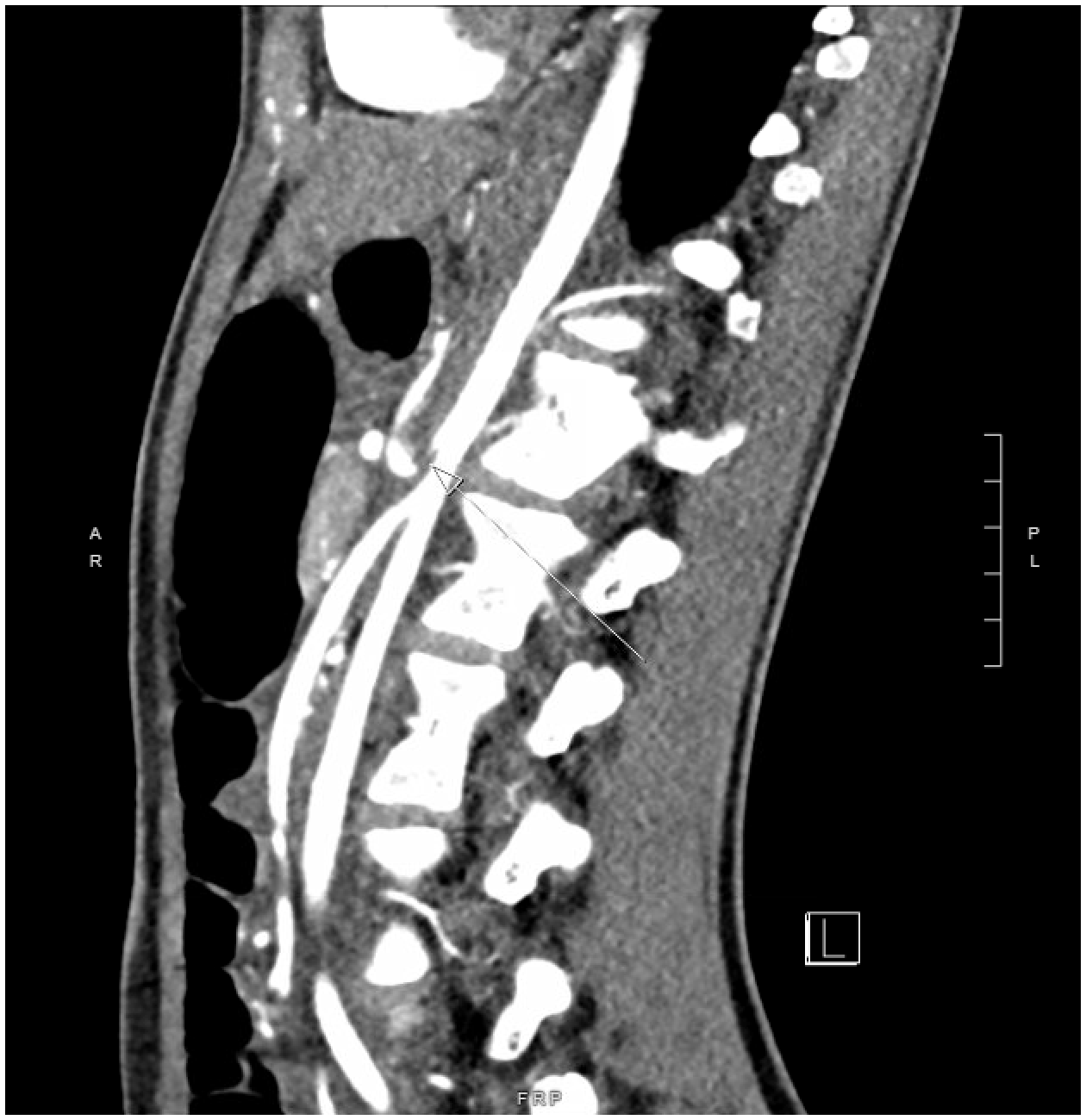
The CT angiography revealed a 4.5 cm segment of moderate narrowing of the abdominal aorta at the intrarenal segment (Figure 2), measuring 0.4 x 0.9 centimetres in diameter, consistent with middle aortic syndrome. Additionally, there was complete fusiform narrowing near the origin of the coeliac artery, with reconstitution of flow distally, suggesting chronic occlusion with collateral formation and restored distal blood flow (Figure 3). Minimal narrowing at the ostium of the superior mesenteric artery and a nondistended hiatal hernia were also noted.
Axial abdominal CT angiogram with intravenous contrast demonstrating aortic narrowing at the intrarenal segment.

Sagittal abdominal CT angiogram with intravenous contrast demonstrating complete fusiform narrowing of the proximal coeliac artery.

Four-point blood pressure measurements did not reveal significant pressure differentials across the extremities. Evaluation by vascular surgery concluded that collateral circulation likely provided adequate flow to compensate for the narrowing near the coeliac artery origin. At the time of admission, the patient exhibited no signs or symptoms of tissue ischaemia. She denied abdominal pain, postprandial nausea or vomiting, or appetite loss, and her weight was stable. On vascular examination, bilateral lower extremities pulses were palpable without any neurological deficits, cyanosis, or oedema.
Given the invasive nature of surgical intervention for middle aortic syndrome, combined with her well-perfused status and absence of clinical signs of mesenteric ischaemia or renal hypoperfusion, vascular surgery determined that the risks of surgery outweighed the potential benefits. The patient was scheduled for outpatient follow-up to monitor her condition, with plans for further evaluation should she develop refractory hypertension, severe claudication, or mesenteric ischaemia that might necessitate surgical intervention.
Discussion
This case report describes a 16-year-old female paediatric patient with Williams syndrome who presented with abdominal pain and hematochezia, ultimately diagnosed with middle aortic syndrome based on CT angiography findings.
The association between middle aortic syndrome and Williams syndrome, though rare, has been documented in the literature. Hall et al (2007) described an infant with rapidly progressive middle aortic syndrome requiring multiple surgical interventions before two months of age, noting that such early presentations are atypical. Reference Hall13 More commonly, middle aortic syndrome associated with Williams syndrome presents in late childhood and early adulthood, consistent with the presentation in this case. Reference Hall13
The true incidence of middle aortic syndrome in Williams syndrome is unknown, largely due to the rarity of Williams syndrome itself and the limited studies exploring its association with other conditions. Despite the paucity of data, it should not be assumed that middle aortic syndrome has a low or negligible incidence in this population. The aetiology of Williams syndrome, involving a microdeletion on chromosome 7 involving the ELN gene responsible for elastin production, provides a compelling and biologically plausible explanation for the development of middle aortic syndrome.
In this case report, the patient’s presentation differed from most cases of middle aortic syndrome in two ways. First, middle aortic syndrome is most commonly found in isolation rather than in association with Williams syndrome. Second, middle aortic syndrome typically presents with severe hypertension as the primary symptom. It is possible that the true prevalence of middle aortic syndrome is significantly higher than currently estimated, as the diagnosis this patient was made subsequently by CT angiography imaging after months of investigating non-specific symptoms, suggesting that CT angiography imaging may be indicated in patients with Williams syndrome. Many patients with Williams syndrome—or otherwise—may exhibit a spectrum of middle aortic syndrome, with only the most advanced cases presenting with severe hypertension or other symptoms leading to diagnostic imaging.
An interesting finding during this patient’s workup was the presence of collagenous gastritis confirmed with biopsy. A thorough literature review and consultation with paediatric gastroenterology at our institution did not identify any previously reported cases of collagenous gastritis in Williams syndrome. There is no established therapy for collagenous gastritis, in part given the rarity of the disease. Reference Kamimura, Kobayashi, Sato, Aoyagi and Terai14 This patient received proton pump inhibitor therapy with omeprazole, which provided only minimal symptom relief. Her symptoms, including chronic abdominal pain persisting for two years, are likely multifactorial in origin, though it remains unclear whether Williams syndrome or middle aortic syndrome predisposed her to the development of collagenous gastritis.
Determining whether this patient’s symptoms were due to collagenous gastritis, middle aortic syndrome, or a combination of the two has significant implications for management. While the relative contributions of these conditions to her symptoms cannot be definitively determined, this case highlights the importance of considering angiography in patients with Williams syndrome who present with hypertension or other symptoms suggesting insufficient blood flow to organs supplied by potentially narrowed vascular segments, especially if an abdominal bruit is auscultated.
Although collagenous gastritis is an uncommon cause of hematochezia, other causes of hematochezia included haemorrhoids, Inflammatory Bowel Disease (IBD), Gastroesophageal Reflux Disease (GERD), and duodenitis, which were ruled out during her admission. Additionally, the patient’s hematochezia resolved with PPI initiation and supportive care. During the months that followed discharge and until the time of writing, the patient’s symptoms have remained quiescent.
Management of middle aortic syndrome may involve medical or surgical approaches, depending on the symptom burden and overall risk to the patient. Surgical intervention carries substantial risks, with reported complication rates of 9% and mortality rates between 2.9 and 4%. Reference Musajee15 The primary goals of surgery are to reverse end-organ damage and control blood pressure in patients with severe hypertension, evidence of end-organ damage, or significant physical debilitation. Reference Musajee15 In paediatric patients, surgical intervention is often deferred until they have reached full adult growth potential. Reference Musajee15 In this case, the patient’s relatively mild symptom burden and reassuring findings—including normal four-point blood pressure measurements, adequate SMA flow and coeliac artery collateralisation, and no signs of tissue ischaemia—supported the decision to defer surgery in favour of outpatient monitoring.
This case report has certain limitations. It describes a single patient, making the findings and management strategies potentially less generalisable. The presence of two coexisting conditions, middle aortic syndrome and collagenous gastritis, complicates the analysis of their independent or combined contributions to the patient’s symptoms. The short-term follow-up limits our understanding of potential long term, including disease progression or complications. Nonetheless, given the rarity of this middle aortic syndrome and the limited studies on its association with Williams, this report adds valuable insight to the modest body of literature available for clinicians managing similar cases.
Conclusion
Patients with Williams syndrome are predisposed to elastin arteriopathies due to a deletion involving the ELN gene at chromosome 7q11.23, providing a plausible aetiology for middle aortic syndrome. This case of a 16-year-old female patient with Williams syndrome and middle aortic syndrome highlights the potential for underdiagnosis, as angiography is often required for detection. While rare, middle aortic syndrome is a serious condition that should be considered in patients with Williams syndrome presenting with signs of arterial insufficiency or hypertension.