Introduction
Stroke is a major risk factor for dementia. The risk of incident dementia in stroke patients is about four-fold relative to controls (Kokmen et al., Reference Kokmen, Whisnant, O'Fallon, Chu and Beard1996) and three times higher in subjects over 75 years than in younger stroke survivors (Lowery et al., Reference Lowery2002). However, the neurobiological basis of this increased risk is unclear, as studies investigating the association between brain changes and the development of post-stroke dementia (PSD) have shown inconsistent results. Besides white matter hyperintensities (WMH), which have been strongly associated with cognitive impairment and dementia in stroke patients (Pasquier et al., Reference Pasquier, Hénon and Leys2000; Jokinen et al., Reference Jokinen, Kalska, Mäntylä, Ylikoski, Hietanen and Pohjasvaara2005), brain infarcts (on the basis of number and size or volume) (Vermeer et al., Reference Vermeer, Prins, den Heijer, Hofman, Koudstaal and Breteler2003), medial temporal (Cordoliani-Mackowiak et al., Reference Cordoliani-Mackowiak, Hénon, Pruvo, Pasquier and Leys2003; Firbank et al., Reference Firbank2007), and hippocampal atrophy (Sachdev et al., Reference Sachdev, Chen, Joscelyne, Wen, Altendorf and Brodaty2007; Firbank et al., Reference Firbank2011) have demonstrated controversial results. Subcortical gray matter structures have also been implicated in cognitive impairment. Thalamic infarcts were associated with memory impairment in a sample of patients with Alzheimer's disease (AD), vascular dementia and mixed dementia (Swartz and Black, Reference Swartz and Black2006). Lacunes in the thalamus and basal ganglia explained 5% and 6% of Clinical Dementia Rating (CDR) Scale variability, respectively, in older people having dementia by clinic-pathological assessment (Gold et al., Reference Gold2005), and putamen and caudate nucleus atrophy has been observed in subjects with AD (de Jong et al., Reference de Jong2008), suggesting a possible role in cognitive decline.
Overall, inconsistent results have been reported in terms of imaging correlates of the increased risk of dementia after stroke. Although we have reported findings of correlates of delayed dementia in older people after stroke, which included a contribution from both hippocampal atrophy and WM lesions (Firbank et al., Reference Firbank2011), we did not examine the possible contribution of subcortical gray matter structures. The aim of this study therefore was to investigate changes in subcortical gray matter structures (thalamus, putamen, and caudate nucleus) in older subjects with PSD, compared to post-stroke no dementia (PSnoD), AD, and healthy controls, by utilizing high-resolution structural magnetic resonance imaging (MRI).
Methods
Participants
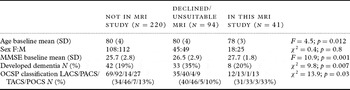
Stroke patients taking part in this imaging investigation were from a previously described cohort (Firbank et al., Reference Firbank2011), which has been followed longitudinally. The subjects were recruited consecutively from hospital-based stroke registers in Tyneside and Wearside (UK), and were aged over 75 years at the time of the stroke, defined using the World Health Organization criteria. Patients were screened at three months to exclude dementia using DSM-IIIR criteria, or those with a Mini-Mental State Examination (MMSE) <24. Subjects underwent annual clinical and cognitive assessments and were invited for an MR scan on average 6.2 years (SD 0.86) after the initial stroke. Of the original 355 stroke subjects, 135 were still undergoing regular study follow-up (153 had died and 67 had refused follow-up). Of these 135 subjects, 41 agreed to an MRI scan (49 refused and 45 had contra-indications to imaging). See Table 1 for details of participants.
Comparison of demographics of those in the current study versus the original cohort

OCSP = Oxfordshire Community Stroke Project, LACS/PACS/TACS/POCS = lacunar/partial anterior/total anterior/posterior cortical stroke.
We also recruited 26 subjects with AD, from a memory clinic and local geriatric medicine services, diagnosed by consensus between two experienced clinicians according to NINCDS-ADRDA criteria (probable) (McKhann et al., Reference McKhann, Drachman, Folstein, Katzman, Price and Stadlan1984), and 30 older controls drawn from the spouses of the stroke subjects and from the community. Controls had no history of stroke, major heart disease, or dementia, and an MMSE score of >25. The appropriate local research ethics committees granted ethical approval for the study. Subjects and/or next of kin gave their consent to participation in the study.
Cognitive and neuropsychological evaluations
Cognitive and neuropsychological tests consisted of a range of cognitive tasks, from global and specific domains. The Cambridge Cognitive Examination (CAMCOG), which incorporates the MMSE and includes a sub-scale for executive function, was used to test the global cognitive performance. The Informant Questionnaire on Cognitive Decline in the Elderly (IQCODE) assessed the cognitive and functional performance, having been administered to a close acquaintance of the subject. The Bristol Activities of Daily Living Scale assessed functional ability, the Full Scale IQ predicted from the NART (National Adult Reading Test) assessed the pre-morbid intellectual ability.
Tests of specific cognitive domains were performed: the Boston Naming Test was used as a language measure, the verbal fluency FAS Test was used to assess language and executive function, and the Rey Auditory Verbal Learning Test (RAVLT) as a memory measure assessed immediate memory plus efficacy of learning (adding first five trials) and recall following a long period (30 minutes).
Dementia diagnosis
In the stroke group, neuropsychological evaluations as per baseline were repeated annually. If the participant was too impaired to complete an MMSE, the MMSE score was ≤24, had fallen by ≥2 points or they had other evidence or suspicion of dementia, the assessor completed the CDR Scale and IQCODE, with an informant. All assessments were reviewed by the assessor and a senior clinician (Old Age Psychiatrist or Geriatrician) to determine whether the participant met the DSM-IIIR criteria (American Psychiatric Association, 1987) that were chosen because they include a general definition for dementia, unlike DSM-IV that presumes etiology, and we wished to include all cases of PSD. Dementia was diagnosed in 8 of the 41 post-stroke subjects.
MRI methods
Acquisition parameters
All subjects were imaged using a 3-Tesla Philips Intera MR system (Philips Medical Systems, Best, The Netherlands). The imaging protocol consisted of turbo spin echo, dual echo (repetition time [TR] = 3,000 msec; echo time [TE] = 18/80 msec; turbo factor = 10; field of view 250 × 187.5 mm2; 50 axial slices; 3-mm thick acquisition; time = 5 minutes), T1-weighted 3D MPRAGE (magnetization-prepared gradient echo) (TR = 9.6 msec; TE = 4.6 msec; flip angle = 8°; inversion delay = 1,250 msec; field of view 240 × 240 mm2; 150 sagittal slices; 1.2-mm slice thickness; time = 3.59 minutes) and FLAIR (fluid-attenuated inversion recovery) (TR = 11,000 msec; inversion time [TI] = 2,800 msec; TE = 125 msec; turbo SE factor = 27; refocus angle = 120; field of view 260 × 207 mm2; 50 axial slices; 3-mm slice thickness; 4:20 minutes).
Image processing
Identification of lacunar infarcts in thalamus, putamen, and caudate nucleus: Assessment of lacunar infarcts was performed in the coronal, axial, and sagittal planes by a single rater who was blinded to clinical information. Rating was performed after a period of training in familiarization of the appearance of lacunes on MR images. Infarcts were defined as between 3 and 10 mm in size and mostly ovoid or wedge shaped (Bokura et al., Reference Bokura, Kobayashi and Yamaguchi1998), being hyperintense on T2 or hypointense on FLAIR (with or without hyperintense rim). T1 was used to identify the location of the lesions. As the perivascular spaces (Virchow Robin spaces) have the same signal as lacunar infarct in the three sequences and are very frequent in basal ganglia, they were defined as lesions smaller than 3 mm in size, with the exception of lesions near to anterior commissure. We assessed reliability by a second observer counting lacunes on 12 cases (six strokes, three AD, three controls). The intra-class correlation coefficient between the two raters was 0.84.
Volumetric assessment of thalamus, putamen, and caudate nucleus: The volume of subcortical structures was measured from the 3D T1 images using the software FIRST, which is a model-based segmentation/registration tool part of FMRIB's Software Library (FSL) (http://www.fmrib.ox.ac.uk/fsl/). The raw volume of the three structures was calculated. In order to check the reliability of the automated segmentation specifically in a post-stroke sample, 11 cases (nine post-stroke and two controls) had manual segmentation of the thalamus, four of them twice. The thalamic boundary delineation was adapted from Portas et al. (Reference Portas1998) and supported by two neuroanatomy atlases. The manual drawing of the thalamus was conducted on 2-mm consecutive slices, in axial and coronal planes from T1-weighted images simultaneously, using the ITK-SNAP software (http://www.itksnap.org). The intra-rater reliability for left and right volumes was 0.93 and 0.91, respectively, calculated as conditional percentage agreement (overlap). The overlap between manual and automated methods was 0.82 and 0.85 for the left and right, respectively.
Volumetric assessment of hippocampus: The volume of the hippocampus was obtained from 3D T1 images using a previously validated automated method (Firbank et al., Reference Firbank2011). Briefly, the images were segmented into gray matter and WM and spatially normalized to MNI space using SPM5 (http://www.fil.ion.ucl.ac.uk/spm/). Following this, hippocampus gray matter was identified within a template in MNI space. Total brain volume (TBV) (gray matter plus WM) and intracranial volume (TBV plus CSF) were also calculated from the segmentation in SPM.
WMH volume: The volumetric assessment of WMH was carried out from FLAIR images using previously validated automated software (Firbank et al., Reference Firbank2011). Briefly, the brain was segmented using SPM5 and an intensity threshold for each slice was applied to segment the WMH. The accuracy of this was checked visually for each subject. A region of interest (ROI) map of the major lobes of the brain in standard space was spatially transformed to each subject using the spatial normalization calculated by SPM5. Finally, the total WMH volume and the WMH volume in each ROI were calculated.
Statistical analysis
The data were analyzed using the statistical package SPSS version 17.0 for Windows. The distribution of demographic, cognitive, and neuropsychological variables across the four groups was explored using ANOVA with Scheffe post-hoc test for age, χ2 test for gender and Mann-Whitney test for cognitive and neuropsychological measures. Regarding the MRI characteristics, the differences between groups were obtained by Mann-Whitney test for lacunes and ANOVA with Fisher least significant difference (LSD) and Scheffe post-hoc tests for volume (the latter correcting for multiple comparisons). WMH measures were log-transformed due to skewness of the raw volumes and the transformed measures were used in all analyses. Additionally, a logistic regression for each pair of comparisons (AD vs. controls and PSD vs. PSnoD) was conducted, using the MRI measures significantly correlated in Fisher LSD test. Specifically in the whole post-stroke sample, three types of analysis were performed: (1) correlation between MRI measures, which was obtained using Spearman's correlation for lacunes and Pearson's correlation for the others; (2) correlation between cognitive/neuropsychological scores and MRI measures, using Spearman's correlation; (3) linear regression analysis that was performed for each cognitive/neuropsychological variable, with the inclusion of variables of age, total brain, and hippocampal volumes and additionally those WMH measures and subcortical volumes significantly correlated in the bivariate analysis (one regression for each significant WMH measure).
Results
Table 1 compares those who participated in this study with those from the original cohort without MRI. The group studied here was slightly older, had a slightly higher baseline cognitive score, and relatively more posterior cortical strokes.
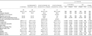
Group demographic and cognitive/neuropsychological features are summarized in Table 2. While the age was very similar across the groups, a higher proportion of females was evident in the AD group when compared with PSnoD group (76.9% vs. 36.4%). The PSD group was, in general, not different cognitively from the AD group, both having consistently lower cognitive scores than controls. In addition, PSnoD and PSD were not significantly different in relation to clinical features, such as smoking (56.3% vs. 50%), hypertension (61.3% vs. 62.5%), diabetes (12.5% vs. 0), and heart disease (41.9% vs. 62.5%).
Demographic, cognitive, and neuropsychological measures for diagnostic groups

NS = not significant.
Age, all cognitive and neuropsychological numbers are mean ± standard deviation. Gender is percentage. NART = full scale IQ estimated from the National Adult Reading Test, RAVLT = Rey Auditory Verbal Learning Test; T6 = trial 6; LOT = learning over trials; OCSP = Oxfordshire Community Stroke Project; LACS/PACS/TACS/POCS = lacunar/partial anterior/total anterior/posterior cortical stroke; APOE = apolipoprotein E; BP = Blood pressure; MMSE = Mini-Mental State Examination; CAMCOG = Cambridge Cognitive Examination; IQCODE = Informant Questionnaire on Cognitive Decline in the Elderly; ADL = activities of daily living; p-value assesses the difference between groups by ANOVA with Scheffe post-hoc test for age, χ2 test for gender and Mann–Whitney test for cognitive and neuropsychological measures.
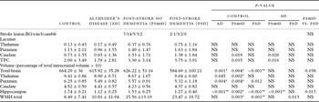
MRI characteristics for diagnostic groups are presented in Table 3. Location and size of the original stroke was not significantly different between dementia and non-dementia groups, although there was a trend (Fisher p = 0.08) for cortical lesions to be larger in PSD (5/6 > 15 mm) compared to PSnoD (14/23 > 15 mm). The number of lacunes was significantly higher only in caudate nucleus, in PSnoD group when compared with controls and AD group. Almost all volumetric measures differentiated control from the other groups. Regarding the comparison between AD and both stroke groups, as we previously reported, hippocampal volume was lower in AD than in the PSnoD group, with no difference observed in relation to the PSD group, which had higher WMH than AD group. Showing statistically weaker results, the PSD group exhibited lower total brain and hippocampal volumes than the PSnoD group. Thus, the data comparing the AD group versus controls and PSnoD versus PSD showed that hippocampus and TBVs were lower in both dementia groups. Moreover, while subcortical gray matter volume was also lower in the AD group, WMH was higher in PSD group. Logistic regressions for both pairs of comparisons showed that reduced hippocampal volume remained independently associated with AD (p = 0.007), with smaller volume of putamen exhibiting a trend of association (p = 0.058), while TBV and thalamus were not significant; no measure (out of TBV, hippocampus, WMH) remained independently associated with PSD group versus PSnoD.
MRI characteristics for diagnostic groups, number of lacunes in subcortical structures, and volume of brain structures and hyperintensities

TPC = thalamus, putamen, and caudate; WMH = white matter hyperintensities; NS = not significant. Lacunes numbers are mean ± standard deviation. Volume is adjusted for intracranial volume and multiplied by 1,000, in mean ± standard deviation.
Stroke lesion = size and location of main stroke lesion, BG/s/m/l/cereblr = Basal ganglia/gray + white matter small (<15mm)/medium/large (>30 mm)/cerebellar.
p-value assesses the difference between groups by Mann–Whitney test for lacunes and ANOVA with Fisher LSD post-hoc test for volume (*p < 0.05 and **p < 0.01 for Scheffe post-hoc test).
WMH measures were log-transformed); p < 0.05 is significant.
The correlations investigated in the whole post-stroke sample are presented in Table 4. With regard to the correlation between MRI and cognitive/neuropsychological measures, data in Table 4 show that hippocampal and WMH volumes were, overall, associated with these measures. The hippocampal volume was positively correlated with cognitive performance in all domains whereas higher WMH volumes were associated with worse cognitive function in global measures. Among the subcortical gray matter measures, only putamen lacunes were negatively correlated with memory test (RAVLT, learning over trials; ρ = −0.365; p = 0.031). In a linear regression model of RAVLT, with age, hippocampus, and TBV volume, the model was not significant (F = 1.6; p = 0.2); the addition of putamen lacunes significantly improved the model (F change = 4.9; p = 0.033).
Correlation between cognitive/neuropsychological measures and MRI measures in the post-stroke sample

Numbers are Spearman correlation coefficients. With the exception of putamen lacunes, all MRI variables are volumetric brain measures. WMH measures were log-transformed (using raw data, the result was the same). No association was observed with thalamus and caudate
lacunes, thalamus, putamen, and caudate volumes.
RAVLT = Rey Auditory Verbal Learning Test; T6 = trial 6; LOT = learning over trials; WMH = white matter hyperintensities;
NS = not significant.
*p < 0.05, **p < 0.01: bivariate analysis. †p < 0.01: linear regression controlling for age, total brain, and hippocampus volume.
Discussion
The results of this study show that subcortical gray matter changes contribute to cognitive impairment after stroke, in addition to participation of WMH. We have previously reported that hippocampal atrophy and reduced global cerebral blood flow (CBF) (gray matter/WM CBF ratio, specifically) were associated with cognitive impairment after stroke in this same cohort (Firbank et al., Reference Firbank2011).
There is previous evidence that vascular lesions and atrophy in the thalamus rather than in caudate–putamen correlate with cognitive impairment in people with dementia and healthy individuals (van der Werf et al., Reference van der Werf, Tisserand, Visser, Hofman, Vuurman and Uylings2001; Swartz et al., Reference Swartz, Stuss, Gao and Black2008), which can be physiologically explained by functions of impulse relay (Herrero et al., Reference Herrero, Barcia and Navarro2002). However, in our study the basal ganglia exhibited a more dominant effect, which may relate to the cognitive abnormalities observed in the early stages of neurodegenerative extra-pyramidal syndromes (Borroni et al., Reference Borroni, Turla, Bertasi, Agosti, Gilberti and Padovani2008). We found increased numbers of lacunes in the thalamus in the PSD group, albeit not significantly, and thalamus volume was significantly reduced in PSnoD with a trend in the AD group, compared to controls. More evidently in the putamen, atrophy was greater in both stroke groups versus controls, and the number of lacunes in the putamen was correlated with worse memory performance in the whole post-stroke sample, even controlling for other MRI measures. Furthermore, the putamen atrophy also occurred consistently in AD subjects, showing involvement of basal ganglia structures in both types of dementia. Few studies have investigated the impact of the putamen on dementia, with contrasting results on atrophy and cognition in AD (Cousins et al., Reference Cousins, Burton, Burn, Gholkar, McKeith and O'Brien2003; de Jong et al., Reference de Jong2008). There is growing evidence that the basal ganglia are involved in cognitive function (Grahn et al., Reference Grahn, Parkinson and Owen2008), with the putamen contributing to learning, and the caudate to planning, and lesions in the basal ganglia or the connecting WM are likely to interfere with these cognitive functions. Findings in the present study are in keeping with a recent report of a correlation between vascular lesions in basal ganglia and impairment in reward-based reversal learning (Bellebaum et al., Reference Bellebaum, Koch, Schwarz and Daum2008). Although it has not investigated the mechanism, this study showed the contribution of basal ganglia changes to cognitive dysfunction. Additionally, the presence of “silent brain infarcts” in basal ganglia and thalamus more than doubled the risk of dementia in a prospective population-based study (Vermeer et al., Reference Vermeer, Prins, den Heijer, Hofman, Koudstaal and Breteler2003).
Confirming the known relation of WMH with PSD (Pasquier et al., Reference Pasquier, Hénon and Leys2000; Wen and Sachdev, Reference Wen and Sachdev2004; Jokinen et al., Reference Jokinen, Kalska, Mäntylä, Ylikoski, Hietanen and Pohjasvaara2005) and vascular dementia (Gootjes et al., Reference Gootjes2004) and the pattern of cognitive domains involved (Cohen et al., Reference Cohen2002), WMH was associated with cognitive impairment after stroke, correlating with global dysfunction in the combined stroke group. Reduced hippocampal volume was present in both PSD and AD, though more evident in AD, which has been previously demonstrated in AD and in ischemic vascular dementia (Du et al., Reference Du2002). Corroborating the participation of the hippocampus in PSD patients, the analysis in the combined stroke group exhibited a strong association between lower hippocampal volume and global cognitive impairment, executive dysfunction, and memory problems.
The original cohort was recruited consecutively from stroke registers throughout a wide geographical region in order to get a relatively representative sample of the study group (stroke survivors aged over 75 without dementia). Unfortunately, we do not have information on those who were eligible for the study but declined to take part. The subjects taking part in this study were relatively representative of our original cohort. The current study was limited by the small number of participants in the PSD group, due to the selection process from an existing cohort of well-characterized stroke patients (Firbank et al., Reference Firbank2007). As a consequence, the survival in stroke patients with dementia probably contributed to the inclusion of the healthiest, thereby underestimating the differences between stroke groups (in the comparison between both stroke groups, no significant difference was found in terms of vascular risk factors, such as hypertension, diabetes, and heart disease). Another difficulty was the small number of lacunes in each group, resulting in large confidence intervals and lower levels of accuracy in findings. We defined lacunes as being 3–10 mm in size. Some perivascular spaces can be larger than 3 mm, and although we tried to exclude perivascular spaces by considering shape and location, it is possible that we included some perivascular spaces in our count of lacunes.
It is probable that the location and size of the original stroke infarct would have an influence on cognitive decline, and it may be that there is an interaction between stroke location and presence of basal ganglia infarcts. We did not investigate this possible interaction due to limited numbers, but it is an important topic for future research.
The principal strength of this study was the approach used to correlate the MRI measures and cognition in a sample with evident vascular disease. First, as the stroke group was from a prospective study, we knew that they did not have dementia at the time and three months after the stroke. Second, we avoided specific etiologic diagnosis, such as “vascular cognitive impairment” and “vascular dementia,” because we wished to explore the role of other brain measures besides vascular parameters in PSD. Furthermore, as well as splitting the stroke group according to dementia, we looked at correlations with cognitive function within the whole stroke group, since our primary assumption considered the existence of a spectrum between normal, cognitive impairment, and dementia after stroke. Additionally, the AD group was essential as a known profile of brain changes, not only to compare with stroke groups but also to validate the imaging procedures and principal findings.
Finally, in order to clarify the actual neurobiological substrates of delayed dementia after stroke, neuropathological examination of such subjects is required.
Conflict of interest
None.
Description of authors' roles
Dr Lopes assessed the MR scans, performed the statistical analysis, and wrote the paper. Ms. Widdrington performed the cognitive evaluations and assisted with data management. Dr Firbank assisted in MR and statistical analysis. Professor Kalaria and Professor O'Brien designed the study, and supervised the data collection. Professor Blamire designed and supervised the MR aspects of the study. All authors critically revised drafts of the paper.
Acknowledgments
We are very grateful to the patients and families for cooperating with the investigation of this study. We thank Carein Todd and Jean Scott for assistance in managing and screening the cohort. We also appreciate the assistance of NE-DeNDRoN staff for help in recruiting the AD subjects and controls. Our work is supported by grants from the Medical Research Council (UK), G0500247 and the Alzheimer's Research Trust (UK) and the UK NIHR Biomedical Research Centre for Ageing and Age Related Diseases award to the Newcastle upon Tyne Hospitals NHS Foundation Trust. Dr Lopes was supported by CNPQ (Conselho Nacional de Desenvolvimento Científico e Tecnológico), Brasil.