Congenital polyvalvular dysplasia is rare, occurring in approximately 1/100,000 people.Reference Bartram, Bartelings, Kramer and Gittenberger-de Groot 1 It is characterised by dysplasia of >2 cardiac valves, with all four valves affected in some. The chordae tendinea, papillary muscles, and valve commissures may also be affected. Multiple genetic syndromes are associated with congenital polyvalvular dysplasia, including Trisomy 13 and 18 and Noonan Syndrome, as well as Marfan and Ehlers–Danlos Syndrome.Reference Bartram, Bartelings, Kramer and Gittenberger-de Groot 1 – Reference Rybczynski, Mir and Sheikhzadeh 3 Congenital polyvalvular dysplasia is associated with other intracardiac anomalies, including ventricular or atrial septal defects, pulmonary vein stenosis, tetralogy of Fallot, and coronary anomalies.Reference Bartram, Bartelings, Kramer and Gittenberger-de Groot 1 Little has been reported about the conduction system in these patients.
Atrial arrhythmias are caused by a variety of different mechanisms. Adults and children with enlarged atria may develop atrial arrhythmias such as atrial flutter or fibrillation.Reference Schotten, Verheule, Kirchhof and Goette 4 In patients with severe mitral valve regurgitation, atrial arrhythmia, in combination with severe left atrial enlargement, is considered an indication for surgical intervention, with the goal of repairing the valve, decreasing the left atrial size, and restoring sinus rhythm.Reference Apostolakis and Shuhaiber 5
Here we report a patient with polyvalvular dysplasia diagnosed as a neonate with severe biatrial enlargement, who was diagnosed with an atrioventricular nodal reentrant tachycardia.
Case report
We present the case of a 10-year-old boy who had been noted on foetal echocardiography to have bilateral atrioventricular valve regurgitation. Echocardiography at birth revealed prominent tricuspid valve prolapse with moderate–severe regurgitation, and mild right ventricular and atrial enlargement. There was also bileaflet mitral valve prolapse with mild mitral regurgitation. On examination, the patient was cyanotic – oxygen sats 60–70% – and hypotensive, but responded well to inotropic support and inhaled nitric oxide. These medications were weaned over several days, and the patient was extubated at 10 days. He required supplemental oxygen for the 1st year of life. The patient was followed up closely with annual evaluation in cardiology clinic, including echocardiography and electrocardiography. He demonstrated normal growth and development, and despite his significant valvular regurgitation, he remained relatively asymptomatic. Evaluation by medical genetics including karyotyping, a full metabolic panel, and FISH studies for 22q11 and Williams Syndrome were all unrevealing. The family history was significant for his mother having three prior miscarriages, with one previous child born with severe mitral stenosis who died at 8 days of life.
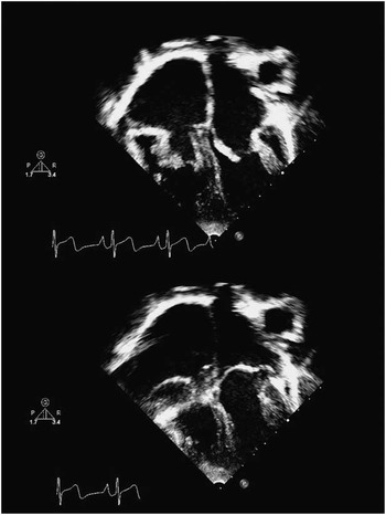
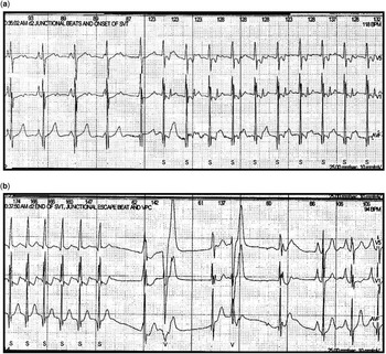
At the age of 8, he presented with a chief compliant of palpitations. Electrocardiography demonstrated a narrow complex tachycardia at 182 bpm. The tachycardia was terminated following administration of facial ice packs. He was managed conservatively, and was started on Nadolol at 10 mg once daily. The patient returned to our institution 1 year later for his yearly evaluation. Chest radiography revealed cardiomegaly with prominent bilateral atrial borders. His echocardiogram revealed stable mitral valve thickening with mild–moderate regurgitation and moderate–severe left atrial enlargement (Fig 1). His tricuspid valve was severely thickened with moderate–severe tricuspid regurgitation and severe right atrial enlargement. Since his initial emergency room visit, he continued to have one to four episodes of self-limited palpations per month, despite increasing his Nadolol to 20 mg daily. These occurred in conjunction with various activities including drinking cold liquids, jumping, swimming, and fear, all of which the patient could use to purposely elicit his tachycardia. The patient had discovered that he could terminate the tachycardia by standing on his head. Ambulatory monitoring during one of his episodes demonstrated both long RP and short RP tachycardia (Fig 2).
Apical echocardiographic image demonstrating bilateral atrioventricular valve thickening – top – and prolapse – bottom.

Holter monitor tracing demonstrating ( a ) an atypical form of atrioventricular nodal reentry tachycardia starting with bradycardia and a junctional escape beat. Note the relatively long RP interval. In the same Holter monitor recording, a more typical form of atrioventricular nodal reentry tachycardia was identified ( b ), ultimately breaking and reverting to sinus rhythm.

The patient was recommended to undergo an electrophysiology study. He was found to have an atypical form of atrioventricular nodal reentry tachycardia occurring between two portions of the slow pathway (Supplementary Fig 1). The inputs to the pathway were located in the posterior septum around his tricuspid valve. Several manoeuvres were performed to confirm the presence of typical and atypical atrioventricular node reentry tachycardia. When tachycardia was induced, premature ventricular beats were placed into the tachycardia. The atrium was not able to be advanced without advancing the His essentially excluding an accessory pathway as a mechanism for the tachycardia. Premature atrial beats were also placed into the tachycardia with a short ventriculoatrial (VA) time. The premature atrial beats were not able to advance tachycardia until the premature atrial contraction was very early, thus making junctional tachycardia very unlikely. With adenosine, the tachycardia slowed, but continued with ventriculoatrial (VA) dissociation, likely representing upper common pathway block.
The patient underwent modification of the slow pathway using cryoablation. At the conclusion of the case, the tachycardia was no longer inducible.
Discussion
Congenital polyvalvular dysplasia is rare and often associated with intracardiac structural abnormalities.Reference Bartram, Bartelings, Kramer and Gittenberger-de Groot 1 Little is available regarding potential conduction system abnormalities and associated arrhythmias in such patients. To the best of our knowledge, this is the first reported case of a successful ablation for atrioventricular nodal reentry tachycardia in a child with true non-syndromic polyvalvular dysplasia.
Bonnet described a series of 22 children with atrioventricular valve dysplasia, of whom three had sustained supraventricular tachycardia.Reference Bonnet, Saygili, Bonhoeffer, Fermont, Sidi and Kachaner 6 In the two patients for whom detail is available, both had dysplasia of the tricuspid, mitral, and pulmonary valves. Interestingly, they were the only patients in the case series not to have been diagnosed with a recognised syndrome.Reference Bonnet, Saygili, Bonhoeffer, Fermont, Sidi and Kachaner 6 These two reported patients are thus similar in presentation to our patient, in that there was no evidence of a specific genetic syndrome, despite the marked valvular abnormalities, with both atrioventricular valves involved.
Daliento et al described two siblings with dysplasia of both the mitral and tricuspid valves.Reference Daliento, Nava, Fasoli, Mazzucco and Thiene 7 These children were both undergoing evaluation for accompanying atrial septal defects and had no history of arrhythmias. During surgical repair of the defects, the younger sibling had difficulty separating from cardiopulmonary bypass, and dying from poor myocardial contractility. Post-mortem anatomic examination of his mitral and tricuspid valves showed both abnormal leaflets and abnormal chordal attachments. The atrioventricular node appeared displaced, closer to the mitral valve. Mahaim-type accessory fibres connected the atrioventricular node to the left side of the ventricular septal crest.Reference Daliento, Nava, Fasoli, Mazzucco and Thiene 7 He had no known clinical history of arrhythmia.
Many patients with valvular dysplasia of a single valve have undergone ablation, particularly tricuspid valve abnormalities such as tricuspid dysplasia and Ebstein’s anomaly. However, within the diagnostic spectrum of congenital polyvalvular dysplasia involving at least two valves, and to the best of our knowledge, this is the first report of successful ablation for atypical atrioventricular nodal reentry tachycardia. We theorise that the abnormal tricuspid valve may create zones of block and abnormal conduction, making a reentrant circuit within the slow pathway more likely.
Conclusions
We report a case of atrioventricular nodal reentry tachycardia in a child with non-syndromic congenital polyvalvular dysplasia. Because of the unusual nature of the tachycardia with two slow pathway inputs separated by a dysplastic tricuspid valve, it is likely that the two are directly related. Evaluation for a reentrant mechanism of supraventricular tachycardia should be considered in patients with valvular dysplasia, and these patients may be amenable to ablation.
Acknowledgements
The authors would like to thank Sarah Scherger, MD for her assistance with this manuscript.
Financial Support
This research received no specific grant from any funding agency, commercial, or not-for-profit sectors.
Conflicts of Interest
Dr Ackerman is a consultant for Transgenomic, Medtronic, Boston Scientific, Biotronik, and St. Jude Medical, Inc. Dr Cannon is a consultant for Medtronic, Inc. However, none of these relationships had any bearing or influence on the case being described.
Supplementary material
To view supplementary material for this article, please visit http://dx.doi.org/10.1017/S1047951114000717