INTRODUCTION
Rhipicephalus (Boophilus) microplus (Acari: Ixodidae), known as the southern cattle tick, is highly prevalent in tropical and subtropical cattle grazing areas around the world and is the vector for lethal bovine diseases such as babesiosis and anaplasmosis (Kocan et al. 2004; Bock et al. Reference Bock, Jackson, De Vos and Jorgensen2004). For these reasons, periodical pesticide treatment of cattle is considered a costly, yet necessary priority for the development of the cattle industry in regions where R. microplus is endemic (George et al. Reference George, Pound and Davey2004). Tick control strategies based on the massive application of toxic pesticides have been compromised by the ever-increasing resistance of R. microplus to different acaricide formulations (George et al. Reference George, Pound and Davey2004). The search for tick control methods other than pesticide treatment of livestock has led to research into biological control procedures based on naturally occurring tick predators such as arthropods like the fire ant Solenopsis geminata (Barre et al. Reference Barre, Mauleon, Garris and Kermarrec1991), parasitoid wasps of the genus Ixodiphagus (Samish et al. Reference Samish, Ginsberg and Glazer2004) and the scuttle fly Megaselia scalaris (Miranda-Miranda et al. Reference Miranda-Miranda, Cossio-Bayugar, Martinez-Ibañez and Bautista-Garfias2011). Scientific research on the biological control of the cattle tick has also focused on entomopathogenic microorganisms that target the cattle tick, including bacteria (Miranda-Miranda et al. Reference Miranda-Miranda, Cossio-Bayugar, Quezada-Delgado, Sachman-Ruiz and Reynaud2010) and fungi (Van der Geest and Bruin, Reference Van der Geest and Bruin2008). However, the entomopathogenic fungi previously tested with R. microplus are species like Bauveria bassiana, Metharizum anisopliae or Lecanicillium sp., which have been found naturally on some other arthropods but later adapted to the cattle tick (Pirali-Kheirabadi et al. Reference Pirali-Kheirabadi, Haddadzadeh, Razzaghi-Abynaeh, Bokaie, Zare, Ghazavi and Shams-Ghahfarokhi2007; Van der Geest and Bruin, Reference Van der Geest and Bruin2008). A more direct approach consists of the isolation of entomopathogenic fungi that occur naturally on Rhipicephalus ticks such as Aspergillus ochraseus which has been reported to infect the dog tick R. sanguineus (Estrada-Peña et al. Reference Estrada-Peña, Gonzalez and Casasolas1990). It was the purpose of this study, to describe an unreported entomopathogenic fungus that naturally infects the cattle tick R. microplus.
MATERIALS AND METHODS
Tick strain
An acaricide-susceptible R. microplus reference strain (Su) was used for all experiments. Ticks were cultured and maintained at the Centro Nacional de Servicios de Constatación en Salud Animal (CENAPA-SAGARPA), Mexico. The Su tick strain was raised by infesting bovines with 2×10⁴ ten to fifteen-day-old larvae according to previously reported procedures (Cossio-Bayugar et al. Reference Cossio-Bayugar, Miranda and Holman2005). Engorged female adult ticks were collected 21 days after infestation and thoroughly inspected to identify those ticks with characteristic symptoms of infection. Adult ticks with obvious signs of infection were placed in sterile Petri dishes and incubated at 32°C in 80% relative humidity.
Fungal isolation
Fungal isolates were obtained from adult ticks showing symptoms of infection (darker colour, absence of oviposition and mycelium or conidiophores growing on the cuticle). Spore samples were taken with a sterile bacteriological loop, inoculated onto Petri dishes containing Sabouraud-Dextrose Agar (SDA) and incubated for 5 days at 28 or 37°C.
Fungal morphological phenotyping and biochemical characteristics
The following morphological and biochemical properties were assessed as previously described: colony and conidiophore morphology (Ahearm et al. Reference Ahearm, Price, Simmons, Mayo, Zhang and Crow2007; Hedayati et al. Reference Hedayati, Pasqualotto, Warn, Bowyer and Denning2007), aflatoxin production (Yabe et al. Reference Yabe, Ando, Ito and Terakado1987), and colloidal chitin culture (St. Leger et al. Reference St. Leger, Staples and Donald1993).
Fungal 18S rRNA gene ribotyping
A plate of potato-dextrose agar medium was inoculated with a representative fungal culture, and then incubated for 72 h at 28°C. The fungal culture was processed for the isolation of total genomic DNA according to a previously reported method (Kuhad et al. Reference Kuhad, Kapoor and Lal2004). 18S ribosomal DNA was amplified by PCR using previously described primers and conditions (Borneman and Hartin, Reference Borneman and Hartin2000). Reactions contained 1×PCR buffer, 100 pg of fungal DNA, 20 pmol of the forward primer nu-SSU-0817 (5′-TTAGCATGGAATATRRAATAGGA-3′), 20 pmol of reverse primer nu-SSU-1536 (3′-ATTGCAATGCYCTATCCCCA-5′), dNTP's at a final concentration of 2 mm, 1·5 mm of MgCl2, and 2 units of Taq polymerase in a total volume of 20 μl. The cycling conditions were 95°C for 5 min, followed by 30 cycles of 95°C for 30 sec, 55°C for 30 sec and 72°C for 45 sec. Amplicons were verified by 1% agarose electrophoresis in 1× TBE buffer (Sambrook and Russel, Reference Sambrook and Russel2001). Amplicons were purified from the agarose gels using a commercial gel extraction kit (Fermentas Cat. No. K0513) and submitted to DNA sequencing using the same forward and reverse PCR primers (nu-SSU-1536 and nu-SSU-1536) at the Institute of Biotechnology of the National Autonomous University of Mexico Terminator Cycle Sequencing Ready Reaction BigDye® terminators v3.1 (PE Applied Biosystems) were used for automated sequencing on an ABI PRISM Model 3730 Xl genetic analyser, with data collection software version 3.0. The sequences were analysed using the online DNA sequence software at the National Center for Biotechnology Information of the National Institutes of Health (NCBI/NIH) website (www.ncbi.nih.gov) using the BLAST similarity search algorithm (Altschul et al. Reference Altschul, Madden, Schaffer, Zhang, Zhang, Miller and Lipman1997).
Koch-Henle postulates experiments
An SDA plate was inoculated with a representative fungal culture, and then incubated for 72 h at 32°C; the fungal spores were suspended in 5 ml of sterile distilled water and sprayed over 100 recently detached, fully engorged female ticks using a sterile glass sprayer. A control group of 100 adult female ticks were sprayed with 5 ml of sterile distilled water alone. Both groups were incubated at 32°C in 80% relative humidity for 20 days. After incubation all ticks and their egg masses were inspected for symptoms of fungal infection or the presence of mycelium and/or conidiophore growth. The survival rate of engorged females in both groups was determined at 20 days post-treatment, and the average weight of egg masses/female, egg eclosion rate and larvae survival rate were measured. Statistical comparisons of egg mass/female and larval survival rate between groups were made by a t-test using GraphPad® software. Mould samples were taken for microscopic morphometric analysis.
Scanning electron microscope (SEM) imaging
Larvae were obtained after collecting egg masses from engorged ticks exposed to experimental infection. The egg masses were incubated in batches of approximately 200 mg in clean cotton-plugged glass vials kept at 32°C with 80% relative humidity until larval hatching. Ten-day-old larvae were killed by freezing at −20°C for 2 h and submitted for SEM imaging at Carl Zeiss de Mexico S.A. de C.V. using a Zeiss EVO model 40 microscope.
RESULTS
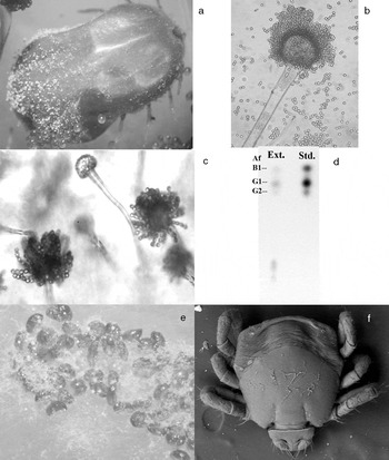
A total of 900 adult engorged females were raised on cattle and 54 of them (6%), exhibited characteristic symptoms of natural fungal infection (Fig. 1a). The spores obtained from infected ticks grew on SDA, yielding yellowish-green cottony colonies, at 28 or 37°C. Microscopic examination of the cultured mycelia showed columnar conidiophores measuring approximately 400 μm in length with spherical vesicles 20–45 μm in diameter, pale conidia with smooth and spherical spores 4–6 μm in diameter (Fig. 1b, c), septated hyphae with dichotomous 45° angle branching, pale green conidiophores bearing phialides that appeared over the entire surface of the colonies after 72 h of culture and chains of spores held in medusa head-like clusters that emerged from the phialides (Fig. 1c). The fungus was able to grow steadily using colloidal chitin as the only source of carbon and thin layer chromatography identified the presence of aflatoxins type B and G in the fungal extracts (Fig. 1d). PCR assays using genomic DNA purified from the isolated fungus and primers specific for the 18S ribosomal RNA gene yielded an amplicon of approximately 750 base pairs (bp), as visualized by agarose gel electrophoresis. The amplicon was subjected to automated DNA sequencing and produced a 710 bp sequence that was electronically submitted to the GenBank sequence database (www.ncbi.nih.gov). GenBank administration assigned the Accession number JF717866. BLAST DNA sequence similarity analysis comparing the 18S rRNA gene from the fungal isolate against the nucleotide database revealed a 99% identity of the fungal isolate 18S rRNA gene to those of A. flavus and A. terreus (708 out of 710 matching bases), as well as a strong identity to 18S rRNA genes of Eurotium herbariorum (Aspergillus glaucus) with 707 out of 710 matching bases.
Different aspects of the fungal infection of Rhipicephalus microplus. (a) Mould growth and dark cuticle colour, symptoms of a fungal disease, were detected on engorged female ticks. (b) Cultured fungus demonstrated columnar conidiophores and smooth 4 to 6 μm diameter spores (40×objective). (c) Medusa head-shaped conidiophores and microscopic morphology of the fungal cultures were consistent with Aspergillus spp. (40×objective). (d) Thin layer chromatography of fungal extracts revealed the presence of aflatoxins type B and G (Af, aflatoxin; Ext., fungal extract; Std., commercial aflatoxin standards). (e) Spores from the cultured fungus induced experimental infection of healthy ticks and their egg masses. (f) Larvae hatching from experimentally infected egg masses showed mycelial growth on their cuticles.

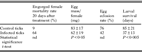
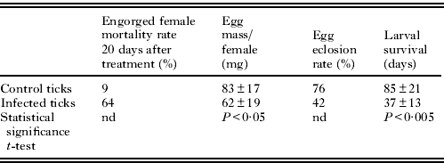
Fungal spores sprayed over healthy ticks and their egg masses produced an infection prevalence of 80% (Fig. 1e), whereas control ticks sprayed with sterile water showed the natural prevalence of approximately 6%. Engorged female mortality rate, average egg mass/female, egg eclosion rate and average larval survival data are summarized in Table 1. Ten-day-old larvae, progeny of experimentally infected ticks showed fungal mycelia growing over their cuticles when imaged by SEM (Fig. 1f).
Effects of fungal experimental infection on engorged females, egg masses and larval offspring
nd, Not determined.
DISCUSSION
This study was motivated by the long-held observations that bovine tick infestation under controlled conditions consistently yielded a small percentage (approximately 6%) of engorged females with obvious symptoms of an infectious disease identifiable by dark colour and mycelial growth. This disease invariably affected oviposition and prevented completion of the life cycle of the infected ticks. This phenomenon was considered worthy of thorough investigation, including the isolation of the fungus which was cultured in mycological medium. The fungal colony properties like mycelia and conidiophore microscopic morphology resembled those of the genus Aspergillus (Ahearm et al. Reference Ahearm, Price, Simmons, Mayo, Zhang and Crow2007; Hedayati et al. Reference Hedayati, Pasqualotto, Warn, Bowyer and Denning2007).
Biochemical tests on the fungus supported the suggestion of the genus Aspergillus as the identity of the isolated mould because the fungus was capable of producing aflatoxin, a property exclusive to moulds belonging to the genus Aspergillus (Yabe et al. Reference Yabe, Ando, Ito and Terakado1987; Hedayati et al. Reference Hedayati, Pasqualotto, Warn, Bowyer and Denning2007). The isolated fungus was able to use colloidal chitin as the only source of carbon, demonstrating that it is an entomopathogenic chitinase-producing fungus. This property narrowed the search to a short list of reported aflatoxin and chitinase-producing entomopathogenic Aspergillus species, including A. flavus, A. parasiticus and A. niger (Hedayati et al. Reference Hedayati, Pasqualotto, Warn, Bowyer and Denning2007). However, conidium shape and colony colour suggested A. flavus as the most likely match for the isolated fungus (Ahearm et al. Reference Ahearm, Price, Simmons, Mayo, Zhang and Crow2007; Hedayati et al. Reference Hedayati, Pasqualotto, Warn, Bowyer and Denning2007).
Identification of A. flavus was supported by 18S rRNA genotyping which showed 99% sequence identity with a 100% query coverage (708 out of 710 compared bases) between the isolated fungus A. flavus strain HKF30 (GenBank Accession no. HM773230) and A. terreus strain HKF20 (GenBank Accession no. HM773230) by BLAST searches (Altschul et al. Reference Altschul, Madden, Schaffer, Zhang, Zhang, Miller and Lipman1997). However, only A. flavus shares the microscopic morphology, colony colour and capability to synthesize aflatoxin among the closest BLAST matches.
A. flavus naturally infected 6% of the tick population; however, under experimental conditions the cultured fungus was capable of inducing 80% prevalence infection, 13 times higher than the observed natural prevalence. Healthy ticks experimentally infected with cultured A. flavus also produced similar symptoms to those originally observed in the natural tick infection, fulfilling the Koch-Henle postulates. High prevalence experimental infection is consistent with the fungal chitinase production found during this study by experimental procedures and suggests that the isolated fungus is entomopathogenic in nature and may have a broad range of arthropod preferences. However, several differences also were evident in the experimentally infected ticks when compared to naturally infected ticks. The most obvious difference was that engorged females were already in the oviposition process when experimentally infected and as a result produced measurable egg masses, whereas naturally infected ticks did not lay eggs at all. Another obvious difference was the absence of the distinctive dark colour on the experimentally infected ticks, which may be attributable to the extended length of the natural infection and probably to the fungus growth within the ticks. Engorged female mortality rate was noticeably increased in experimentally infected ticks presumably due to the fungal infection. But the most important difference found in experimentally infected ticks was the opportunity to observe and measure the effect of A. flavus on the tick egg masses and their larval progeny, since most of the eggs oviposited by experimentally infected ticks also became infected, showing a statistically significant effect on the weight of the egg mass when compared to the control group (P<0·05 t-test). Likewise, the larval progeny of experimentally infected ticks were affected by A. flavus and exhibited a substantial reduction in the eclosion rate and fungal growth on the larval cuticles, as well as a statistically significant decrease of their survival after hatching (P<0·005 t-test).
Under certain natural circumstances, A. flavus is able to produce a lethal infection in R. microplus engorged ticks and their progeny. The precise mechanism of fungal infection must be clarified to comprehensively assess the potential use of A. flavus as an alternative control method for cattle ticks and tick-transmitted diseases. Further studies on this subject should be considered carefully if alternative control methods are implemented, especially because of the ever-increasing acaricide resistance of R. microplus spreading worldwide.
ACKNOWLEDGEMENTS
We would like to thank to Carl Zeiss de México S.A. de C.V. and Dr Marco A. Ramirez O. for their invaluable assistance with the SEM imaging analysis. We also thank E. Lopez, S. Becerra, J. Yañez and P. Gaytan of the Sequencing Unit at the IBT UNAM. This research was partially funded by: SEP-CONACYT; grant numbers: 58609 and 103026.