The survival of patients with single-ventricle physiology has steadily improved over the past several decades.Reference Newburger, Sleeper and Bellinger 1 , Reference Pasquali, Jacobs and He 2 The risks leading to morbidity and mortality are concentrated in the acute post-operative period following the stage 1 palliation. During this period, variations in temperature, systemic vascular resistance, bleeding, sedation, and ventricular function all combine to increase the risks of low cardiac output syndrome and sudden cardiac arrest.Reference Tabbutt, Ghanayem and Ravishankar 3 , Reference Tweddell, Hoffman and Fedderly 4 These risks can be reduced by standardising practice surrounding the use of vasodilators, inotropes, sedatives, as well as temperature and bleeding management, which has led to improvement in short-term outcomes in several studies.Reference Mills, Kaza and Walsh 5 , Reference Hoffman, Mussatto and Brosig 6 The use of delayed sternal closure has also been frequently used as a technique in the post-operative care of patients following the stage 1 palliation.Reference Hakimi, Walters, Pinsky, Gallagher and Lyons 7 , Reference Tabbutt, Duncan, McLaughlin, Wessel, Jonas and Laussen 8
Although the theoretical physiologic benefits of delayed sternal closure have been well described, few studies have systematically detailed the physiologic changes that occur following sternal closure. Without approximation of the sternal edges, the volume of the thorax increases. This increases the compliance of the chest wall, such that intra-thoracic, pleural, and pericardial pressures are minimised during temporary increases in intra-thoracic volume caused by tissue oedema, bleeding, and foreign objects in the chest.Reference Shore, Capuani and Lincoln 9 Improved thoracic compliance also improves compliance of the respiratory system, diminishing mean airway pressure and improving minute ventilation.Reference Bacha, Larrazabal and Pigula 11 In the end, these changes minimise intra-thoracic pressure, which may improve cardiac filling and expand the intra-thoracic volume and may in turn improve ventricular compliance.Reference Tyberg, Grant and Kingma 10
The purpose of this study was to describe the physiologic changes that occur following delayed sternal closure in a contemporary cohort from a single centre. Further, we attempted to define haemodynamic patterns associated with failure of sternal closure in order to identify patients at proximate risk of sternal closure failure.
Materials and methods
Study design and setting
We conducted a single-centre, retrospective cohort study of infants who had undergone stage 1 palliation using a delayed sternal closure approach at Boston Children’s Hospital.
Study population
Following approval by the Institutional Review Board at Boston Children’s Hospital with a waiver for informed consent, we identified all patients through screening of our institution’s surgical database. Patients were included if they underwent stage 1 palliation at our centre between January 2010 and June 2017.
Intra-operative and peri-operative management
Before the operation, all infants were intubated with a cuffed endotracheal tube, and central venous catheters were placed ~1 cm above the superior caval vein and right atrium junction, and venous oximetery took place using co-oximetry on blood sampled from the proximal port. Infants underwent a stage 1 palliation with either a modified Blalock–Taussig shunt or Sano modification – i.e. right ventricle to pulmonary artery conduit. Cerebral, but not flank, near infrared spectroscopy (cNIRS; Casmed Foresight Elite; Branford, Connecticut, United States of America) was continuously measured in the post-operative period through sternal closure. Our ventilation strategy uses large tidal volumes (~10 ml/kg), a short inspiratory time, and a low rate respiratory rate in an effort to minimise mean airway pressure, maximise venous return, and maximise lung recruitment. In a sub-set of patients (n=22 patients), oxygen consumption was measured and used to calculate systemic blood flow by the modified Fick principle as previously described (Oxygen Consumption Based Assessment of Hemodynamics in Newborns, NCT02184169).Reference Mills, Kaza and Walsh 5
Surgical variables, including anatomic sub-types, ischaemic times, and listing of subsequent operations, were extracted from an internal surgical database. Technical performance scores were assessed at the time of discharge or reintervention before discharge, as previously described.Reference Bacha, Larrazabal and Pigula 11 The precise time of sternal approximation was obtained from the operative and nursing notes. For 24 hours before and after the time of sternal closure, we extracted hourly values for the following variables from an electronic data warehouse (BCH360; Netezza, IBM, Armonk, New York, United States of America): haemodynamics – arterial blood pressures, central venous pressure, heart rate, and pulse oximetry saturation; markers of perfusion – pulse pressure, superior caval vein oxyhaemoglobin saturation, cerebral near infrared spectroscopy, urine output, and serum lactic acid; ventilator parameters – respiratory rate, peak inspiratory pressure, positive end expiratory pressure, tidal volume, end-tidal CO2, mean airway pressure, minute ventilation, and carbon dioxide elimination; and arterial blood gas – pH, carbon dioxide tension, and oxyhaemoglobin saturation. Physiologic dead space fraction was calculated as (arterial partial pressure of carbon dioxide−end tidal carbon dioxide) divided by the arterial partial pressure of carbon dioxide. Compliance of the respiratory system was calculated as tidal volume divided by the difference between peak inspiratory pressure and positive end expiratory pressure. Ventilation index was calculated as the product of arterial partial pressure of carbon dioxide, peak inspiratory pressure, and respiratory rate divided by 1000.Reference Horvath, Shore, Schultz, Rosenkranz, Cousins and Ricci 16 The inotrope score was calculated as dopamine (mcg/kg/minute)+dobutamine (mcg/kg/minute)+(100×epinephrine (mcg/kg/minute)) and the vasoactive-inotrope score was calculated as IS + (100×norepinephrine (mcg/kg/minute)) + (10×milrinone (mcg/kg/minute)) + (10,000×vasopressin (units/kg/minute)).Reference Gaies, Gurney and Yen 12 , Reference Wernovsky, Wypij and Jonas 13 In addition, the clinical indication for sternal reopening was summarised from the operative note.
Statistical analysis
Categorical variables were summarised as frequencies and percentages. Comparisons between categorical variables were made using the chi-square analysis or Fisher’s exact test, as appropriate. Continuous variables were presented as mean ± standard deviation or median with interquartile ranges [25th%, 75th%]. Comparisons among continuous variables were made either by the Student’s t-test or the Wilcoxon rank-sum test, depending upon the normality of distribution. In all analyses, a two-sided p-value of <0.05 was considered statistically significant. We performed three sets of analysis. First, haemodynamic and respiratory data from 24 hours before and after sternal closure were averaged per patient and compared between groups – i.e. before versus after delayed sternal closure – by paired samples t-test. Second, haemodynamic and respiratory differences at the time of initial sternal closure were assessed between patients who subsequently underwent failed versus successful delayed sternal closure and were compared by taking hourly means 24 hour before and after sternal closure and comparing them using linear mixed effects modelling to account for within-subject correlation with failure versus success assignment as the fixed effect (R version 3.4.1; R Foundation for Statistical Computing). Third, in patients who failed sternal closure, changes in haemodynamics surrounding the time of sternal reopening were compared in an 8:1 ratio of contemporaneous patients at the same post-operative time. In this analysis, acute haemodynamic data were extracted every 5 seconds from a central server (Analytics Platform; Etiometry, Inc., Boston, Massachusetts, United States of America), binned into 1-minute medians, and compared between groups for 2 hours before and after sternal reopening as described above.
Results
Patient characteristics and clinical outcomes
During the study period, 193 patients underwent stage 1 palliation. Of these, 25 underwent primary sternal closure during the S1P, of whom four patients (16%) underwent sternal reopening – i.e. failed primary sternal closure. Of the 168 patients whose sternum was left open in the operating room, 12 (7.6%) died before an attempt at sternal closure. Of the remaining 156 patients who underwent delayed sternal closure, 11 (7.1%) underwent reopening of the sternotomy, a rate similar to patients undergoing primary sternal closure (p=0.13). Cumulative fluid balance from post-Norwood ICU admission to the time of sternal closure was similar between patients undergoing failed versus successful delayed sternal closure (−95 versus −96 ml/kg, p=0.96). The fluid volume administered in the 48 hours following delayed sternal closure exceeded that administered following primary sternal closure (252 versus 201 ml/kg over 48 hours, p=0.0011). The duration of post-operative mechanical ventilation was significantly shorter in patients undergoing primary sternal closure (4 versus 9 days, p<0.0001), as was post-operative ICU length of stay (8 versus 15 days, p=0.0001) and post-operative hospital length of stay (21 versus 38 days, p=0.0011). Survival to hospital discharge was similar in patients undergoing delayed (143/156, 92%) versus primary (24/25, 96%) sternal closure (relative risk 0.95 [95% confidence interval 0.89–1.14], p=0.69). Survival to hospital discharge was higher in patients with a successful (136/145, 94%) versus failed (7/11, 64%) delayed sternal closure (relative risk 1.47 [1.10–2.65], p=0.007), but it was similar in patients with a successful primary (20/21, 95%) versus failed primary (4/4, 100%) sternal closure (relative risk 0.95 [0.77 to 1.87], p>0.999). The timing of delayed sternal closure is shown by post-operative day in Figure 1.
Histogram of timing of delayed sternal closure in patients following stage 1 palliation. Relative frequency (solid bars, left axis) and cumulative frequency (grey circles, right axis) of the postoperative day of the delayed sternal closure. For patients who subsequently failed sternal closure, only the first closure is listed.

Differences in operative technique and residual lesions
As a group, patients who failed delayed sternal closure had a longer aortic cross-clamp time (123±42 versus 99±35 minutes, p=0.029) and circulatory arrest time (39±28 versus 19±17 minutes, p=0.0009) than those who did not fail. On the basis of post-bypass epicardial echocardiogram, there were no significant differences in the fraction of patients with more than moderate tricuspid regurgitation, more than moderate ventricular dysfunction, or more than trivial residual aortic arch obstruction in patients who failed delayed sternal closure (Table 1). However, the failure of delayed sternal closure was closely associated with Technical Performance Score, assessed at the time of discharge: 1.3% of patients with a Technical Performance Score of 1 failed sternal closure compared with 18.9% of patients with a score of 3 (p=0.0028). Patients who failed primary sternal closure exhibited a similar trend, although this did not reach statistical significance (p=0.083, Table 1).
Comparison of patient, surgical, and postoperative characteristics between patients with delayed and primary sternal closure.

ACC=aortic cross clamp; CA=circulatory arrest; CPB=cardiopulmonary bypass; DSC=delayed sternal closure; EGA=estimated gestational age; ICU LOS=ICU length of stay; LOS=length of stay; LV=single left ventricle – including double inlet left ventricle, tricuspid atresia, left dominant CAVC; POD=post-operative day; PSC=primary sternal closure; RV-PA=right ventricle-pulmonary artery; RV=right ventricle – includes hypoplastic left ventricle, right dominant complete atrioventricular canal defect [CAVC]; S1P=stage 1 palliation; SC=sternal closure; TPS=technical performance score; TR=tricuspid valve regurgitation
Data are presented in mean ± standard deviation or median [25th%, 75th%] as appropriate
Differences between patients with successful and failed delayed sternal closure
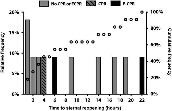
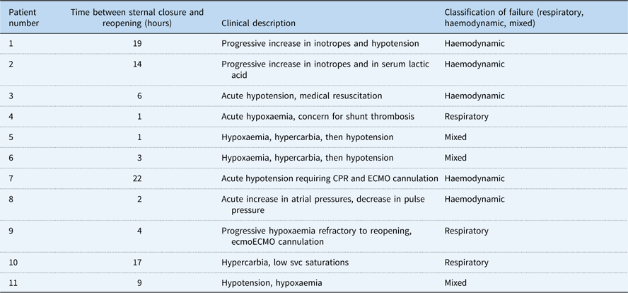
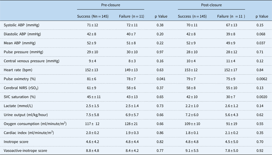
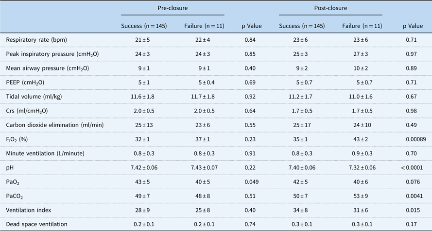
Among the 11 patients who failed delayed sternal closure, five were reopened within 4 hours following the original sternal closure and all were reopened within 24 hours, three in the setting of cardiopulmonary resuscitation (Fig 2). The clinical indications for sternal reopening were primarily respiratory in three, primarily haemodynamic in five, and a combination in three (Table 2). Superior caval vein saturation was lower following sternal closure in patients who subsequently failed sternal closure (30±7 versus 42±10%, p=0.002). Of the remaining haemodynamic (Table 3) and respiratory parameters (Table 4), there were no clinically significant differences between patients who did and did not fail delayed sternal closure, although several parameters met statistical significance. Our power to detect differences in oxygen consumption and derived cardiac index was limited given that only three of the 22 patients in whom oxygen consumption was measured failed sternal closure. In the 2 hours following sternal reopening, there were no clinically important haemodynamic differences before and after reopening, nor in comparison with time-matched controls who did not fail sternal closure (Supplementary Table S1).
Histogram of timing of failed sternal closure. Relative frequency (bars, left axis) and cumulative frequency (circles, right axis) of the number of hours from the time of delayed sternal closure to sternal reopening. Dashed bars represent sternal reopening in the setting of cardiopulmonary resuscitation (CPR), solid black bars represent sternal reopening with CPR and extracorporeal life support (ECPR), and grey bars represent sternal reopening without CPR or ECPR.

Clinical descriptions and classifications of the indications for reopening of the sternum in the 11 patients undergoing failure of sternal closure.

CPR=cardiopulmonary resuscitation; ECMO=extracorporeal membrane oxygenation; SVC=superior caval vein
Here, respiratory denotes a circulatory problem manifest in gas exchange, and haemodynamic denotes a circulatory problem with hydrodynamic manifestations
Haemodynamic differences over the 24 hours prior to and following sternal closure, stratified by eventual successful and failed sternal closures.

ABP=arterial blood pressure; bpm=beats per minute; NIRS=near-infrared spectroscopy; SVC=superior caval vein
Data in the failure group were censored at the time of sternal reopening. Data presented in mean±SD format
Respiratory differences over the 12 hours prior to and following sternal closure, stratified by eventual successful and failed sternal closures.

bpm=breaths per minute; FiO2=fraction of inspired oxygen; PaO2=partial pressure of oxygen in arterial blood; PaCO2=partial pressure of carbon dioxide in arterial; PEEP=positive end-expiratory pressure
Data presented in mean±SD format
Changes in haemodynamic and ventilatory parameters surrounding delayed sternal closure
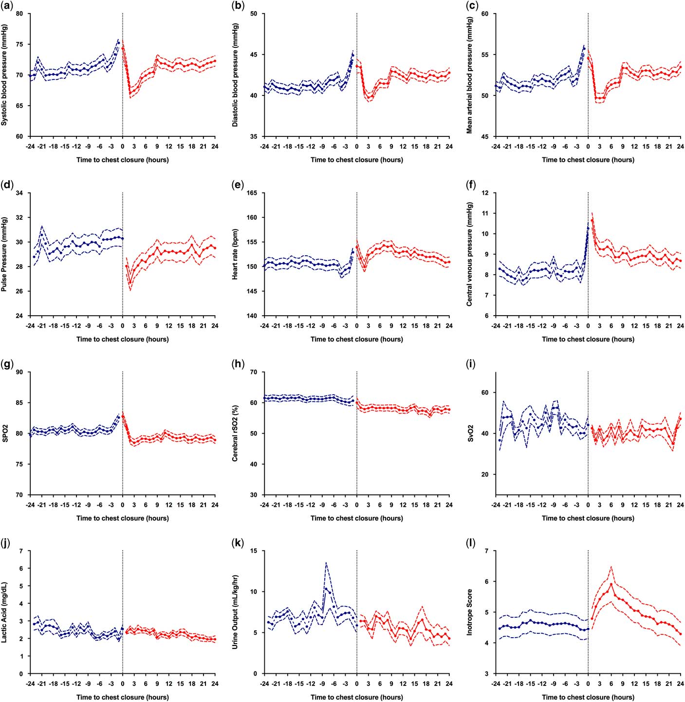
When examined in aggregate over the 24 hours before and after sternal closure, there were few clinically significant changes in haemodynamic and ventilatory parameters, although many reached statistical significance. The most important finding was an approximately 4-unit decrease in cerebral regional oxygenation index and 4% point decrease in superior caval vein oxyhaemoglobin saturation following sternal closure. As depicted in Figure 3 and analysed in Supplementary Table S2, there were also statistically significant but clinically insignificant decreases in systolic blood pressure, pulse pressure, and pulse oximetry and increases in central venous pressure and heart rate in the 24 hours following sternal closure. There was no significant change in urine output following delayed sternal closure. In the subset of patients included in the OxyCAHN study, there were no significant changes in measured oxygen consumption or calculated systemic blood flow. Vasoactive inotrope score in aggregate increased for the first several hours following sternal closure.
Haemodynamic variables over time before and after delayed sternal closure. ( a ) Systolic blood pressure, ( b ) diastolic blood pressure, ( c ) mean arterial blood pressure, ( d ) pulse pressure, ( e ) heart rate, ( f ) central venous pressure, ( g ) pulse oximetry (SpO2), ( h ) cerebral regional oxyhaemoglobin saturation (rSO2) as assessed by near infrared spectroscopy, ( i ) superior caval vein oxyhaemoglobin saturation (SvO2), ( j ) serum lactic acid, ( k ) hourly urine output (UOP), and ( l ) inotrope score for 24 hours before (red) and after (blue) delayed sternal closure. Solid lines are means and dotted lines are standard error of the mean (SEM).

After delayed sternal closure, patients were ventilated with ~1.5 breaths per minute higher respiratory rate, and 1 cm H2O higher average mean airway pressure, accounting for a significantly higher ventilation index. After sternal closure, tidal volume and compliance of the respiratory system were lower, and ventilation index was higher. Arterial pH and arterial oxygen tension were lower and arterial carbon dioxide tension was higher following sternal closure. Although measurable, all of these changes were of small clinical magnitude (Fig 4).
Respiratory variables over time before and after delayed sternal closure. ( a ) Peak inspiratory pressure, ( b ) mean airway pressure, ( c ) positive end expiratory pressure, ( d ) tidal volume, ( e ) compliance of the respiratory system, ( f ) minute ventilation, ( g ) dead space ventilation, and ( h ) carbon dioxide elimination (VCO2) for 24 hours before (blue) and after (red) delayed sternal closure. Solid lines are means and dotted lines are SEM.

Discussion
We have described the physiologic impact of sternal closure when performed according to our current practice. In patients undergoing delayed sternal closure following stage 1 palliation in this way, most of the haemodynamic perturbations were predictable and of clinically negligible magnitude. We identified a temporary decrease in pulse pressure and sustained, at least for 24 hours, rise in central venous pressure, as well as a temporary increase in inotrope score, without a negative impact urine output or lactate level. Patients who failed sternal closure often exhibited perturbations in haemodynamics, oxygenation, or ventilation, highlighting that low cardiac output in this population may manifest as a hydrodynamic failure, as hypoxaemia, or as hypercarbia and increased dead space ventilation. In addition, we identified important associations distinguishing patients who did and did not fail sternal closure, including longer operative ischaemia times, and a lower Technical Performance Score. Taken together, these findings might provide a framework for the generation of an algorithm to assess patients for earlier timing of sternal closure.
It is generally appreciated that sternal closure causes transient perturbations in haemodynamic and respiratory function. Most descriptions of such effects are more significant than those we describe here. Some studies identify a decrement in blood pressure and cardiac index by as much as 25%,Reference Vojtovič, Reich and Selko 14 although we did not note changes of this magnitude. Nonetheless, we did note immediate decreases in blood pressure and pulse pressure, as well as a consistent increase in central venous pressure. We hypothesise that the combination of an increase in central venous pressure and a decremental decline in pulse pressure suggests that delayed sternal closure is likely to impair ventricular diastolic compliance, which may impair venous drainage and diminish cardiac output.Reference Kay, Brass and Lincoln 15 This probably also explains the decrement in cerebral regional oxygenation index and superior caval vein oxyhaemoglobin saturations, findings that have been previously associated with sternal closure.Reference Horvath, Shore, Schultz, Rosenkranz, Cousins and Ricci 16 Other studies have described a decrease in compliance of the respiratory system as high as 19% following sternal closure,Reference Bacha, Larrazabal and Pigula 11 whereas the change in our cohort was less prominent. Patient selection in each of these studies was mixed between patients with univentricular and biventricular disease, although it may be expected that patients following the stage 1 palliation would be the most vulnerable to physiologic perturbations in preload. The small magnitude of these changes may reflect our institution’s aggressive use of diuretics, as well as our tendency for later delayed sternal closure when compared with other institutions, which together may result in less body wall oedema and improved thoracic compliance at the time of sternal closure, minimising its effects on preload and respiratory system compliance. It is also possible that a component of the haemodynamic changes following sternal closure was related to procedural anaesthesia. Unfortunately, our practices surrounding assessment for sternal closure were not protocolised, and this represents an important area of future work in order to better inform the timing of sternal closure. It is possible that sternal closure could safely take place sooner than described in our cohort,Reference Riphagen, McDougall and Tibby 17 a question that will need to be investigated prospectively.
Patients who subsequently failed sternal closure exhibited a clinically similar pre-sternal closure haemodynamic and respiratory profiles to those who did not, making it difficult for these data to inform a prospective algorithm to assess readiness for sternal closure. It should be noted that owing to the low number of patients who failed sternal closure, we had limited power to detect any but the most consistent and large differences between groups. Patients who subsequently underwent sternal reopening exhibited significantly lower superior caval vein oxyhaemoglobin saturations than those whose sternal closures were successful, highlighting the importance of venous oximetry in the care of these patients. This finding is likely to represent the presence of cerebral hypoxia due to lower cerebral blood oxygen delivery. We note that, in these same patients, the cerebral oxygen saturation index was not significantly lower between patients who did and did not fail sternal closure, probably because of the insensitivity of cerebral near infrared spectroscopy to detect cerebral hypoxia;Reference Rescoe, Tang and Perry 18 thus, this technique should not be used in isolation for the post-operative monitoring of cerebral hypoxia. We found it remarkable that patients who failed delayed sternal closure did not exhibit significant changes in haemodynamics following initial sternal closure. Such findings were probably obscured by the low number of patients in the failure group, as well as by resuscitative measures that are often targeted at normalising haemodynamics.
Haemodynamic compromise following sternal closure may manifest in a number of ways, each likely representing a different manifestation of the same physiology. The most phenotypically obvious one is hydrodynamic: the onset of hypotension, elevated atrial pressures, and narrowed pulse pressures – so-called tamponade physiology – as a manifestation of impaired ventricular filling, diminished stroke volume, and low cardiac output. If this physiology persists, it results in venous oxyhaemoglobin desaturation, arterial hypoxaemia, and dead space ventilation owing to low cardiac output. Another manifestation is one of diminished pulmonary blood flow and increased dead space ventilation, including hypoxaemia, hypercarbia, and increased minute ventilation. Such findings may represent an anatomic obstruction to pulmonary blood flow – e.g. alteration in the lie of or compression of a Sano conduit – obstructed blood flow to one of the branch pulmonary arteries, or elevated total pulmonary resistance – more likely if the atrial septum was restrictive preoperatively. Alternatively, diminished pulmonary blood flow may be a manifestation of diminished total cardiac output, which includes pulmonary blood flow. In other patients, hydrodynamic and ventilatory manifestations of haemodynamic impairment occurred together. Whatever the manifestation, the target of resuscitation should be an improvement in pulmonary blood flow or total cardiac output, so long as primary respiratory pathology is ruled out by chest radiograph.
Although the majority of patients in this data set underwent delayed sternal closure, there are important findings in the comparisons between patients undergoing primary and delayed sternal closure. Patients undergoing delayed sternal closure were heavily diuresed, with an average post-operative cumulative fluid balance of −95 ml/kg before sternal closure. This volume was partially re-administered following sternal closure: an average of 252 ml/kg was given in the 48 hours following delayed sternal closure, higher than following primary sternal closure (201 ml/kg/48 hours, p=0.0011). Further, patients undergoing primary sternal closure exhibited a shorter duration of mechanical ventilation (by an average of 5 days), shorter ICU length of stay, and shorter hospital length of stay. Although there is certainly a selection bias to partly account for these associations – sicker patients are more likely to undergo delayed sternal closure – the differences between these groups are striking and merit careful consideration.
We believe that the next step for this work is the creation of an algorithm to inform the timing of sternal closure, be it primary closure or delayed. It is important to note that many groups have described more significant haemodynamic changes with sternal closure than we have in this our cohort in which the timing of sternal closure was conservative, such that earlier closure may result in more significant perturbations. Our work also highlights the vital importance of operative events on the timing of sternal closure. It is possible that longer ischaemic times primarily lead to worsened post-operative cardiac function, oedema, and failure of sternal closure, such that a time threshold might exist beyond which delayed sternal closure would reduce the incidence of adverse events. Finally, the association between Technical Performance Score and failure of delayed sternal closure raises the possibility of risk-stratifying patients on the basis of echocardiographic findings. The Technical Performance Score reported here included findings – namely aortic arch, coronary artery, pulmonary artery, or atrial septal obstruction – that may be manifest following operative recovery rather than immediately post-operatively. Eight of the 12 patients who failed delayed sternal closure had a Technical Performance Score of 3. Unfortunately, the Technical Performance Score is defined by the need for a reintervention – rather than an echo finding – so it cannot be determined a priori to risk-stratify patients. Findings that could be identified at the time of sternal closure, such as aortic arch obstruction in the operating room, were not significantly different between groups in our cohort, perhaps owing to loading conditions, inotropic support, anaesthetic effect, and others that may create different physiology during recovery than is present in the operating room.
Limitations
For the duration of the study, the care that these patients received was according to routine clinical practice patterns and therefore was not standardised. Variations in surgical technique, shunt types, timing of sternal closure, choice and dosing of vasoactive medications and administration of volume, titration of mechanical ventilation, and other aspects of intensive care may have a significant effect on haemodynamics following sternal closure. Similarly, it is difficult to exclude the possibility of an era effect over this seven-year study, as several aspects of care changed during that time, including our approach to systemic vasodilator use.Reference Mills, Kaza and Walsh 5 Our understanding of the physiologic effects of delayed sternal closure and the ability to predict failed sternal closures may improve if a larger number of patients can be included under a homogeneous care environment. This may be possible by protocolising care and completing this study at several large centres simultaneously. Second, in this study, we tested a large number of variables over a repeated number of times. Although we used a linear mixed model to account for within-subject repeated measures over time, each variable was independently evaluated, raising the possibility that some of the significant findings are false discoveries. However, as outlined above, many of the significant associations are physiologically related, making their truth as associations plausible. These parameters also exhibited the lowest p values, which increases their likelihood as true findings.Reference Benjamini and Hochberg 19
Conclusions
In a cohort of neonates following stage 1 palliation, delayed sternal closure caused transient and mild changes in haemodynamic and ventilatory parameters. In this retrospective study, the incidence of failed closure was similar between primary and delayed closures. Venous oxyhaemoglobin saturation was significantly lower in patients who failed sternal closure than in those who did not, and monitoring of this parameter for at least 24 hours following sternal closure may be important to the early identification of these patients.
Acknowledgements
The authors thank the nursing staff, nurse educators, respiratory therapy, cardiac anesthesia, operating room, and cardiac surgical staff at Boston Children’s Hospital for assistance with this work, and Ellen McCusty for assistance with automated data extraction.
Financial Support
The authors acknowledge funding for this work from the Gerber Foundation (K.I.M., J.N.K.), Hess Family Cardiac Innovation Fund (J.N.K.), and the Boston Children’s Hospital Heart Center Strategic Investment Fund (J.N.K.).
Conflicts of Interest
None.
Ethical Standards
This retrospective study was approved by the Institutional Review Board at Boston Children’s Hospital.