

Introduction
Apathy is not only a symptom of depression but can occur independently (Marin et al., Reference Marin, Firinciogullari and Biedrzycki1994, Levy et al., Reference Levy1998; Brodaty et al., Reference Brodaty, Sachdev, Withall, Altendorf, Valenzuela and Lorentz2005). Disorders characterized by high rates of apathy include stroke (Starkstein et al., Reference Starkstein, Federoff, Price, Leiguarda and Robinson1993; Brodaty et al., Reference Brodaty, Sachdev, Withall, Altendorf, Valenzuela and Lorentz2005), Alzheimer's disease (Marin et al., Reference Marin, Firinciogullari and Biedrzycki1994; Landes et al., Reference Landes, Sperry and Strauss2005), and subcortical dementias such as Parkinson's disease and progressive supranuclear palsy (Litvan et al., Reference Litvan, Mega, Cummings and Fairbanks1996; Kirsch-Darrow et al., Reference Kirsch-Darrow, Fernandez, Marsiske, Okun and Bowers2006). In a number of these studies, apathy and depression were shown to correlate only modestly (Levy et al., Reference Levy1998) or apathy was reported in the absence of depression (Brodaty et al., Reference Brodaty, Sachdev, Withall, Altendorf, Valenzuela and Lorentz2005, Kirsch-Darrow et al., Reference Kirsch-Darrow, Fernandez, Marsiske, Okun and Bowers2006).
There is a growing body of research supporting the independence of apathy and depression. Evidence for the independence of apathy and depression comes from differences in neuropathological correlations and associated psychiatric symptomatology. Regarding neuropathology, apathy has been linked to pathology in the left orbitofrontal cortex (Holthoff et al., Reference Holthoff2005), right anterior cingulate gray matter (Lavretsky et al., Reference Lavretsky, Ballmaier, Pham, Toga and Kumar2007) and right frontosubcortical circuit (Brodaty et al., Reference Brodaty, Sachdev, Withall, Altendorf, Valenzuela and Lorentz2005) while depression has been linked to pathology in left dorsolateral prefrontal cortex (Holthoff et al., Reference Holthoff2005) and orbitofrontal gray matter (Lavretsky et al., Reference Lavretsky, Ballmaier, Pham, Toga and Kumar2007). Regarding psychiatric symptoms, apathy has been associated with older age, disinhibition, lower cognitive score, faster cognitive and functional decline and aberrant motor behaviors, whilst depression has been linked to anxiety, agitation, irritability and hallucinations (Levy et al., Reference Levy1998, Starkstein et al., Reference Starkstein, Jorge, Mizrahi and Robinson2006).
Apathy is a commonly reported symptom in dementia. It is related to decreased functional and cognitive performance and is a significant source of distress for caregivers (Landes et al., Reference Landes, Sperry, Strauss and Geldmacher2001). Depression is also relatively common in people with dementia although to a lesser extent than apathy (Migliorelli et al., Reference Migliorelli, Teson, Sabe, Petracchi, Leiguarda and Starkstein1995). Although there has been some investigation of the relationship between depression and dementia following stroke (Kauhanen et al., Reference Kauhanen1999, Brodaty et al., Reference Brodaty, Withall, Altendorf and Sachdev2007), how apathy contributes to this has not been shown.
We aimed to examine, for the first time, the longitudinal relationship between apathy and depression following stroke – in particular, the differences or commonalities in their risk factors and clinical courses and their relationships to dementia. We hypothesized that apathy and depression would occur independently following stroke and would not share the same risk factors but that both would be related to post-stroke dementia.
Methods
Subjects
Consecutive eligible patients hospitalized for ischemic stroke and volunteer comparison subjects (controls) were asked to participate (see Procedure, below). An ischemic stroke was defined as “rapidly developing clinical signs of focal (or global) disturbance of cerebral function, with symptoms lasting 24 hours or longer, with no apparent cause other than of vascular origin” in which a brain computerized tomography (CT) or magnetic resonance imaging (MRI) scan does not show intracranial hemorrhage (Kunitz et al., Reference Kunitz1984). The following exclusion criteria were applied: age over 85 years, hemorrhagic stroke; transient ischemic attack (TIA); impairment of consciousness persisting for greater than seven days at time of stroke; inability to give informed consent; insufficient fluency in English to complete testing; past history of dementia or other neurological disease known to affect cognition; current alcohol or drug abuse; a retrospective score on the Informant Questionnaire on Cognitive Decline in the Elderly (IQCODE; Jorm, Reference Jorm1994) ≥3.31 (indicating possible dementia prior to the index stroke); a Diagnostic and Statistical Manual of Mental Disorders, 4th edition (DSM-IV) diagnosis of mental retardation; severe aphasia; and lack of an appropriate informant. Controls were excluded if they had a history of stroke or any of the above exclusion criteria.
From consecutive admissions to the inpatient stroke units at Prince of Wales and St George Hospitals in Sydney, Australia over a 38-month period between May 1997 and June 2000, 205 patients met inclusion and exclusion criteria and gave consent for entry into the study. By the time of detailed index assessment – on average, four months following the cerebrovascular event – 37 were lost to follow-up (27 withdrew from the study, three died, four relocated outside Sydney, two could not be contacted and one was excluded for baseline dementia/aphasia missed earlier) leaving 168 participants. The follow-up assessment occurred approximately 12 months after the index assessment.
Of the 168 stroke patients who received an index interview, 134 were interviewed for a DSM diagnosis of depression (see below) at both index and follow-up assessments, and ratings for apathy were completed for 107 patients at both time points. In total, 106 people had post-stroke ratings for both depression and apathy at both index and follow-up assessments.
Control participants were recruited through advertisement and from local community groups. Of the 130 controls who met eligibility criteria, 107 agreed to participate. The control and stroke patient groups were matched for sex and age. The control sample was used to derive z-scores for neuropsychological data and to derive the apathy cut-off score of 36/37 on the AES (see Brodaty et al., Reference Brodaty, Sachdev, Withall, Altendorf, Valenzuela and Lorentz2005) and no further data are provided here. The study had institutional ethics committee approval and subjects provided written informed consent after receiving a complete description of the study.
Demographic and clinical measures
Age, gender, education, marital status and whether the person was living with a family member (as a proxy for social support) were recorded. Stroke diagnoses were made according to the International Statistical Classification of Diseases and Related Health Problems, 10th revision (ICD-10) criteria and confirmed by consensus at team clinical meetings. Severity was established with the European Stroke Scale (ESS; Hantson et al., Reference Hantson1994). Measures of functional ability included Activities of Daily Living (ADL; Katz and Akpom, Reference Katz and Akpom1976) and Instrumental Activities of Daily Living (IADL; Lawton and Brody, Reference Lawton and Brody1969). Vascular risk factors (i.e. hypertension, diabetes mellitus, myocardial infarction, angina, coronary artery disease, atrial fibrillation, hypercholesterolemia and smoking) and alcohol use were recorded.
Psychiatric measures
Psychiatric measures included the Structured Clinical Interview for DSM-IV (SCID-I; First et al., Reference First, Spitzer, Gibbon and Williams1997), informant-rated version of the Apathy Evaluation Scale (AES; Marin et al., Reference Marin, Biedrzycki and Firinciogullari1991) and details of past psychiatric history. Participants were subdivided into those with diagnoses of major and minor or no depression according to DSM-IV criteria. Participants were also subdivided into those with or without apathy and this was defined as a score of ≥37 on the AES (Brodaty et al., Reference Brodaty, Sachdev, Withall, Altendorf, Valenzuela and Lorentz2005).
Neuropsychological Measures
Global cognitive function was assessed with the Mini-mental State Examination (MMSE; Folstein et al., Reference Folstein, Folstein and McHugh1975) and premorbid intelligence with the National Adult Reading Test-Revised (NART-R; Nelson, Reference Nelson1983). The IQCODE was rated retrospectively for the five years prior to the stroke as an indicator of premorbid cognitive decline. We report results from three domains chosen from a full neuropsychological battery performed by trained psychologists based on findings from our earlier study (see Brodaty et al., Reference Brodaty, Sachdev, Withall, Altendorf, Valenzuela and Lorentz2005 for details). These domains and their component tests are: (i) Attention and Concentration – Wechsler Adult Intelligence Scale – Revised Digit Span – forwards and Wechsler Memory Scale – Revised Mental control subtest; (ii) Executive Function – Color Form Sorting Test, Verbal Fluency phonemic (FAS) and semantic (animals), and Trail Making Test Part B; and (iii) Speed of Information Processing – Trail Making Test Part A and Symbol Digit Modalities Test – oral. Z-scores for the neuropsychological domains were derived from the means and standard deviations for the control group at index assessment. Neuropsychological assessments of subjects judged to be significantly, clinically depressed at clinical interview were deferred until patients improved. Improvement was defined as a GDS score of <5, a reduction in self- or informant-reported symptoms and/or a further psychiatric assessment of improvement. Subjects were tested in their own homes and given breaks during testing to minimize fatigue. Those with right- or left sided paresis were not tested on motor tasks requiring the use of the affected side.
Neuroimaging
All patients received either a CT or MRI scan at admission. A subset of patients (n = 68) had an MRI scan at the index assessment, three months after admission at approximately the same time as their neuropsychological assessment. As no MRI scans were performed at the follow-up assessments only index MRI data are presented here.
MRI was performed on a 1.5 T Signa GE scanner (GE Systems, Milwaukee, WI, U.S.A.) using the following protocol: a scout mid-sagittal cut (2D, repetition time TR 300 ms, echo time TE 14 ms; 5 mm thick, number of excitations 1.5); 1.5 mm thick T1-weighted contiguous coronal sections through the whole brain using a FSPGR sequence (TR 14.3 ms, TE 5.4 ms); and 4 mm thick (0 skip) T2-weighted fluid attenuation inversion recovery (FLAIR) coronal slices through the whole brain (TR 8900, TE 145, TI2200, FOV 25, 256r192). Trained staff scored these with good inter-rater (κ scores from 0.7 to 0.9 on various measures) and intra-rater (κ = 0.8–0.9) reliability. ANALYZE (Mayo Foundation, Rochester, MI, U.S.A.) software was used for all ratings. Cortical atrophy was rated on a 0–2 scale in the frontal, temporal and mid-parietal regions, the sum of which gave the total cortical atrophy score (higher scores indicate more atrophy, maximum score = 6). Deep white-matter hyperintensities (DWMH) were rated on a 0–3 scale, with higher score representing more pathology (Fazekas et al., Reference Fazekas, Chawluk, Alavi, Hurtig and Zimmerman1987). For the DWMH rating, the frontal, temporo-parietal and occipital white matter, and the internal capsules were rated separately, and the scores for both sides added to give a total DWMH score (maximum score = 24). In rating DWMHs, peri-infarct hyperintensities were disregarded. The DWMHs are, therefore, non-infarct lesions on FLAIR T2-weighted imaging that are not related to an actual infarct as seen on either T1- or T2-weighted imaging.
The small numbers of people in individual cells at index MRI scan (total n = 68) precluded between-group statistical analyses with MRI data.
Consensus dementia diagnoses
The diagnosis was assigned to each subject in a case conference at which all medical, psychiatric, neuropsychological and neuroimaging data (including CT or MRI scan reports performed on nearly all patients at time of stroke) were presented and a consensus was reached. A neuropsychiatrist, psychogeriatrician, neurologist and one or more research psychologists attended the meeting. For dementia diagnoses, subjects were required to have definite impairment in two or more cognitive domains (impairment in memory was not necessary) and demonstrate evidence of functional decline because of the cognitive deficits. Vascular dementia diagnoses required evidence of cerebrovascular disease on MRI or CT scan judged to be sufficient to account for cognitive impairment. Impairment in a cognitive domain was defined as definite impairment (<5th percentile) on at least one test or marginal impairment (5th–10th percentile) on at least two tests in that domain. Functional decline was a failure on one ADL item or two IADL items due to cognitive deficits as judged by consensus. Side of stroke was determined by consensus from clinical and neuroimaging data obtained at time of current stroke.
Statistical analyses
The Statistical Package for the Social Sciences (SPSS) version 17 for Windows (SPSS Inc., Chicago, IL) was used for all statistical analyses. Independent-samples t-tests or analyses of covariance were employed for between-group comparisons on continuous variables. Bonferroni corrections were used for multiple comparisons. Univariate analyses of the association between categorical variables in both groups were performed using χ2 analysis. For 2 × 2 tables, Yates’ Continuity Correction is reported. Odds ratios (with 95% confidence intervals) were calculated for the relative risk of psychiatric disorders for each group. For all analyses, probability levels reported were two-tailed and the level of significance was set at 0.05. Logistic regression was used to test the hypothesis that apathy and depression are associated with different risk factors. Predictors were entered collectively unless otherwise specified. For the analyses investigating relationships between apathy, depression and dementia, as small cell sizes precluded analysis using odds ratio patterns, logistic regression was used to test the predictive value of each disorder at index assessment on the outcome of the other disorders at follow-up.
Results
The sample
Of 205 stroke patients recruited at baseline, there were no significant differences between the 168 patients who completed the index assessment and the 37 who did not with respect to age (t = −1.13, p = 0.26), education (t = −1.21, p = 0.23) or sex (χ2 = 0.00, p = 0.99). At index assessment, 129 patients had complete apathy and depression scores. Of these, 106 patients also had a complete follow-up assessment. There were no significant differences as regards rates of apathy (p = 0.52), dementia (p = 0.41), or depression (p = 1.00) between the 106 patients who received a follow-up assessment and the 23 who did not. There were also no statistical differences between the 68 patients with MRI ratings and the 38 without these ratings with respect to age (p = 0.61), gender (p = 0.85), MMSE score (p = 0.65), stroke severity (p = 0.40) or rates of dementia (p = 0.40), apathy (p = 0.70) or depression (p = 0.09) at index assessment.
Frequency of depression and apathy at index and follow-up assessments
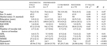
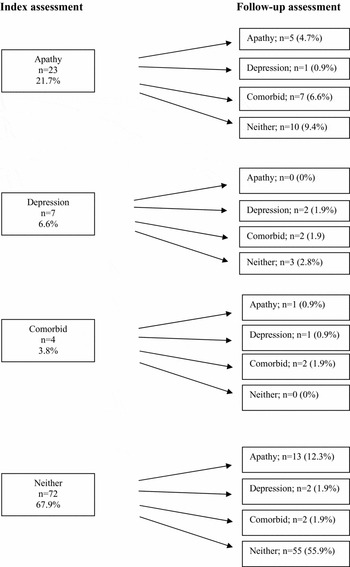
Socio-demographic and clinical characteristics of this sample are shown in Table 1. Index rates of DSM depression diagnosis and AES-defined apathy for the 106 patients rated on both at both time points are shown in Figures 1.
Demographic and clinical characteristics of apathetic, depressed, co-morbid and non-symptomatic stroke patients at index assessment (n = 106)

Note: Sample size varies according to variable.
aOne way ANOVA; bχ2 analysis; suggestions for Bonferroni corrections: cognitive and physical functioning at index: p = 0.01 (0.05/5).
NART-R IQ = National Adult Reading Test Intelligence Quotient; IQCODE = Informant Questionnaire on Cognitive Decline in the Elderly; ESS = European Stroke Scale; ADL = Activities of Daily Living; IADL = Instrumental Activities of Daily Living; MMSE = Mini-Mental State Examination; AES = Apathy Evaluation Scale.
Progression of apathy and depression from index to follow-up assessment (n = 106).

There were no significant differences between groups (apathy only, depression only, comorbid apathy and depression and neither) according to age, gender, marital status, education, premorbid IQ, premorbid cognitive decline, stroke severity or total number of vascular risk factors at baseline. With respect to cognitive and functional performance, there were significant differences between groups for both the ADL and IADL scales, with the apathetic and comorbid groups performing the poorest, but not for global cognition following a Bonferroni correction. There were also significantly higher rates of dementia in the apathetic and comorbid groups.
Vascular risk factors, MRI and stroke variables
There were no significant differences as regards number of vascular risk factors between the 17 patients in the neither disorder group at index who developed apathy, depression or comorbid disorder at follow-up and the 55 patients who remained apathy and depression free (2.73 [1.10] vs 2.64 [1.19], t = −0.26, df = 58.0, p = 0.80).
There were also no significant differences between the subjects who developed apathy, depression, or comorbid disorder and those who did not with respect to frontal atrophy (mean [SD] 0.61 [0.78] versus 0.30 [0.58], t = 1.55, df = 24.16, p = 0.13) or DWMH (4.08 [2.76] versus 3.29 [2.26], t = 1.20, df = 66.0, p = 0.23) at index assessment.
There were 13 patients at index with apathy or depression who had neither disorder at follow-up (7 of these had MRI ratings at index) and 21 patients who had these disorders at both index and follow-up (11 of these had index MRI ratings). There were no significant differences between these groups with respect to total atrophy (2.43 [2.51] versus 2.09 [2.30], t = −0.29, df = 16.0, p = 0.77) or DWMH (4.68 [3.19] versus 3.70 [2.53], t = −0.72, df = 16, p. 048). Unfortunately, the small sample sizes precluded between-group statistical analyses of MRI data.
Finally, there were no significant differences between the 38 patients who had developed apathy, depression or comorbid disorder at follow-up and the 55 patients who did not as regards having had more than one stroke (t = 1.01, df = 100, p = 0.32) or interval stroke (t = −1.17, df = 46.92, p = 0.25).
Performance on neuropsychological domains at index and follow-up assessments
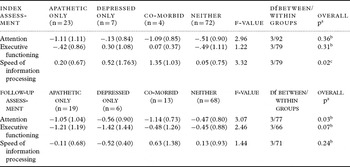
For descriptive purposes, neuropsychological domain performance at both index and follow-up assessments are presented in Table 2. Sample sizes are too small to draw statistical conclusions.
Z-scores for neuropsychological domains for apathetic, depressed, co-morbid and non-symptomatic stroke patients at index and follow-up assessments (n = 106)

*Note: Sample size varies according to variable.
aOne way ANOVA; bBonferroni correction: no significant differences between groups. cTamhane correction: no significant differences between groups.
Overlap and associations between apathy and depression at index and follow-up assessments
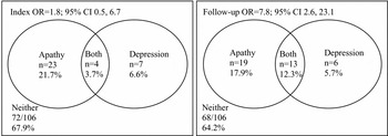
The overlap between apathy and depression was not significant at index assessment (OR = 1.79, 95% CI 0.48, 6.66) but was significant one year later (OR = 7.75, 95% CI 2.60, 23.13; see Figure 2).
Overlap between apathy and depression at index and at follow-up (n = 106).

After correcting for index depression, apathy at index was associated with depression at follow-up (OR = 7.22, 95% CI 2.16, 24.10). By contrast, after correcting for index apathy, index depression was not found to be associated with apathy at follow-up (OR = 1.87, 95% CI 0.49, 7.21).
Associations of apathy and depression at follow-up with cognitive and physical functioning and with dementia
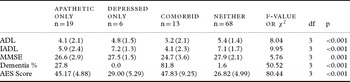
At follow-up, there were significant differences between groups on all variables measuring functional performance, global cognition and prevalence of dementia (Table 3). As at index, the poorest performances were for those in the apathetic and comorbid groups. These groups also had the highest prevalence of dementia; interestingly, no-one in the depressed only group was diagnosed with dementia at follow-up.
Clinical characteristics of apathetic, depressed, comorbid and non-symptomatic stroke patients at follow-up (n = 106)

Note: Sample size varies according to variable.
aOne way ANOVA; bχ2 analysis; suggestions for Bonferroni corrections for cognitive and physical functioning at follow-up: p = 0.01 (0.05/4).
ADL = Activities of Daily Living; IADL = Instrumental Activities of Daily Living; MMSE = Mini-mental State Examination; AES = Apathy Evaluation Scale.
Dementia at index was shown to predict apathy (OR = 12.5, 95% CI 3.0, 52.0) and depression at follow-up (OR = 10.35, 95% CI 2.84, 37.72; correcting for index apathy and depression, respectively). However, neither apathy (OR = 0.74, 95% CI 0.07, 7.55) nor depression (OR = 1.94, 95% CI 0.15, 24.53) at index was found to be a predictor of dementia at follow-up (correcting for dementia at index assessment).
Risk factor analysis
Univariate logistic regression was used to test the hypothesis that different risk factors were associated with apathy and depression at follow-up. Apathy at follow-up was significantly associated with a diagnosis of dementia at index (OR = 18.15, 95% CI 4.70, 70.02) but not age (OR = 1.01, 95% CI 0.96, 1.06), gender (OR = 0.73, 95% CI 0.31, 1.72), stroke severity (OR = 0.99, 95% CI 0.96, 1.02), previous psychiatric history (OR = 1.59, 95% CI 0.61, 4.19) or having a family member living with the patient (OR = 0.74, 95% CI 0.32, 1.72).
Depression at follow-up was significantly associated with a diagnosis of dementia at index assessment (OR = 8.78, 95% CI 2.76, 27.93) and not having a family member living with the patient (OR = 0.22, 95% CI 0.08, 0.64) but not age (OR = 1.06, 95% CI 0.99, 1.13), gender (OR = 1.91, 95% CI 0.70, 5.19), stroke severity (OR = 1.00, 0.95, 1.04) or previous psychiatric history (OR = 1.31, 95% CI 0.42, 4.12).
Discussion
This is the first paper to examine longitudinally the relationship between apathy and depression following stroke. The key finding here is that apathy and depression initially appear to be independent neuropsychiatric sequelae three months after stroke but to have a common predictor and become overlapping constructs approximately one year later. The overlap might be due to cumulative vascular pathology and because of the relationship of each of these syndromes to dementia, which was an important predictor, or possibly causal, factor for both.
Apathy was more common than depression in this sample at both the index and follow-up assessments. Rates are comparable to those reported in other studies investigating people 3–4 months after being admitted to hospital for stroke (Starkstein et al., Reference Starkstein, Federoff, Price, Leiguarda and Robinson1993, Kauhanen et al., Reference Kauhanen1999). Over time, the proportion of people with either pure apathy or depression decreased but the number of people with comorbid apathy and depression increased. Those in the apathetic and comorbid groups presented with the lowest scores on the MMSE, ADL and IADL scales at both index and follow-up, indicating that people with apathy have significant global cognitive and functional impairment.
The relationship between apathy and depression is more complex than we hypothesized. Both share a relationship with dementia, with the odds ratio numerically greater for apathy than for depression. Not living with a family member was a predictor of depression, but not apathy, at follow-up, suggesting confirmation of an additional psychosocial aspect to the development of depression after stroke (Astrom et al., Reference Astrom, Adolfsson and Asplund1993; Gainotti et al., Reference Gainotti, Azzoni and Marra1999). We did not calculate between-group differences in neuropsychological performance or key MRI measures as the small numbers make statistical analyses unreliable. Other neuroimaging studies have supported different etiological pathways that are associated with apathy and depression (Holthoff et al., Reference Holthoff2005; Lavretsky et al., Reference Lavretsky, Ballmaier, Pham, Toga and Kumar2007). We suggest that over time advancing brain pathology results in the merging of these disorders and this is reflected by more people being diagnosed with comorbid apathy and depression at follow-up. It is of note, however, that there were no differences in the number of vascular risk factors between those with new onset apathy and/or depression.
Dementia at index predicted both apathy and depression at follow-up but the reverse did not hold. This finding, in addition to all groups having no evidence of cognitive decline prior to study entry, supports the theory that both apathy and depression result from cumulative brain pathology and that dementia emerges first and then leads to these neuropsychiatric symptoms. This might occur in a dose effect fashion. This theory also fits with the finding that a large majority (82%) of people with comorbid apathy and depression at follow-up were also diagnosed with dementia, in contrast to just over one-quarter of those diagnosed with apathy alone and none with depression alone. Longitudinal studies with a sufficiently large sample, as well as neuropsychological and neuroimaging data, are needed to examine this theory.
Significant numbers of people manifested either pure depression or apathy at both index and follow-up assessments. Distinguishing apathy from depression clinically has important treatment implications especially since the former is under-recognized and/or misdiagnosed (Brodaty et al., Reference Brodaty, Sachdev, Withall, Altendorf, Valenzuela and Lorentz2005, van Reekum et al., Reference van Reekum, Stuss and Ostrander2005). Whereas antidepressants have been shown to have reasonable efficacy in depression after stroke and have been used prophylactically with some success (Andersen et al., Reference Andersen, Vestergaard, Riis and Lauritzen1994, Niedermaier et al., Reference Niedermaier, Bohrer, Schulte, Schlattmann and Heuser2004), there is no accepted treatment regimen for apathy. Preliminary findings in other disorders, however, have suggested that methylphenidate, acetylcholinesterase inhibitors and dopaminergic agents may be useful and specific pharmacological treatments for persons with apathy (van Reekum et al., Reference van Reekum, Stuss and Ostrander2005, Padala et al., Reference Padala, Petty and Bhatia2005). Modafinil has also been proposed as a possible treatment to aid wakefulness in post-stroke depression and this might also have utility for those with apathy (Sugden and Bourgeois, et al., Reference Sugden and Bourgeois2004). Identification of apathy and introduction of psychosocial strategies, such as use of routines and prompts, may ease caregiver distress and in turn benefit the patient (van Reekum Reference van Reekum, Stuss and Ostrander2005). Our findings also stress the importance of monitoring patients’ psychosocial circumstances, as poor social support – as measured by living with a family member after stroke – was identified as a risk factor for the development of depression.
The strengths of this study include a longitudinal study design, the distinguishing of subgroups with apathy only, depression only and comorbid apathy and depression, and the exclusion of persons with pre-stroke dementia at recruitment. Several methodological issues, however, bear comment. Patients recruited for the study included those with first and repeat strokes. Secondly, data were not available on the presence of apathy or depression prior to stroke nor were informants asked when the apathy and/or depression had improved in the period between index and follow-up assessments. MRI scans were not performed at the follow-up assessment. Overlap between apathy and depression may be partly attributable to common items required for their definition in the AES or DSM. Finally, the small sample sizes in each group affected the power of the study and precluded further analyses of neuroimaging and neuropsychological data and of odds ratio patterns. Further longitudinal studies with much larger sample sizes are needed to resolve these issues.
We conclude that apathy and depression may be independent in the months immediately following a stroke, possibly arising from the disruption of different cortical circuits. However, over time these syndromes overlap; this may result from cumulative vascular brain pathology and is associated with dementia. The implications of these results are that practitioners should make every effort to prevent further cerebrovascular disease and related neuropsychiatric sequelae.
Conflict of interest
None.
Description of authors’ roles
A. Withall helped to formulate the research question and collect the data, assisted with consensus diagnosis, and with the analysis and interpretation of the data, and co-wrote the paper. H. Brodaty helped to formulate the research question, coordinated the study, helped in the collection of data and with consensus diagnosis, assisted with the interpretation of data and co-wrote the paper. A. Altendorf helped to formulate the research question and collect the data, and helped with the analysis and interpretation of data. P. S. Sachdev helped to formulate the research question, coordinated the study, helped with the collection and interpretation of data, and assisted with consensus diagnosis.
Acknowledgments
The authors thank the neurologists and staff at the Stroke Units of the Prince of Wales and St George Hospitals, Ron Shnier and MRI staff, SEALS Laboratories, Karen Berman, David Gillies, Lesley Howard, Teresa Lee, Jeff Looi, Lisa Lorentz, Eveline Milne, Amy Ross, Claire Thompson, Michael Valenzuela, Wei Wen and Alessandro Zagami who contributed to this study.
The study was supported by grants from the National Health and Medical Research Council of Australia (970922, 222842), the Rebecca Cooper Foundation, and Fairfax Family Foundation, and by a Fellowship from the NSW Institute of Psychiatry.