Introduction
In the pig breeding industry, embryo transfer is a technique expected to significantly change the export and import of genetics in the future (Martinez et al., Reference Martinez, Cuello, Parrilla, Martinez, Nohalez, Vazquez, Vazquez, Roca and Gil2016). In addition to increasing genetic gain, the main advantage of embryo transfer is biosecurity, as it leads to a minimal risk of pathogen introduction on farms when embryos instead of live animals can be distributed (Stringfellow and Givens, Reference Stringfellow and Givens2009). Furthermore, less transport of live animals will be needed, which enhances animal welfare, is better for the environment and decreases transportation and quarantine costs (Martinez et al., Reference Martinez, Cuello, Parrilla, Rodriguez-Martinez, Roca, Vazquez, Vazquez and Gil2013; Fowler et al., Reference Fowler, Mandawala, Griffin, Walling and Harvey2018).
Commercial application of embryo transfer is dependent on the optimal storage of embryos, for multiple reasons. Embryo transfer technique using non-surgical deep uterine transfer (NsDU-ET), requires embryos to be at the morula or pre-hatching blastocyst stages (Martinez et al., Reference Martinez, Cuello, Parrilla, Rodriguez-Martinez, Roca, Vazquez, Vazquez and Gil2013, Reference Martinez, Martinez, Cambra, Maside, Lucas, Vazquez, Vazquez, Roca, Rodriguez-Martinez, Gil, Parrilla and Cuello2019b). During in vitro embryo production (IVEP) it can be difficult to obtain enough embryos at the desired stage of development. Therefore it may be necessary to store embryos to group several days of IVEP for an embryo transfer. In addition, transportation worldwide or even throughout one country takes time and therefore embryos must be stored under the best possible conditions prior to embryo transfer.
The most common preservation technique is cryopreservation, but porcine embryos are very sensitive to reduced temperatures mainly due to their high concentration of intracellular lipids (Mandawala et al., Reference Mandawala, Harvey, Roy and Fowler2016). The lipid content decreases as the embryos develop and blastocysts contain a lower amount of lipids compared with embryos at earlier developmental stages and thereby tolerate preservation better (Sanchez-Osorio et al., Reference Sanchez-Osorio, Cuello, Gil, Almiñana, Parrilla, Caballero, Garcia, Vazquez, Roca and Martinez2008; Cuello et al., Reference Cuello, Martinez, Nohalez, Parrilla, Roca, Gil and Martinez2016; Martinez et al., Reference Martinez, Martinez, Cambra, Maside, Lucas, Vazquez, Vazquez, Roca, Rodriguez-Martinez, Gil, Parrilla and Cuello2019b). Promising results have earlier been reported for in vivo-derived porcine blastocysts regarding vitrification (Berthelot et al., Reference Berthelot, Martinat-Botté, Locatelli, Perreau and Terqui2000; Gomis et al., Reference Gomis, Cuello, Sanchez-Osorio, Gil, Parrilla, Angel, Maside, del Olmo, Vazquez, Roca and Martinez2012; Cuello et al., Reference Cuello, Martinez, Nohalez, Parrilla, Roca, Gil and Martinez2016). However, exposure to altering oxygen levels, light and other non-optimal in vitro culture conditions (Chen et al., Reference Chen, Redel, Kerns, Spate and Prather2021), are affecting in vitro embryo development. Therefore, in vitro-produced (IVP) embryos are, in general, of inferior quality compared with in vivo-derived embryos (Sudano et al., Reference Sudano, Paschoal, Maziero, Rascado, Crocomo, Magalhães, Monteiro, Martins, Machado and Landim-Alvarenga2013; Marsico et al., Reference Marsico, de Camargo, Valente and Sudano2019). Limited research has been conducted on this subject, but lower survival and hatching rates have been demonstrated after the vitrification of IVP porcine embryos compared with in vivo-derived embryos (Mito et al., Reference Mito, Yoshioka, Noguchi, Yamashita, Misumi, Hoshi and Hoshi2015; Nohalez et al., Reference Nohalez, Martinez, Parrilla, Roca, Gil, Rodriguez-Martinez, Martinez and Cuello2018) and more research is therefore needed to achieve high-quality IVP embryos upon storage.
Establishing specific pathogen-free (SPF) swine farms contributes substantially to reducing the climate footprint, as healthier animals will need less use of antibiotics and show a higher feed conversion rate (Bonesmo and Enger, Reference Bonesmo and Enger2021). In establishing and maintaining SPF farms, embryo transfer is an invaluable tool. The embryos would often need to be transported for short distances only, and embryo transfer of fresh embryos would be advantageous.
Liquid storage offers an alternative to cryopreservation for shorter storage periods. It has practical advantages as no liquid nitrogen is required, nor CO2 gas when using HEPES-buffered medium. In addition, when using liquid storage, the embryos can be kept closer to their physiological temperatures. Martinez et al. (Reference Martinez, Angel, Cuello, Sanchez-Osorio, Gomis, Parrilla, Vila, Colina, Diaz, Reixach, Vazquez, Vazquez, Roca and Gil2014) studied the effect of a 24 h liquid storage of in vivo-derived porcine embryos, and found that the tested temperature of 25°C had a negative effect on embryo quality, while both 24 h and 48 h storage at 37°C with medium supplemented with bovine serum albumin (BSA) gave similar viability as their untreated control groups (Martinez et al., Reference Martinez, Angel, Cuello, Sanchez-Osorio, Gomis, Parrilla, Vila, Colina, Diaz, Reixach, Vazquez, Vazquez, Roca and Gil2014, Reference Martinez, Nohalez, Parrilla, Lucas, Sanchez-Osorio, Roca, Cuello, Rodriguez-Martinez, Martinez and Gil2018). Liquid storage for 24 h did, however, show negative effects on both survival and further development when applying IVP embryos (Lin et al., Reference Lin, Le, Takebayashi, Hirata, Tanihara, Thongkittidilok, Sawamoto, Kikuchi and Otoi2022).
The aim of this study was to gain knowledge on how liquid storage at 37°C for 3 h in CO2-free medium affected IVP embryo quality by evaluating morphology, in vitro developmental capacity and apoptosis. A 3 h preservation regime was chosen as it is the time span that enables handling and transportation to a local recipient porcine farm for planned embryo transfers.
Materials and methods
Chemicals and media
All chemicals and reagents were purchased from Sigma-Aldrich (Oslo, Norway) unless otherwise stated. Washing of cumulus–oocyte complexes (COCs) was performed using porcine X medium (PXM), maturation using porcine oocyte medium (POM), fertilization using porcine gamete medium (PGM) and embryo culture using porcine zygote medium-5 (PZM-5) (Yoshioka et al., Reference Yoshioka, Suzuki and Onishi2008) and from day 4 following fertilization porcine blastocyst medium (PBM) was used (Mito and Hoshi, Reference Mito, Hoshi and Herrick2019). Polyvinyl alcohol in the original medium was replaced by 0.4% BSA in POM, PZM-5 and PBM medium and 0.6% BSA in PGM medium. Minor changes were made to the POM medium and the final composition was: 108 mM NaCl, 10 mM KCl, 0.35 mM KH2PO4, 0.4 mM MgSO4·7H2O, 25 mM NaHCO3, 5.0 mM glucose, 0.2 mM Na-pyruvate, 2.0 mM Ca-(lactate)2·5H2O, 2.0 mM l-glutamine, 5.0 mM hypotaurine, 20 ml/l BME amino acids, 10.0 ml/l MEM non-essential amino acids, 0.6 mM L-cysteine, 0.01 mg/ml gentamicin, 4.0 mg/ml BSA, serum substitute, 10 ng/ml epidermal growth factor, and 50 µM β-mercaptoethanol (Gibco). NCSU-23 medium (Petters and Wells, Reference Petters and Wells1993) supplemented with 10 mM HEPES and 0.4% BSA as described in Martinez et al. (Reference Martinez, Angel, Cuello, Sanchez-Osorio, Gomis, Parrilla, Vila, Colina, Diaz, Reixach, Vazquez, Vazquez, Roca and Gil2014) (NCSU–HEPES–BSA) was used as the storage medium.
Animal material and ethics
Sow ovaries were collected at a commercial abattoir, originating from random herds. As the material was collected from animals that were routinely slaughtered, no ethical approval was required. In Norway, swine are cared for according to internationally recognized guidelines and regulations for keeping pigs in Norway (Animal Welfare Act, 10 July 2009, https://www.regjeringen.no/en/dokumenter/animal-welfare-act/id571188/ and Regulations for keeping pigs in Norway, 18 February 2003, https://lovdata.no/dokument/LTI/forskrift/2003-02-18-175). Data were collected from May to August 2021. Semen was collected from boars under regular semen production at Norsvin SA Alfa semen station and cryopreserved as described in Waterhouse et al. (Reference Waterhouse, Hofmo, Tverdal and Miller2006).
Oocyte collection and in vitro maturation (IVM)
In vitro embryo production was conducted as described by Jochems et al. (Reference Jochems, Gaustad, Zak, Grindflek, Zeremichael, Oskam, Myromslien, Kommisrud and Krogenæs2022). Sow ovaries in different phases of the oestrus cycle were collected and transported to the laboratory in 0.9% NaCl at 32–38°C within 2 h of slaughter. Upon arrival, ovaries were washed with 0.9% NaCl containing 2.5 µg/ml kanamycin and placed in a beaker in a water bath at 34–35°C until follicle aspiration. Follicles with a diameter of 3–8 mm were aspirated with an 18-gauge needle and a 10 ml syringe 4 h after slaughter. Oocytes with a compact cumulus and evenly granulated cytoplasm were selected and washed three times in PXM and once in POM medium. Groups of ∼30 oocytes were transferred into each well of a Nunc® four-well dish containing 500 µl of pre-equilibrated POM medium. For the first 20 h, COCs were matured in POM supplemented with 0.05 IU/ml porcine FSH and LH (Insight Biotechnology Ltd, Wembley, UK), and 0.1 mM dibutyryl-cAMP (dbcAMP). Subsequently, COCs were matured for another 24 h in POM without hormones and dbcAMP. Oocytes were cultured for, in total, 44 h at 38.8°C in an humified atmosphere containing 6% CO2 in air.
In vitro fertilization (IVF) and culture (IVC)
Fertilization was performed by applying cryopreserved sperm originating from a single ejaculate from one Landrace boar. Each 2.5 ml straw was thawed at 50°C for 50 s (Waterhouse et al., Reference Waterhouse, Hofmo, Tverdal and Miller2006) and diluted in 40 ml TriXcell (IMV technologies, L’Aigle, France) at room temperature (RT). Sperm cells were washed and selected at RT using Percoll® density gradient centrifugation by layering 2 ml of 45% Percoll on top of 2 ml 90% Percoll. Finally, 1 ml of semen was carefully placed on top and the sample was centrifugated at 700 g for 20 min. The supernatant was removed by aspiration; the pellet was resuspended in 4 ml PGM without BSA and centrifuged at 500 g for 5 min. The pellet was then resuspended in 200 µl PGM without BSA. Sperm motility and concentration were measured using computer-assisted sperm analysis (CASA) and a Sperm Class Analyzer® version 6.1 (Microptic SL, Barcelona, Spain), together with a phase-contrast Eclipse Ci-S/Ci-L microscope (Nikon, Japan) and Basler digital camera (Basler Vision Technologies, Ahrensburg, Germany). The sperm sample was diluted to 6 × 10⁵ progressively motile sperm cells/ml in 300 µl pre-equilibrated PGM with BSA.
The COCs were carefully washed once in PGM and groups of 30 oocytes were co-incubated at a ratio of 1 oocyte:600 progressively motile sperm cells (3.6 × 10⁴ progressive motile sperm cells/ml) in a final volume of 500 µl pre-equilibrated PGM. To remove an excess of sperm cells, oocytes were transferred to a new well with 500 µl PGM after 2 h of co-incubation. After, in total, 4 h fertilization, presumptive zygotes were denuded of cumulus cells by vortexing for 1 min in 2 ml PXM in a 15 ml tube. The presumptive zygotes were washed twice in PXM medium and once in PZM-5 before culture in 500 µl PZM-5 under 400 µl mineral oil (IVF Biosciences, Falmouth, UK) at 38.8°C in an humified atmosphere containing 6% CO2 and 7% O2. On day 4 of culture (fertilization day being day 0) the presumptive embryos were moved from PZM-5 medium to PBM medium with no mineral oil for the remaining culture time. Suitable blastocysts were taken out on days 5 and 6 for the liquid storage experiment.
Experimental design
Suitable blastocysts developed on day 5 post-fertilization were selected and allocated randomly to the storage or control group, which received the same procedure throughout the experiment. The process was repeated for embryos that did not reach the blastocyst stage until day 6 post-fertilization. For documentation of the blastocyst developmental stage and quality, photographs were taken using a Huawei Android Smartphone through the lens of a Leica DM IL inverted microscope at each assessment point.
As IVP embryos would be expected to be more sensitive to liquid storage compared with their in vivo counterparts, the optimal conditions from Martinez et al. (Reference Martinez, Nohalez, Parrilla, Lucas, Sanchez-Osorio, Roca, Cuello, Rodriguez-Martinez, Martinez and Gil2018) were used in the current study: 37°C and BSA applied in the storage medium. Prior to the 3 h storage period, blastocysts were washed through drops of the respective medium and up to 10 blastocysts were transferred to 2-ml cryotubes (Greiner Bio-One, Oslo, Norway) containing 1 ml of medium. Blastocysts in the storage group were cultured in NCSU–HEPES–BSA medium in a portable embryo transport incubator (Minitube, Tiefenbach, Germany) at 37°C without CO2 buffering. The control group was cultured conventionally in PBM medium in an incubator at 38.8°C in a humified atmosphere containing 6% CO2 and 7% O2.
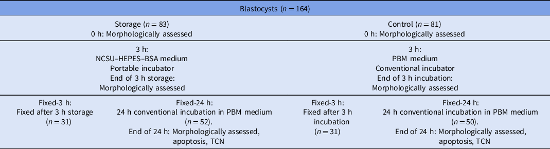
Following the 3 h storage period, blastocysts were morphologically assessed. To elucidate the degree of apoptosis at the end of the 3 h storage period, a group of the blastocysts was fixed and subsequently stained at this time point (from this point forwards referred to as Fixed-3 h), while further developmental potential and apoptosis after further incubation was assessed for the rest (from this point forwards referred to as Fixed-24 h; Table 1). The latter blastocysts (Fixed-24 h) were washed through two drops of PBM medium and incubated for 24 h in 500 µl of pre-equilibrated PBM medium in a conventional incubator at 38.8°C in an humified atmosphere containing 6% CO2 and 7% O2. After culture, blastocysts were assessed, pictured, and fixed.
Experimental design. On days 5 and 6 following fertilization, blastocysts classified as excellent or good were randomly split in two groups; storage and control. NCSU–HEPES–BSA was used as storage medium, while porcine blastocyst medium (PBM) was used in the conventional incubator. At the end point, blastocysts were fixed in paraformaldehyde, stained for assessment of apoptosis and total cell number (TCN), and analyzed using confocal microscopy, either straight after the 3 h storage period (Fixed-3 h) or after a further 24 h conventional incubation (Fixed-24 h)

The blastocysts were of different stages at the start of the experiments, and total cell number was therefore only used to calculate the proportion of apoptotic cells. Because of the relatively low numbers in each experimental group, blastocysts developed on day 5 and day 6 post-fertilization were merged when comparing the storage and control groups. As there were few significant differences between the storage and control groups, these groups were merged to explore general embryo quality between early versus later developed blastocysts.
Evaluation of blastocyst morphology and in vitro developmental capacity
Embryos were evaluated using a Leica DM IL inverted microscope (Leica Microsystems GmbH, Wetzlar, Germany). Only blastocysts classified as excellent or good as described in Bó and Mapletoft (Reference Bó and Mapletoft2013) were used in the experiment. Throughout the experiment blastocyst developmental stage was scored using the following criteria:
-
1. Young (blastocysts with an initially visible blastocoel filling less than 50% of the blastocysts).
-
2. Normal (blastocysts with a blastocoel filling 50% or more of the blastocysts).
-
3. Expanded [full blastocysts with increased outer diameter and thinned zona pellucida (ZP)].
-
4. Pre-hatching (expanded blastocysts with an extremely thin ZP).
-
5. Hatching/hatched (expanded blastocyst with fractured or lost ZP).
During the experiment, blastocysts that progressed to a more advanced developmental stage with excellent or good morphology were classified as progressed. At the start of the experiment the blastocysts were at different developmental stages. When evaluating progression, it was assumed the blastocysts developed in a stepwise manner. For illustration, if there were two blastocysts at the start, one normal and one expanded, and at the end there were one expanded and one pre-hatching blastocyst, it was assumed both had progressed; the normal to expanded and the expanded to pre-hatching. This was done in a consistent manner to minimize variation in scoring between the groups. The maximum was set at 10 blastocysts in each cryotube, but the average number was 4.1 and only five out of 40 had numbers above five, which made it realistic to keep track of the different blastocysts. Collapsed blastocysts were not categorized by their developmental stage as this could be difficult to judge. They were instead classified as partly collapsed (90–75% of normal size) or collapsed (<75% of normal size). Degenerated (highly fragmented) was only observed on two occasions during the experiment and was for that reason included in the collapsed group.
Evaluation of apoptosis
Blastocysts were fixed in 4% paraformaldehyde for 30 min at RT and stored in phosphate-buffered saline (PBS) containing 0.1% BSA (PBS/BSA) at 4°C until the staining procedure. Before staining, the blastocysts were permeabilized in 0.3% Triton X-100 supplemented with 0.1% sodium citrate in PBS/BSA for 1 h at RT, then rinsed three times in PBS/BSA.
For assessment of apoptosis, cells were labelled using the in situ cell death detection kit terminal deoxynucleotidyl transferase dUTP nick end labelling (TUNEL), TMR red (Roche, Mannheim, Germany). Up to 10 blastocysts were transferred to an Eppendorf tube containing 20 µl of TUNEL reaction mixture composed of enzyme solution and label solution in 1:9 v/v ratio and incubated at 37°C for 1 h in the dark and afterwards rinsed three times in PBS/BSA. TUNEL assay will label fragmented DNA, the endpoint of both intrinsic and extrinsic apoptosis as well as necrosis. As all are measures of reduced embryo quality, all TUNEL-positive nuclei were included in the present study. The TUNEL staining procedure was optimized and fluorescence microscope settings were determined by incubating positive and negative TUNEL controls in RQ1 RNase-free DNase (Promega, Oslo, Norway) for 45 min at 37°C in the dark before TUNEL staining. Positive controls were subsequently incubated in a conventional TUNEL mixture, while the negative control was incubated in label solution only.
For visualization of total cell number, blastocysts were stained for 10 min at RT with 10 µg/ml Hoechst 33342. Finally, they were mounted in 6 µl fluorescence mounting medium (Dako, Glostrup, Denmark) on a glass slide under a coverslip and stored at −20°C. Slides were assessed by fluorescence microscopy using a Leica SP8 laser scanning confocal microscope (Leica Microsystems GmbH, Wetzlar, Germany). Hoechst staining (blue) was evaluated with a 405 nm excitation laser and a 425–465 nm emission filter and TUNEL-positive nuclei (red) with a 552 nm excitation laser and a 570–620 nm emission filter. The proportion of apoptotic cells was calculated by dividing the number of TUNEL-positive nuclei by the total cell number.
Statistical analysis
Statistical analysis was performed using RStudio version 4.1.2 (2021–11–01). Data were tested for normality using the Shapiro–Wilk test. As the data were not normally distributed even after log transformation, the original data for apoptosis were analyzed using the Mann–Whitney U-test. Data for progressed and collapsed were analyzed using Fisher’s exact test when the expected values in any of the cells of the contingency table were <5, and the chi-squared test was used when the expected values were >5. The results were considered statistically significant when the P-value was ≤ 0.05. Graphs were plotted using Microsoft Excel (16.0.1).
Results
Blastocyst development
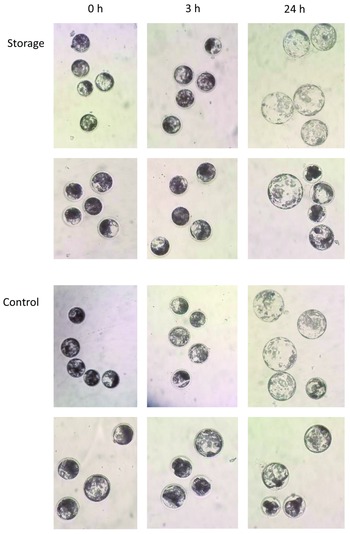
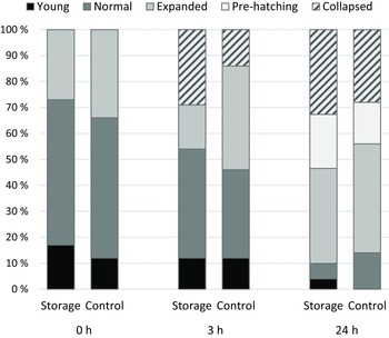
Images of the IVP porcine blastocysts throughout the liquid storage experiment are shown in Figure 1, illustrating variation in both the storage and control groups. The distribution of the different blastocyst developmental stages at the start of the experiment (0 h) after the 3 h storage period (3 h) and after 24 h conventional incubation post-storage (24 h) are shown in Figure 2. For the 3 h storage period a proportion of the blastocysts collapsed. During the 24 h conventional incubation post-storage more blastocysts collapsed, but a high proportion of the blastocysts progressed, so less young and normal blastocysts were observed and more expanded as well as pre-hatching.
Liquid storage of porcine in vitro-produced blastocysts. Representative images for the liquid storage and control Fixed-24 h groups at three different time points: at the start of the experiment (0 h), after the 3 h storage period (3 h), and after a further 24 h conventional incubation (24 h).

Blastocyst developmental stages for the liquid storage (n = 52) and control (n = 50) Fixed-24 h groups, in which the blastocysts were stored for 24 h in a conventional incubator after the 3 h storage period. Developmental stages were recorded at the start of the experiment (0 h), after the 3 h storage period (3 h), and after a further 24 h conventional incubation (24 h). The blastocyst development categories ‘pre-hatching’ and ‘hatching’ were merged to ‘pre-hatching’ and ‘collapsed’ and ‘partly collapsed’ was merged to ‘collapsed’.

Effect of treatment
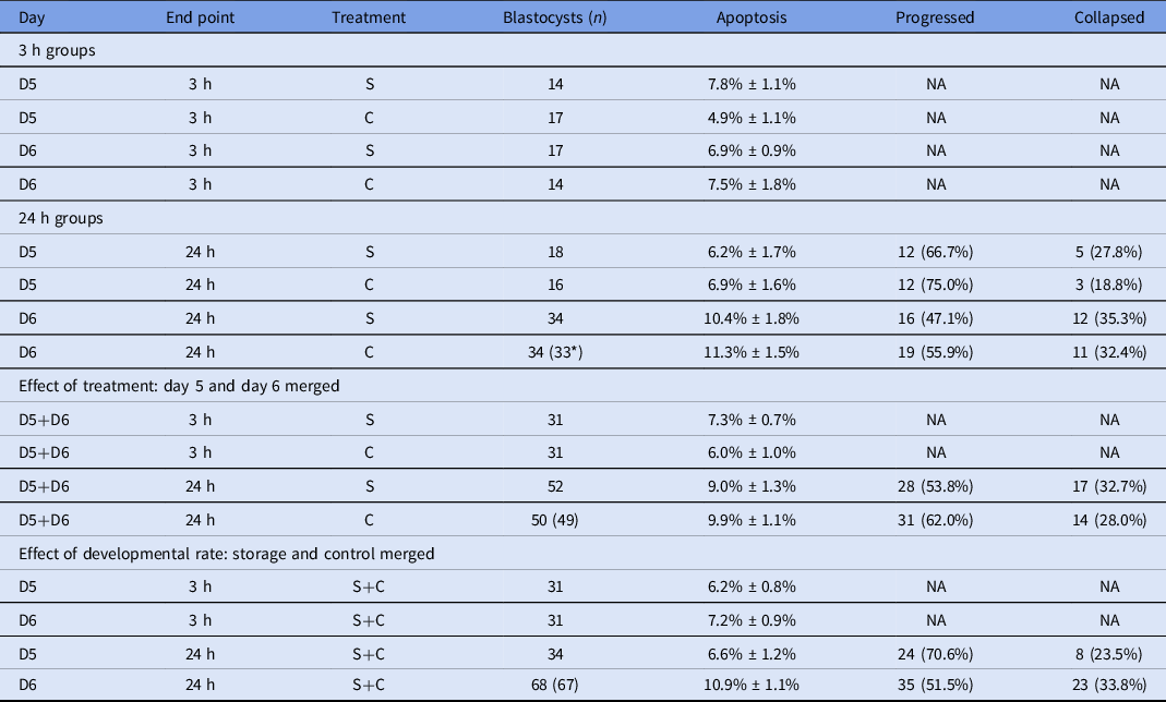
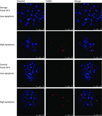
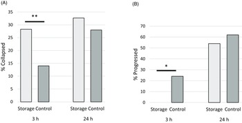
The proportions of apoptosis, progressed and collapsed blastocysts for the different treatment groups are shown in Table 2, and representative images of different levels of apoptosis are shown in Figure 3. The purpose of the Fixed-3 h groups was to assess apoptosis at that time point. Comparisons between the proportion of collapsed and progressed blastocysts between the 3 h and 24 h time points were made within the Fixed-24 h group, and the effect of this treatment is shown in Figure 4.
Proportion of apoptosis, progressed, and collapsed porcine blastocysts after incubation in storage (S) or control (C) medium and respective incubators without or with 6% CO2 for 3 h (3 h) and an additional 24 h (24 h) conventional incubation following storage. D5 embryos reached the blastocyst stage and were incubated at day 5, while D6 embryos reached the blastocyst stage at day 6. Results are shown as number of blastocysts (n), mean percentage apoptosis ± SE, and number (percentage) of progressed and collapsed blastocysts

* One blastocyst was lost during staining, so for total cell number and apoptosis n is 33 while for progression and collapsed n is 34.
Hoechst staining combined with TUNEL TMR red of blastocysts after liquid storage or control treatment. Images on the left are cell nuclei stained with Hoechst (blue colour), middle images are cell nuclei stained with TUNEL TMR red assay (red colour), and images on the right are the Hoechst and TUNEL TMR images merged.

(A) The percentage collapsed in the Fixed-24 h storage and control groups at the time points 3 h and 24 h. (B) The percentage progressed in the Fixed-24 h storage and control groups at the time points 3 h and 24 h. *Indicates significant difference between bars (P < 0.05), **indicates a trend (P < 0.10).

There was no significant difference between the storage and the control at the 24 h time point for any of the parameters, nor for apoptosis in the Fixed-3 h groups. However, in the Fixed-24 h groups there was a trend for more collapse at the 3 h time point in the storage group compared with the control group (28.3% vs 14%, P = 0.092; Figure 4A), but not after an additional 24 h conventional incubation (32.7% vs 28.0%, P = 0.670). This was not due to re-expansion of the collapsed blastocysts in the storage group, but through more blastocysts collapsing in the control group. In the whole study, only two of the total number that collapsed re-expanded fully. Also, significantly fewer blastocysts progressed during the 3 h storage period in the storage groups compared with the control groups (0% vs 24%, P = 0.000). Still, after a further 24 h conventional incubation there was no significant difference observed (53.8% vs 62.0%, P = 0.429; Figure 4B).
There was no significant difference between the proportion of apoptosis seen in the Fixed-3 h and the Fixed-24 h storage groups (7.3% ± 0.7% vs 9.0% ± 1.3%, P = 0.281), whereas there was significantly less apoptosis in the Fixed-3 h versus the Fixed-24 h control group (6.0% ± 1.0% vs 9.9% ± 1.1%, P = 0.014).
Effect of developmental rate on blastocyst quality
Overall, embryos that reached the blastocyst stage at day 5 after fertilization showed significantly less apoptosis (6.6% ± 1.2% vs 10.9% ± 1.1%, P = 0.010) and a trend for more progression (70.6% vs 51.5%, P = 0.089) than embryos reaching the blastocyst stage on day 6. There was no significant difference in the proportion collapsed between day 5 and day 6 blastocysts.
For the day 5 blastocysts, there was no significant difference in apoptosis between the Fixed-3 h and Fixed-24 h groups (6.2% ± 0.8% vs 6.6% ± 1.2%, P = 0.818). However, for the day 6 blastocysts the proportion of apoptosis was significantly higher in the Fixed-24 h group (10.9% ± 1.1%) than for the Fixed-3 h group (7.2% ± 0.9%, P = 0.015).
Discussion
In this study, we found that in vitro-produced porcine embryos could be successfully stored in transportable incubators using CO2-free medium. There was no significant difference from the control medium in development after 24 h re-cultivation under conventional conditions, nor in the proportion of apoptotic cells.
Effect of treatment: progression
There was a developmental delay in the storage group as less blastocysts progressed during the 3 h storage period in the storage group compared with the control group. However, after 24 h conventional culture after storage, there was no significant difference between the storage and control groups regarding the number of embryos that progressed to more advanced developmental stages.
The transition to blastocysts is an indicator of the embryo developmental capacity and blastocysts are consequently more likely to develop further (Rubio Pomar et al., Reference Rubio Pomar, Ducro-Steverink, Hazeleger, Teerds, Colenbrander and Bevers2004). For this reason, and because of the increased tolerance to oxidative stress and preservation, only blastocysts were used in this study. Most of the blastocysts that did not collapse during storage did progress to the expanded or pre-hatching category during the conventional culture; but in line with other similar studies, e.g. Martinez et al. (Reference Martinez, Cambra, Nohalez, Parrilla, Roca, Vazquez, Rodriguez-Martinez, Gil, Martinez and Cuello2019a), some of the blastocysts with good morphology did not develop further. This emphasizes the need for additional quality parameters in addition to morphology and a period of conventional culture following storage to assess developmental capacity. Martinez et al. (Reference Martinez, Cambra, Nohalez, Parrilla, Roca, Vazquez, Rodriguez-Martinez, Gil, Martinez and Cuello2019a) discussed that 24 h conventional culture after 72-h liquid storage did not give the embryos enough time to compensate for the developmental delay but, for the short storage time of 3 h, 24 h conventional culture appeared to be sufficient. This is most probably due to a less significant developmental delay after the shorter storage time.
To the best of our knowledge there has only been one other study on liquid storage of porcine IVP embryos in which Lin et al. (Reference Lin, Le, Takebayashi, Hirata, Tanihara, Thongkittidilok, Sawamoto, Kikuchi and Otoi2022) tested different storage media and storage temperatures over 24 h liquid storage and concluded that 25°C was the best storage temperature. Compared with our study, not unexpectedly, more blastocysts developed to more advanced developmental stages after 3 h storage (53.8%) than after 24 h storage (11.2%). Converting our measure of collapsed blastocysts to Lin et al.’s measure of survived blastocysts (re-expanded), more blastocysts survived after 3 h storage (67.3%) than after 24 h (31.8%). Despite differences in the experimental set-up, 24 h storage seems to have a marked negative effect on embryo quality compared with a shorter storage time.
There are contradicting results regarding the optimum storage temperature for porcine embryos. Martinez et al. (Reference Martinez, Angel, Cuello, Sanchez-Osorio, Gomis, Parrilla, Vila, Colina, Diaz, Reixach, Vazquez, Vazquez, Roca and Gil2014) found that storing blastocysts at 25°C had a negative effect on embryo viability compared with 37°C. However, Lin et al. (Reference Lin, Le, Takebayashi, Hirata, Tanihara, Thongkittidilok, Sawamoto, Kikuchi and Otoi2022) found apoptosis to be higher with increased temperature (30°C versus 25°C), whereas Rubio Pomar et al. (Reference Rubio Pomar, Ducro-Steverink, Hazeleger, Teerds, Colenbrander and Bevers2004) saw no significant difference in the number of apoptotic cells between storage at 25°C and at 38°C. Different storage media and embryo sources used in the different studies do, however, make it difficult to compare these results.
Storage time will also affect the optimum storage temperature. In our study of 3 h liquid storage, arresting development was not important and the embryos could be stored at 37°C. However, for a longer storage time, further development has to be prevented, and the embryos would need to be stored at a lower temperature. Martinez et al. (Reference Martinez, Cambra, Nohalez, Parrilla, Roca, Vazquez, Rodriguez-Martinez, Gil, Martinez and Cuello2019a) showed that storing in vivo-derived porcine embryos at 20°C arrested development, but it also had a negative effect on embryo viability. Still, after 72-h liquid storage at 20°C and further 24 h conventional incubation, 80% of the in vivo-derived blastocysts seemed viable. Lin et al. (Reference Lin, Le, Takebayashi, Hirata, Tanihara, Thongkittidilok, Sawamoto, Kikuchi and Otoi2022) investigated storing IVP porcine embryos at 20°C, with only 13% being viable and none developing further after 24 h storage and 48 h further conventional incubation. Even though different storage media were used, this illustrates the higher sensitivity of IVP porcine embryos compared with in vivo-derived embryos. More studies are needed to investigate how IVP embryos could undergo liquid storage for longer time periods without compromising their quality.
Effect of treatment: collapsing
In this study there was a trend for more blastocysts to collapse in the storage group compared with the control group after the 3 h storage period. This trend was not seen after a further 24 h conventional incubation, due to more blastocysts collapsing in the control group. Weak collapsing (also called contractions) is a normal element during blastulation (Viñals Gonzalez et al., Reference Viñals Gonzalez, Odia, Cawood, Gaunt, Saab, Seshadri and Serhal2018) and prior to hatching (Niimura, Reference Niimura2003); conversely, strong collapse may inhibit hatching (Niimura, Reference Niimura2003). Blastocyst collapse is caused by the outflow of blastocoel fluid and, although the molecular mechanisms behind blastocyst collapse are not known, it can be caused by different stressors, e.g. change in temperature, incubation medium or mechanical stress (Martinez et al., Reference Martinez, Cambra, Nohalez, Parrilla, Roca, Vazquez, Rodriguez-Martinez, Gil, Martinez and Cuello2019a).
In the present study the lower quality embryos were probably most susceptible to collapse. Several studies have suggested that collapse is a consequence of reduced embryo quality (Marcos et al., Reference Marcos, Pérez-Albalá, Mifsud, Molla, Landeras and Meseguer2015; Bodri et al., Reference Bodri, Sugimoto, Yao Serna, Kawachiya, Kato and Matsumoto2016) caused by compromised oocyte or sperm quality or suboptimal in vitro culture conditions (Marcos et al., Reference Marcos, Pérez-Albalá, Mifsud, Molla, Landeras and Meseguer2015). In addition, aneuploid embryos have a higher frequency of collapse than euploid embryos (Viñals Gonzalez et al., Reference Viñals Gonzalez, Odia, Cawood, Gaunt, Saab, Seshadri and Serhal2018; Sciorio et al., Reference Sciorio, Thong and Pickering2020). Re-expansion requires considerable energy for active transport of Na+ into the blastocoel cavity (Sciorio et al., Reference Sciorio, Thong and Pickering2020) and a strong collapse following re-expansion may reduce embryo viability, while embryos of low quality may not be able to re-expand. Frequent collapsing events and strong undergoing collapse have been shown to lead to reduced implantation rate (Marcos et al., Reference Marcos, Pérez-Albalá, Mifsud, Molla, Landeras and Meseguer2015; Viñals Gonzalez et al., Reference Viñals Gonzalez, Odia, Cawood, Gaunt, Saab, Seshadri and Serhal2018; Sciorio et al., Reference Sciorio, Thong and Pickering2020). Blastocyst collapse is therefore suggested to be an indicator of embryo quality and developmental capacity (Niimura, Reference Niimura2003) and blastocysts showing a strong collapse are not recommended to be chosen for embryo transfer.
Embryos in both the control and storage groups were subjected to mechanical stress through pipetting, extra time outside the incubator and thereby temperature and oxidative stress and slight fluctuations in pH. The additional stress for the embryos in the storage group through change of medium and the conditions in the transport incubator was most probably the cause of the earlier collapse seen in this group.
Very little re-expansion was seen in this study. As part of normal developmental events bovine blastocysts needed 6–10 h to re-expand (Gonzales et al., Reference Gonzales, Jones, Pinyopummintr, Carnevale, Ginther, Shapiro and Bavister1996), but Martinez et al. (Reference Martinez, Cambra, Nohalez, Parrilla, Roca, Vazquez, Rodriguez-Martinez, Gil, Martinez and Cuello2019a) argued that 24 h may not be sufficient time for the blastocysts to re-expand after storage. Increasing the post-storage culture period to examine the embryo re-expansion and further developmental potential could therefore have been beneficial. This might, however, have led to some complications with more of the embryos deteriorating after hatching. The phenomenon of collapse and other methods to assess their further developmental potential do need further investigation.
Effect of treatment: apoptosis
Apoptosis, programmed cell death, has the function of removing abnormal or irreversibly damaged cells in preimplantation embryos (Ramos-Ibeas et al., Reference Ramos-Ibeas, Gimeno, Cañón-Beltrán, Gutiérrez-Adán, Rizos and Gómez2020; Cambra et al., Reference Cambra, Martinez, Rodriguez-Martinez, Gil and Cuello2021). It has a low incidence in in vivo-derived, compared with in vitro-produced, porcine embryos (Rubio Pomar et al., Reference Rubio Pomar, Ducro-Steverink, Hazeleger, Teerds, Colenbrander and Bevers2004) and the degree of apoptosis can reliably predict human embryo quality (Haouzi and Hamamah, Reference Haouzi and Hamamah2009) and porcine embryo further developmental capacity (Chen et al., Reference Chen, Dai, Wu, Zhang, Sun and Zhang2018).
Apoptosis was evaluated after the 3 h storage period to investigate how far the process of apoptosis had reached at that time point and how it progressed during the 24 h conventional incubation. There was no significant difference in the degree of apoptosis between the storage and control groups at any time point, indicating that change to the storage medium and the conditions in the transport incubator did not cause a noteworthy increase in apoptosis.
Both groups did however show more apoptosis than previously observed in our laboratory (unpublished data) and, in agreement with other studies, with porcine embryos (Hao et al., Reference Hao, Lai, Mao, Im, Bonk and Prather2004; Chen et al., Reference Chen, Dai, Wu, Zhang, Sun and Zhang2018). The extra stressors subjected to both the storage and control groups, as discussed above, appeared to have caused DNA damage and led to the activation of apoptosis, which demonstrates the sensitivity of the IVP porcine embryos. The current experimental set-up had the benefit of purely elucidating the effect of the storage medium and transport incubator, however a control group that remained in the incubator for the duration of the experiment could have been beneficial.
Effect of developmental rate on blastocyst quality
In this study, embryos that reached the blastocyst stage at day 5 post-fertilization showed significantly less apoptosis after storage and further conventional incubation, and a trend for more progression than embryos reaching the blastocyst stage on day 6. This is in agreement with previous studies that showed that early developing blastocysts were of better quality than later developing blastocysts. Early developing bovine embryos showed lower levels of apoptosis (Ramos-Ibeas et al., Reference Ramos-Ibeas, Gimeno, Cañón-Beltrán, Gutiérrez-Adán, Rizos and Gómez2020) and a higher probability of leading to live offspring (Van Soom et al., Reference Van Soom, Ysebaert and De Kruif1997; Rubio Pomar et al., Reference Rubio Pomar, Ducro-Steverink, Hazeleger, Teerds, Colenbrander and Bevers2004; Bó and Mapletoft, Reference Bó and Mapletoft2013; Marsico et al., Reference Marsico, de Camargo, Valente and Sudano2019), while fast developing human and porcine embryos showed a lower incident of aneuploidy (Campbell et al., Reference Campbell, Fishel, Bowman, Duffy, Sedler and Hickman2013) and porcine embryos a better resistance to cryopreservation (Morató et al., Reference Morató, Castillo-Martín, Yeste and Bonet2016; Ohlweiler et al., Reference Ohlweiler, Mezzalira and Mezzalira2019).
Applying NsDU-ET, Martinez et al. (Reference Martinez, Angel, Cuello, Sanchez-Osorio, Gomis, Parrilla, Vila, Colina, Diaz, Reixach, Vazquez, Vazquez, Roca and Gil2014) transferred 30 blastocysts that had progressed during their study of 24 h liquid storage of in vivo-derived porcine embryos. However, it has been suggested that, when applying NsDU-ET, the actual number of transferred embryos is important, with the transfer of 40 embryos giving better farrowing rates and litter sizes than transferring 30 embryos (Martinez et al., Reference Martinez, Martinez, Cambra, Maside, Lucas, Vazquez, Vazquez, Roca, Rodriguez-Martinez, Gil, Parrilla and Cuello2019b). In addition, as IVP embryos are in general of lower quality than in vivo-derived embryos, it would be advised to store ∼60 IVP porcine blastocysts developed on day 5 post-fertilization to transfer 40 blastocysts with in vitro developmental capacity after 3 h liquid storage by NsDU-ET.
For the day 6 blastocysts, the degree of apoptosis increased during the conventional culture post-storage, while there was no significant increase seen with the day 5 blastocysts. The day 6 blastocysts may have reduced innate quality that rendered them susceptible to increased damage during the storage procedure, and this damage may not have resulted in apoptosis until after further incubation. It is also possible that day 5 blastocysts were able to repair some of the damage caused during the storage procedure and, consequently, the proportion of apoptosis did not increase for this group after further incubation. Consequently, although the end point of apoptosis can be measured at the end of the 3 h storage period, further incubation should be included to assess the potential progression of apoptosis. In addition, more quality parameters should be applied to elucidate the different processes leading to damage in the embryos throughout the storage experiment.
In conclusion, in vitro-produced porcine blastocysts can be stored for 3 h at physiological temperature in transportable incubators using a CO2-independent medium without compromising quality. In this study, morphology, further development, and apoptosis were used to assess embryo quality after liquid storage and we found that blastocysts developed on day 5 post-fertilization were most suitable for short-term liquid storage. The 3 h storage period can possibly identify embryos of lower quality as these are most likely to be more susceptible to undergoing a strong collapse. As such, combining the non-invasive measures of timing of development and morphology can aid in deselecting embryos of inferior quality for embryo transfer. To transfer 40 blastocysts with in vitro developmental capacity using NsDU-ET, 60 day-5 blastocysts should be placed into liquid storage, and blastocysts that have collapsed during the storage period should not be expected to be of good quality.
Additional quality parameters are however needed to assess the underlying mechanisms leading to collapsing blastocysts and to determine the reasons for embryo damage during liquid storage. Furthermore, studies regarding increased incubation time and storage temperature are essential to enable the transport of in vitro-produced embryos over longer distances.
Availability of data and materials
The data that support the findings of this study are available from the corresponding author upon reasonable request.
Acknowledgements
The authors thank Rune Heggelund (Nortura Rudshøgda) for arrangements at the slaughterhouse and Teklu Tewoldebrhan Zeremichael for technical assistance.
Financial support
This work was funded by an internal scholarship from the Inland Norway University of Applied Sciences. In addition, the study received financial support from Norsvin SA.
Conflict of interest declaration
The authors declare no conflict of interest.
Ethical standards
Oocytes were collected from routinely slaughtered animals, a procedure that did not require ethical approval.