Introduction
Low success rate in the production of in vitro-derived pig embryos is due to the deficiency of cytoplasmic maturation leading to high incidence of polyspermy and a low development rate and low quality of blastocysts for in vitro-produced compared with in vivo-produced embryos (Yoshida et al., Reference Yoshida, Bamba and Kojima1989; Abeydeera & Day, Reference Abeydeera and Day1997; Gil et al., Reference Gil, Cuello, Parrilla, Vazquez, Roca and Martinez2010).
It has been shown previously that the use of dibutyryl cyclic adenosine monophosphate (dbcAMP) to temporarily inhibit meiotic resumption and allow pig oocytes to fully complete mRNA synthesis can increase developmental competent of the gametes (Funahashi et al., Reference Funahashi, Cantley and Day1997; Bagg et al., Reference Bagg, Nottle, Grupen and Armstrong2006; Kim et al., Reference Kim, Cho, Song, Wee, Park, Cho, Yu, Lee, Han and Koo2008; Nascimento et al., Reference Nascimento, Albornoz, Che, Visintin and Bordignon2010; Cayo-Colca et al., Reference Cayo-Colca, Yamagami, Phan and Miyano2011).
One of important markers in oocyte maturation is the level of glutathione (GSH), as GSH is related to the cytoplasmic maturation of oocytes (de Matos et al., Reference de Matos, Gasparrini, Pasqualini and Thompson2002; Gasparrini et al., Reference Gasparrini, Sayound, Negali, Matos, Donnay and Zicarelli2003). GSH protects oocytes from reactive oxygen species (ROS) (Meister, Reference Meister1983; Yoshida et al., Reference Yoshida, Ishigaki, Nagai, Chikyu and Pursel1993), which has been shown to compromise developmental potential of bovine oocytes. Oxidative stress is affected by ROS, and this stress results in an imbalance of the intracellular redox state (Deleuze & Goudet, Reference Deleuze and Goudet2010). Increased levels of ROS in bovine oocytes during in vitro maturation (IVM) have a detrimental effect on embryonic development (Hashimoto et al., Reference Hashimoto, Minami, Yamada and Imai2000). Moreover, the GSH content of an oocyte is correlated with the presence of cumulus cells (CCs) (Sawai et al., Reference Sawai, Funahashi and Niwa1997; Nagai, Reference Nagai2001). CCs support oocyte competence, which is the ability of an oocyte to complete nuclear maturation, fertilize and develop to blastocyst (Suzuki et al., Reference Suzuki, Jeong and Yang2000; Albertini et al., Reference Albertini, Combelles, Benecchi and Carabatsos2001). Therefore, CC apoptosis affects oocyte competence (Lee et al., Reference Lee, Joo, Na, Yoon, Choi and Kim2001). However, studies on the effect of dbcAMP on porcine oocyte maturation have focused primarily on meiotic progression and subsequent embryonic development (Funahashi et al., Reference Funahashi, Cantley and Day1997; Bagg et al., Reference Bagg, Nottle, Grupen and Armstrong2006; Kim et al., Reference Kim, Cho, Song, Wee, Park, Cho, Yu, Lee, Han and Koo2008). There is limited information on the relationships between glutathione levels of oocytes and ROS levels of oocytes and CC apoptosis as related to dbcAMP.
The main purpose of this study was to determine the effects of dbcAMP on GSH and ROS levels in oocytes in relation to CC apoptosis. In addition, the effects of dbcAMP on subsequent embryonic development and apoptosis levels in blastocysts following in vitro fertilization (IVF) or parthenogenetic activation (PA) were also investigated.
Materials and methods
Chemicals
Unless otherwise noted, all chemicals and reagents used were purchased from Sigma-Aldrich (St. Louis, MO, USA).
Oocyte collection and in vitro maturation (IVM)
Porcine ovaries were collected from a local abattoir and transported to the laboratory at 34–36°C in 0.9% saline supplemented with 100 IU/ml penicillin G and 100 μg/ml streptomycin. Cumulus–oocyte complexes (COCs) were aspirated through an 18-gauge needle. Oocytes with compact cumulus mass and a dark, homogenous cytoplasm were washed three times in Tyrode's lactate–HEPES–polyvinyl alcohol (TL–HEPES–PVA: 114 mM NaCl, 3.2 mM KCl, 0.4 mM Na2H2PO4, 2 mM CaCl2·2H2O, 0.5 mM MgCl2·6H2O, 5 mM NaHCO3, 20 mM HEPES, 16.6 mM sodium lactate (60% syrup), 0.5% PVA, 10 IU/ml penicillin, and 10 μg/ml streptomycin). COCs were cultured in NCSU-23 medium supplemented with 10% porcine follicular fluid (PFF), 0.6 mM cysteine, 10 ng/ml epidermal growth factor (EGF), 10 IU/ml pregnant mare serum gonadotropin (PMSG), and 10 IU/ml human chorionic gonadotropin (HCG) for 22 h and then for another 22 h in maturation medium without hormones at 39°C in a humidified atmosphere of 5% CO2 in air. PFF was collected from ovarian follicles 3–6 mm in diameter by centrifugation at 1600 g for 30 min and filtration through a 1.2 μm syringe filter.
Assessment of nuclear maturation
Meiotic stages were examined as described previously (Yu, Reference Yu2011). Oocytes were stained with aceto-orcein [1% (w/v) orcein in 45% (v/v) acetic acid] followed by aceto-glycerol (1:1:3 glycerol:acetic acid:distilled water), and then evaluated under a light microscope (DM 2500, Leica, Wetzlar, Germany) at ×400 magnification. Percentages of germinal vesicle (GV) at 22 h of culture and metaphase II (MII) rates at 44 h of culture were determined.
Measurement of intracellular ROS and GSH levels of oocytes
The levels of ROS in the oocytes were measured by dichlorohydrofluorescein diacetate (DCHFDA). After maturation, oocytes were transferred into in vitro culture 1 medium (IVC1: d-glucose-free NCSU-23 supplemented with 0.17 mM sodium pyruvate, 2.73 mM sodium lactate, and 0.4% BSA) that contained 10 μM DCHFDA. After 30 min of culture, oocytes were washed in DPBS–PVA. The oocytes were then placed on a glass slide with a 10 μl drop of DPBS–PVA. The fluorescent emissions from the oocytes were recorded as .tiff files using a cooled charge-coupled device (CCD) camera attached to a fluorescence microscope (Axio-Observer A1; Charles Zeiss, Goettingen, Germany) with filters (excitation: 450–490 nm, emission: 515–565 nm). The recorded fluorescent images were analysed using ImageJ software 1.33u (National Institutes of Health, Bethesda, MD, USA) by the intensity of fluorescence in each oocyte picture.
Cell Tracker Blue CMF2HC (4-chloromethyl-6,8-difluoro-7-hydroxycoumarin; Invitrogen) was used to detect GSH levels in oocytes as a blue fluorescence. After maturation, oocytes were incubated for 30 min in TL–HEPES–PVA supplemented with 10 μM Cell Tracker. The oocytes were then washed with DPBS–PVA and placed into a 10 μl droplet. Fluorescence was observed using a fluorescence microscope with an ultraviolet (UV) filter (370 nm). The fluorescence density was measured as described above.
Detection of apoptosis of cumulus cells
CCs were removed by pipetting gently into TL–HEPES–PVA supplemented with 0.1% hyaluronidase. CCs were washed in phosphate-buffered saline (PBS)–PVA without Ca2+ and Mg2+ (–), pelleted by centrifugation three times, then fixed in 4% paraformaldehyde in PBS at 4°C. CCs were then washed three times in PBS (–)/PVA and permeabilized by incubation in 0.5% Triton X-100 in PBS for 1 h at 4°C. After permeabilization, CCs were washed three times in PBS (–)/PVA and incubated with terminal deoxynucleotidyl transferase nick end labeling (TUNEL) assay kit (In Situ Cell Death Detection Kit, Roche, Mannheim, Germany) in the dark for 1 h at 39°C. CCs were then counterstained with 40 μg/ml propidium iodide (PI). The numbers of TUNEL-positive cells and the total CCs were determined from optical images of whole-mount CCs taken using fluorescence microscopy. The percentage of TUNEL-positive cells is described as the percentage of TUNEL-positive cells relative to the total number of cells.
Parthenogenetic activation
After IVM, CCs were removed as described above. For chemical activation (CA), oocytes were incubated in IVC 1 medium containing 5 μM ionomycin for 5 min, washed twice, and incubated for 3 h in IVC 1 medium supplemented with 2.0 mM 6-dimethylaminopurine (6-DMAP). For electrical activation (EA), oocytes were transferred to pulsing medium consisting of 0.3 M d-mannitol, 0.1 mM MgSO4, 0.05 mM CaCl2, and 0.01% PVA, washed three times, and then transferred to a chamber containing two electrodes overlaid with pulsing medium. The oocytes were then stimulated with a direct-current pulse of 1.5 kV/cm for a duration of 100 μs using a BTX Electro-Cell Manipulator 2001 (BTX, San Diego, CA, USA).
In vitro fertilization (IVF)
Extended spermatozoa supplied by Irae Yangdon were maintained at 17°C. Percoll solutions and gradients were prepared as described previously (Yu, Reference Yu2011). The pellet recovered after aspiration of the supernatant was washed twice by centrifugation at 350 g for 3 min with 5 ml D-PBS supplemented with 0.1% BSA, 10 IU/ml penicillin, and 10 μg/ml streptomycin. After the supernatant was discarded, motile spermatozoa were collected. The sperm concentration (spermatozoa/ml) was diluted to 10 × 105 with Tris-buffered medium (mTBM: 113.1 mM NaCl, 3 mM KCl, 7.5 mM CaCl2·2H2O, 20 mM Tris, 11 mM d-glucose, 5 mM sodium pyruvate, 2 mM caffeine, and 0.2% BSA). After IVM, CCs were removed as described above. Denuded oocytes were washed three times with mTBM and transferred to an mTBM insemination drop (45 μl). A 5 μl volume of spermatozoa was added to each insemination drop to give a final concentration of 1 × 10⁵ spermatozoa/ml. Oocytes and spermatozoa were co-cultured for 6 h at 39°C in a humidified atmosphere of 5% CO2 in air.
IVC
Following either PA or IVF, presumptive zygotes were washed three times, transferred to IVC1 medium and incubated for 2 days at 39°C in a humidified atmosphere of 5% CO2 in air. After 2 days of embryo culture, cleavage formation was assessed. Embryos were then washed twice, transferred to in vitro culture 2 medium (IVC 2: NCSU-23 containing 0.4% BSA), and incubated for 6 days, after which time blastocyst formation was assessed.
Detection of apoptosis of blastocysts
Apoptosis of blastocysts were determined by TUNEL assays as described above. The percentage of TUNEL-positive nuclei is described as the percentage of TUNEL-positive nuclei relative to the total cell number (PI, positive nuclei) of the blastocyst.
Experimental design
Experiment 1: Effect of dbcAMP on nuclear maturation
This experiment was conducted to assess the effect of dbcAMP on nuclear maturation. Oocytes (n = 1376) were cultured in NCSU-23 supplemented with 0 mM (control), 0.5 mM, 1 mM, 5 mM, or 10 mM dbcAMP and hormones for 22 h, and were then cultured in NCSU-23 without dbcAMP and hormones for another 22 h. The GV rate at 22 h of culture and the MII rate at 44 h of culture were evaluated. Five replicates were conducted for this experiment.
Experiment 2: Effect of dbcAMP on levels of GSH and ROS, and CC apoptosis
This experiment was carried out to determine levels of GSH and ROS in oocytes, and CC apoptosis. Oocytes (n = 331) were cultured as described in Experiment 1. GSH and ROS levels of oocytes at 44 h of culture were assessed. In addition, apoptosis of CCs (n = 3743) was determined at 44 h of culture. Each experiment was replicated four times.
Experiment 3: Effect of dbcAMP on embryonic development and the levels of apoptosis in blastocysts following PA or IVF
This experiment was designed to evaluate the effect of dbcAMP during IVM on embryonic development and the level of apoptosis in blastocysts following PA or IVF. Oocytes (n = 1747) were cultured in NCSU-23 supplemented with 0 mM (control), 0.5 mM, or 1 mM dbcAMP, then fertilized in vitro or activated parthenogenetically. Cleavage rates on day 2 of culture and blastocyst formation on day 8 of culture were examined. The cell numbers and apoptosis levels in blastocysts were assessed. Five replicates were conducted for this experiment.
Statistical analysis
Percentage data were subjected to an arcsin transformation before analysis. All data and data sets are presented as the mean ± standard error of the mean (SEM), and were analysed by Duncan's multiple range test using the Statistical Analysis System ver. 8x (SAS, Cary, NC, USA). For all analyses, a P-value < 0.05 was considered to be significant.
Results
The effect of dbcAMP on nuclear maturation of oocytes
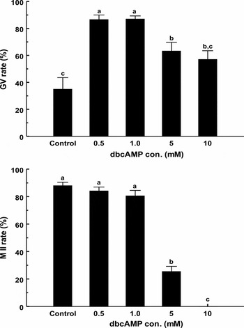
At the start of culture, the GV percentage was 89.9% (data not shown). The proportion of oocytes that remained at the GV stage after 22 h of exposure to 0.5 mM (86.5%) and 1 mM dbcAMP (87.0%) were higher than those of oocytes cultured in 5 mM (63.2%), 10 mM dbcAMP (57.0%) and in the absence of dbcAMP (34.9%) (Fig. 1; P < 0.05). After an additional 22 h of culture without removal of dbcAMP, the metaphase I (MI) rates were higher in the control group (87.9%) and those that were treated with 0.5 mM (84.1%) or 1 mM dbcAMP (80.5%) (Fig. 1; P < 0.05).
Effect of dbcAMP on germinal vesicle (GV) rates (top) of oocytes at 22 h of culture and metaphase II (MII) rates (bottom) of oocytes at 44 h. Data are expressed as mean ± standard error of the mean (SEM). a–cDifferent superscripts indicate significant differences among groups (P < 0.05).

The effects of dbcAMP on GSH and ROS levels of oocytes
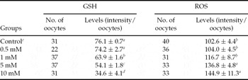
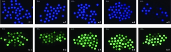
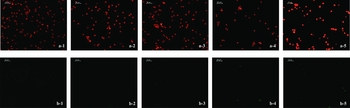
GSH levels of oocytes in 0.5 mM dbcAMP or in the absence of dbcAMP were higher than levels in other treatment groups (Table 1 and Fig. 2(a); P < 0.05). ROS levels were lower in oocytes exposed to 0.5 mM and 1 mM dbcAMP and the control group (Table 1 and Fig. 2(b); P < 0.05).
The effects of dbcAMP concentrations on glutathione (GSH) and reactive oxygen species (ROS) levels of porcine oocytes cultured for 44 h

a –dWithin the column, values with different letters are different (P < 0.05).
eControl: absence of dbcAMP.
Data are expressed as mean ± standard error of the mean (SEM).
Representative images of the intracellular glutathione (GSH) and reactive oxygen species (ROS) levels of porcine oocytes by staining with Cell Tracker Blue CMF2HC and DCHFDA, respectively. (a) GSH in groups treated without dbcAMP (control, a-1), or with 0.5 mM dbcAMP (a-2), 1 mM dbcAMP (a-3), 5 mM dbcAMP (a-4), or 10 mM dbcAMP (a-5). (b) ROS in groups treated without dbcAMP (control, b-1), or with 0.5 mM dbcAMP (b-2), 1 mM dbcAMP (b-3), 5 mM dbcAMP (b-4), or 10 mM dbcAMP (b-5). The oocytes expressed higher levels of intracellular GSH (blue) in control, and 0.5 mM dbcAMP groups. The oocytes showed higher levels of intracellular ROS, appearing as a brighter green colour in 5 mM and 10 mM dbcAMP groups.

The effect of dbcAMP on apoptosis levels of CCs
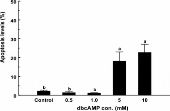
CC apoptosis levels were lower in 0.5 mM (1.4%), 1 mM dbcAMP (1.1%), and control (2.2%) than in other groups (18.1% and 22.7% in 5 mM and 10 mM dbcAMP, respectively) (Fig. 3; P < 0.05). CCs in 5 mM and 10 mM dbcAMP groups showed higher levels of nuclear DNA fragmentation than those in 0.5 mM and 1 mM dbcAMP and the control groups (Fig. 4).
The apoptosis levels of cumulus cells (CCS) at 44 h of culture. Data are expressed as mean ± standard error of the mean (SEM). a,bDifferent superscripts indicate significant differences among groups (P < 0.05).

Representative apoptosis images of porcine cumulus cells (CCs). (a) propidium iodide (PI) stains the nucleus (red) in oocytes treated without dbcAMP (a-1), or with 0.5 mM dbcAMP (a-2), 1 mM dbcAMP (a-3), 5 mM dbcAMP (a-4), or 10 mM dbcAMP (a-5). (b) TUNEL staining (green) shows fragmented DNA in oocytes treated without dbcAMP (b-1), or with 0.5 mM dbcAMP (b-2), 1 mM dbcAMP (b-3), 5 mM dbcAMP (b-4), or 10 mM dbcAMP (b-5). Higher TUNEL staining is seen in 5 mM and 10 mM dbcAMP treatment groups indicating a higher level of nuclear DNA fragmentation of the CCs.

The effects of dbcAMP on embryonic development and blastocyst cell numbers and apoptosis following IVF or PA
The effect of dbcAMP on the cleavage rate, blastocyst rate, and cell numbers of blastocysts following IVF was not significantly different among groups (Table 2). The blastocyst apoptosis levels in 1 mM dbcAMP were two-fold higher than in 0.5 mM dbcAMP (P < 0.05). Following PA, regardless of electrical or chemical methods, embryonic development, including cleavage rate and blastocyst rate, was not significantly different among groups, although the cleavage rate in oocytes exposed to 0.5 mM dbcAMP was higher than in those exposed to 1 mM dbcAMP following CA (Tables 3 and 4). Cell numbers and apoptosis levels of blastocysts were not significantly different among groups.
Effects of dbcAMP in maturation medium on developmental competence and apoptosis in porcine embryos following in vitro fertilization (IVF)

aControl: absence of dbcAMP.
bCleavage rates were assessed on day 2 of culture.
cBlastocyst rates and apoptosis levels were assessed on day 8 of culture.
d,eWithin the column, values with different letters are different (P < 0.05).
Data are expressed as mean ± standard error of the mean (SEM).
Effects of dbcAMP in maturation medium on developmental competence and apoptosis in porcine embryos following chemical activation (CA)

aControl: absence of dbcAMP.
bCleavage rates were assessed on day 2 of culture.
cBlastocyst rates and apoptosis levels were assessed on day 8 of culture.
Data are expressed as mean ± standard error of the mean (SEM).
Effects of dbcAMP in maturation medium on developmental competence and apoptosis in porcine embryos following electrical activation (EA)

aControl: absence of dbcAMP.
bCleavage rates were assessed on day 2 of culture.
cBlastocyst rates and apoptosis levels were assessed on day 8 of culture.
Data are expressed as mean ± standard error of the mean (SEM).
Discussion
Dibutyryl cAMP has been used as an effective meiosis-inhibiting agent to maintain meiotic arrest and enhance meiotic competence in pig oocytes (Bagg et al., Reference Bagg, Nottle, Grupen and Armstrong2006; Kim et al., Reference Kim, Cho, Song, Wee, Park, Cho, Yu, Lee, Han and Koo2008; Nascimento et al., Reference Nascimento, Albornoz, Che, Visintin and Bordignon2010). In the present study, significant numbers of oocytes in the control group resumed meiosis after 22 h in vitro culture, but MII rate at 44 h was the same as 0.5 mM and 1.0 mM dbcAMP. It seems that temporary inhibiting meiotic resumption does not improve nuclear maturation. Funahashi et al. (Reference Funahashi, Cantley and Day1997) showed that oocytes cultured in the absence of dbcAMP had the similar MII rate to dbcAMP group at 44 h of culture. Sugimura et al. (Reference Sugimura, Yamanaka, Kawahara, Wakai, Yokoo and Sato2010) also indicated that dbcAMP treatment did not affect the proportion of oocytes that attain nuclear maturation of pig oocytes at 44 h.
On the other hand, ROS levels in oocytes were elevated by an increase in dbcAMP concentration, although the results with 0.5 mM and 1 mM dbcAMP were not significantly different compared with the control group in this study. We suspect that higher concentrations of dbcAMP might act as an ROS inducer and generate excess ROS during IVM. An increase in GSH levels in oocytes was concomitant with a decline in ROS levels. The function of GSH in oocytes is mainly related to antioxidative properties to protect oocytes from toxic ROS activity (Meister, Reference Meister1983; Yoshida et al., Reference Yoshida, Ishigaki, Nagai, Chikyu and Pursel1993). GSH also has important roles in cellular defense against oxidative aggregation and redox homeostasis that are critical for proper functioning of cellular processes, including apoptosis (Circu & Aw, Reference Circu and Aw2008).
CCs play an important role in the oocyte's meiotic and developmental competence in supplying energy substrates such as glucose (Tanghe et al., Reference Tanghe, Van Soom, Nauwynck, Coryn and de Kruif2002; Thompson et al., Reference Thompson, Lane and Gilchrist2007). CCs are involved in the cytoplasmic maturation of oocytes and the synthesis of GSH by oocytes during IVM (Chian et al., Reference Chian, Niwa and Sirad1994; de Matos et al., Reference de Matos, Furnus and Moses1997; Sawai et al., Reference Sawai, Funahashi and Niwa1997). Ultimately, apoptosis of CCs may contribute to the maturation failure of oocytes in vitro (Nabenishi et al., Reference Nabenishi, Ohta, Nishimoto, Morita, Ashizawa and Tsuzuki2011). The degree of apoptosis of CCs may be predictive of low developmental potential (Van Soom et al., Reference Van Soom, Vandaele, Goossens, de Kruif and Peelman2007; Yuan et al., Reference Yuan, Hao, Liu, Wu, Yang, Liu, Tian, Zhu and Zeng2008). Based on our findings, oocyte maturation is highly related to the rate of CC apoptosis; MII rates and GSH levels in oocytes in 0.5 M and 1 mM dbcAMP and in control conditions were significantly higher than those in other groups, while apoptosis levels of CCs were decreased significantly concomitantly with a reduction of ROS levels in oocytes. CCs can protect pig oocytes from oxidative stress during IVM (Tatemoto et al., Reference Tatemoto, Sakurai and Muto2000). GSH synthesis has been shown to occur simultaneously in both oocytes and their closed CCs, and during meiotic maturation these cell types have a similar profile of changes (Luberda, Reference Luberda2005).
Funahashi et al. (Reference Funahashi, Cantley and Day1997) indicated that porcine pre-pubertal oocytes treated with dbcAMP for the first 20 h of culture improved the efficiency of in vitro production by increasing blastocyst rates. Bagg et al. (Reference Bagg, Nottle, Grupen and Armstrong2006) demonstrated that dbcAMP treatment increased subsequent blastocyst formation rates of pre-pubertal oocytes, whereas blastocyst formation rates of adult oocytes remained unchanged. cAMP deficiency in oocytes of immature gilts may account for their asynchronous meiotic progression. In the present study, oocytes were collected randomly from ovaries of both pre-pubertal and adult pigs. The effect of dbcAMP on adult oocytes might be attenuated and was less effective on subsequent embryonic development, as dbcAMP treatment during IVM did not improve oocytes maturation under our study's parameters compared with the control condition.
Nascimento et al. (Reference Nascimento, Albornoz, Che, Visintin and Bordignon2010) demonstrated that dbcAMP and PFF had a synergistic effect in promoting development of swine oocytes collected from pre-pubertal gilts and enhancing embryo development to the blastocyst stage. Follicular fluid has an effect on oocyte meiosis and follicular fluid from large follicles had a less inhibitory effect on oocyte maturation than fluid from small and medium follicles (Ayoub & Hunter, Reference Ayoub and Hunter1993; Dostal & Pavlok, Reference Dostal and Pavlok1996). In both previous studies and in our present study, PFF was aspirated from 3–6-mm diameter follicles but the effects on embryonic development were different between the studies. One possible explanation could that the differences were due to the volume of PFF. The typical volume of PFF used during porcine IVM is 10%, while Nascimento et al. (Reference Nascimento, Albornoz, Che, Visintin and Bordignon2010) cultured oocytes in maturation medium supplemented with 25% PFF. Follicular fluid includes a group of unknown factors, including growth factors, steroid hormones, and anti-oxidants (Byskov et al., Reference Byskov, Andersen and Leonardsen2002; Monget et al., Reference Monget, Mazerbourg, Delpuech, Maurel, Maniere, Zapf, Lalmanach, Oxvig and Overgarrd2003; Tatemoto et al., Reference Tatemoto, Muto, Sunagawa, Shinjo and Nakada2004), that could affect the standardization of the IVM method and prevent the exact identification of components regulating the process (Marques et al., Reference Marques, de Barros, Goissis, Cavalcanti, Viana, Assumpção and Visintin2012). It might be unrealistic to expect to observe a precise effect of dbcAMP in undefined media. Therefore, we suggest that the effect of dbcAMP be investigated in a defined medium to reduce variability among laboratories caused by inconsistency in follicular fluid or serum.
In conclusion, the present study demonstrates that dbcAMP did not enhance developmental competence of pig oocytes. However, the presence of high concentration of this compound increased ROS production and reduced GSH synthesis. We suggest that future studies are required to investigate the effect of dbcAMP in a defined media to reduce variability associated with follicular fluid or other protein supplements.
Acknowledgements
The authors are thankful to the local slaughterhouse for the donation of porcine ovaries. This research received no specific grant from any funding agency, commercial or not-for-profit sectors.