Systemic ventricular outflow tract obstruction can occur in patients with single-ventricle morphology who require Fontan procedure. The timing of the development of the obstruction and its mechanism varies amongst various cardiac diagnoses. There is some data on pre-existing systemic ventricular outflow tract obstruction and associated poor outcomes in patients with Fontan procedure. Reference Franken, Admiraal and Verrall1 Surgical intervention for systemic ventricular outflow tract obstruction is considered at any stage of Fontan procedure if clinically necessary. Reference Freedom2 However, the data are lacking about the impact of systemic ventricular outflow tract obstruction following the completion of Fontan procedure.
Case report
The patient is an 8-year-old male who was born with pulmonary atresia with an intact ventricular septum. He underwent a right modified Blalock–Taussig shunt as a neonate, followed by the hemi-Fontan procedure with bilateral pulmonary artery patch angioplasty, atrial septectomy, and ligation of the modified Blalock–Taussig shunt at the age of 7 months. He underwent completion of lateral tunnel, fenestrated Fontan procedure at the age of 2.5 years. The peak and mean preoperative pressure gradients across the left ventricular outflow tract prior to the Fontan procedure were 14 mmHg and 7 mmHg, respectively. At the time of discharge, the peak and mean pressure gradients on the echocardiogram were 7 mmHg and 3 mmHg, respectively.
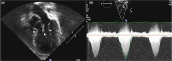
Approximately 6 months after the Fontan procedure, a routine follow-up echocardiogram revealed a peak pressure gradient of 42 mmHg (mean 18 mmHg) across the left ventricular outflow tract. A prominent muscle ridge of the ventricular septum and chordal attachments from the mitral valve caused dynamic obstruction (Fig 1a, 1b). There was also a systolic anterior motion of the anterior mitral valve leaflet (supplementary video). There was associated development of mild mitral valve regurgitation. He was asymptomatic and observed without intervention at that time.
(a ) Apical three-chamber view of transthoracic echocardiography during his follow-up. There was a systemic ventricular outflow tract obstruction with a prominent muscle ridge of ventricular septum (*) and chordal attachments from the mitral valve (arrow). (b ) Doppler echocardiogram across SVOT. There was a peak pressure gradient of 42 mmHg.

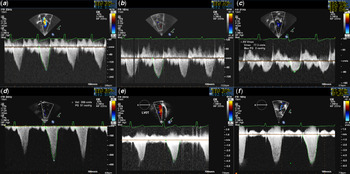
Over the next 5 years, the gradient gradually increased across the left ventricular outflow tract (range 40–80 mmHg) (Fig 2a–2f) with stable mild mitral regurgitation. Despite the increase in gradient, he remained clinically asymptomatic.
Longitudinal changes in Doppler echocardiogram across systemic ventricular outflow tract. (a ) pre-Fontan procedure (a peak pressure gradient of 13 mmHg), (b ) at the time of discharge after FP (6 mmHg), (c ) 3 months after FP (2 mmHg), (d ) 6 months after FP (50 mmHg), (e ) at 4 years of age (71 mmHg), (f ) at 5 years of age (120 mmHg).

At 8 years old, he started to complain of fatigue and difficulty in keeping up with his siblings. The echocardiogram showed a peak pressure gradient of 145 mmHg (mean gradient 60–70 mmHg) across the left ventricular outflow tract. There was moderate mitral valve regurgitation with normal left ventricular systolic function. The Fontan pathway was widely patent. Since his significant systemic ventricular outflow tract gradient coupled with his clinical symptoms, he underwent surgical repair to relieve his left ventricular outflow tract obstruction. Operative findings showed a discrete subaortic membrane, abnormal accessory tissue of the anterior leaflet of the mitral valve with abnormal attachment to the ventricular septum, and abnormal attachment of the anterior papillary muscle to the septum. He underwent resection of the subaortic membrane and resection of abnormal accessory tissue of the anterior mitral valve leaflets with a detachment of abnormal attachment to the ventricular septum. Separation of the anterior papillary muscle of the interventricular septum to the base of the papillary muscle with debulking the papillary muscle was also performed. The post-operative left ventricular outflow tract pressure gradient on the echocardiogram was 40 mmHg. He was discharged on post-operative day 15. The mitral valve regurgitation improved to mild and left ventricular systolic function remained normal. At his 2 months follow-up visit, there was no residual gradient across the left ventricular outflow tract with a peak gradient of 12 mmHg and a mean of 5 mmHg. He is clinically doing better and has no complaint of fatigue.
Discussion
Systemic ventricular outflow tract obstruction is rarely seen in the functionally single-ventricle patient. The mechanisms of systemic ventricular outflow tract obstruction are related to the type of ventricular morphology. For the patient with a dominant left ventricle, a rudimentary right ventricle, and transposition of the great arteries, the systemic ventricular outflow tract obstruction results from a restrictive ventricular septal defect or a narrowed subaortic right ventricular infundibulum, or both. For the patient with a dominant right ventricle, the subaortic area of the right ventricle is wedged between the infundibular septum medially and the ventriculoinfundibular fold laterally. Reference Razzouk, Freedom and Cohen3
The timing of the development of systemic ventricular outflow tract obstruction varies amongst the reported cases. It is known that the presence of systemic ventricular outflow tract obstruction prior to the Fontan procedure is associated with poor outcomes. Reference Freedom2 Therefore, patients generally undergo surgical correction of the ventricular outflow tract obstruction during the interstage period, most often prior to the Fontan procedure. There are only a few retrospective studies that investigated the development of systemic ventricular outflow tract obstruction after the Fontan procedure. Finta et al reported seven patients who did not have systemic ventricular outflow tract obstruction before the Fontan procedure, but developed progressive obstruction after the Fontan procedure. In their cohort, the incidence of the systemic ventricular outflow tract obstruction was 12% (7 out of 57 patients). The mean pressure gradient across the systemic ventricular outflow tract before the Fontan procedure was 6.3 ± 2.9 (SD) mmHg, which was significantly higher than the group of patients who did not develop systemic ventricular outflow tract obstruction during the follow-up period (0.6 ± 0.3 mmHg, p < 0.001). The authors suggest that the pressure gradient across the systemic ventricular outflow tract should not be ignored, even if it was very mild. Reference Finta, Beekman, Lupinetti and Bove4 Razzouk et al reported a case series of 12 patients who developed subaortic stenosis after the Fontan procedure. The patient with pulmonary atresia with an intact ventricular septum in their report developed left ventricular outflow tract obstruction immediately after the Fontan procedure. The left ventricular outflow tract was due to a septal bulge and abnormal systolic anterior motion of the mitral valve. Reference Razzouk, Freedom and Cohen3
In our case, the mechanism of left ventricular outflow tract obstruction was the combination of subaortic membrane, abnormal accessory tissue of the mitral valve, and abnormal orientation of the anterior papillary muscle. Although no significant left ventricular outflow tract obstruction was evident prior to the Fontan procedure, the obstruction gradually became worse over 5 years. Based on our case, we propose that monitoring for systemic ventricular outflow tract obstruction is an important aspect of management after the Fontan procedure even in the absence of obstruction prior to the Fontan procedure.
Supplementary material
To view supplementary material for this article, please visit https://doi.org/10.1017/S1047951121001888.
Acknowledgement
None.
Financial support
None.
Conflicts of interest
The authors declare that they have no conflict of interest.
Ethical standards
There is no research involving this patient.
Author contributions
D.T.: writing the manuscript; all authors: critically reviewing the manuscript.