INTRODUCTION
Echinococcus granulosus, the causative agent of cystic echinococcosis (CE), causes an important parasitic infection that is widely prevalent throughout Iran, where a variety of animals act as intermediate hosts. Carnivores harbour the adult worms while sheep, cattle, camels, goats, pigs, buffalo and horses harbour the metacestode stage in their livers and lungs (Thompson and McManus, Reference Thompson and McManus2002). E. granulosus is of veterinary importance because infection with the metacestode may cause severe illness in animals (Jenkins et al. Reference Jenkins, Roming and Thompson2005). Hydatid disease is a well-known zoonosis in Iran and causes extensive economic losses in the livestock industry (Rokni, Reference Rokni2009). E. granulosus shows a wide range of intra-specific variation in relation to host specificity, epidemiology, morphology, biology and genetics (Sweatman and Williams, Reference Sweatman and Williams1963; Thompson and Kumaratilake, Reference Thompson and Kumaratilake1985; Thompson and Lymbery, Reference Thompson and Lymbery1988; Thompson et al. Reference Thompson, Lymbery and Constantine1995). A total of 10 strains (G1–G10) of E. granulosus have been genotypically characterized so far (Thompson, Reference Thompson2008). Some of these strains show marked differences in morphology and biology. There is little known about cystic echinococcosis in buffaloes in the Middle East. Three strains of E. granulosus namely G1 (sheep strain), G3 (buffalo strain) and G6 (camel strain) have been identified in Iran (Hosseini and Eslami, Reference Hosseini and Eslami1998; Fasihi Harandi et al. Reference Fasihi Harandi, Hobbs, Adams, Mobedi, Morgan-Ryan and Thompson2002; Ahmadi and Dalimi, Reference Ahmadi and Dalimi2006; Sharbatkhori et al. Reference Sharbatkhori, Mirhendi, Fasihi Harandi, Rezaeian, Mohebali, Eshraghian, Rahimi and Beigom Kia2010; Amin Pour et al. Reference Amin Pour, Hosseini and Shayan2011a).
Researchers have found E. granulosus strains in Middle Eastern buffaloes (Utuk et al. Reference Utuk, Simsek, Koroglub and McManus2008; Simsek and Eroksuz, Reference Simsek and Eroksuz2008; Vural et al. Reference Vural, Baca, Gauci, Bagci, Gicik and Lightowlers2008; Šnábel et al. Reference Šnábel, Altintas, Amelio, Nakao, Romig, Yolasigmaz, Gunes, Turk, Busi, Hüttner, Ševcová, Ito, Altintas and Dubinský2009; Amin Pour et al. Reference Amin Pour, Hosseini and Shayan2011a). The water buffalo (Bubalus bubalisis) an important intermediate host is mainly susceptible to the G3 strain, although the sheep strain (G1) and cattle strain (G5) can also infect buffaloes (Thompson and McManus, Reference Thompson and McManus2002; Pedenkar et al. Reference Pedenkar, Gatne, Thompson and Trub2009). Recent analysis showed that the prevalence and fertility rates of hydatid cysts in buffaloes in different parts of Iran were 9% and 7·3%, respectively (Amin Pour et al. Reference Amin Pour, Hosseini and Shayan2011b).
The adult and larval morphology plus the biological situation have been considered as valid criteria for E. granulosus strain identification and these have been used by many investigators (Kumaratilake and Thompson, Reference Kumaratilake and Thompson1984; Kumaratilake et al. Reference Kumaratilake, Thompson and Eckert1986; Said et al. Reference Said, Abdel-Hafez and Al-Yaman1988; Worbes et al. Reference Worbes, Thompson and Eckert1989; Eckert et al. Reference Eckert, Thompson, Michael, Kumaratilake and El-Sawah1989, Reference Eckert, Thompson, Micheal, Lymbery, Paulowski, Gottstin and Morgan1993; Constantine et al. Reference Constantine, Thompson, Jenkins, Hobbs and Lymbery1993; Ponce Gordo and Cuesta Bandera, Reference Ponce Gordo and Cuesta Bandera1997; Hosseini and Eslami, Reference Hosseini and Eslami1998; Fasihi Harandi et al. Reference Fasihi Harandi, Hobbs, Adams, Mobedi, Morgan-Ryan and Thompson2002; Tashani et al. Reference Tashani, Zhang, Boufana, Jegi and McManus2002; Ahmadi, Reference Ahmadi2004; Ahmadi and Dalimi, Reference Ahmadi and Dalimi2006; Thompson et al. Reference Thompson, Boxell, Ralston, Constantine, Hobbs, Shury and Olson2006). It is important to determine the strain variation of E. granulosus in endemic regions such as Iran, where several species of animals of the intermediate host are infected with hydatid cysts (Thompson, Reference Thompson, Thompson and Lymbery1995; Fasihi Harandi et al. Reference Fasihi Harandi, Hobbs, Adams, Mobedi, Morgan-Ryan and Thompson2002).
According to a report of the Iranian Veterinary Organization, the population of buffaloes is estimated to be 447 000, of which 435 320 are in West Azerbaijan, East Azerbaijan, Ardabil, Gilan and Khuzestan provinces. To determine the host range of strains existing in Iran, the present study evaluated the morphological characteristics of E. granulosus isolates of buffaloes from the above-mentioned areas, where more than 95% of Iranian buffaloes are found.
MATERIALS AND METHODS
Parasite materials
Liver and lung hydatid cysts from naturally infected buffaloes were obtained from Urmia (West Azerbaijan), Tabriz (East Azerbaijan), Ardabil (Ardabil), Rasht (Gilan) and Ahvaz (Khuzestan) abattoirs. After removing the cyst fluid, the protoscoleces were aspirated under sterile conditions and rinsed twice with sterile saline. These protoscoleces were checked for viability using the Eosin dye-exclusion test and by observing flame cell activity. The protoscoleces used for the experimental infection had a mean viability of approximately 75%. Protoscoleces were then stored in 10% buffered formalin at room temperature until they were used for morphological analysis.
Experimental study
Three puppies (3–4 months old) were first de-wormed using piperazine (65 mg/kg) and praziquantel (5 mg/kg) to eliminate any intestinal helminth infections. The dogs were fed manually and prior to experimental infection, they were vaccinated against distemper adenoviruses, parvoviruses and leptospirosis. Approximately 10 000 viable protoscoleces from 3 different random isolates were administered orally to each animal (each dog was infected with 1 isolate). On days 35 and 41 post-infection (p.i) dogs were euthanized (2 dogs on day 35 and 1 on day 41 p.i) in narcosis status using an overdose injection of sodium pentobarbital. After autopsy, the intestines were incubated for 1·5 h in phosphate-buffered saline (PBS) at 37°C. Finally the contents of the intestines were washed and the infection rate determined by the count of the total worms. Fixation and staining procedures were carried out as previously described by Eckert et al. (Reference Eckert, Thompson, Michael, Kumaratilake and El-Sawah1989) and Hosseini and Eslami (Reference Hosseini and Eslami1998). The animal experiments were performed under the University of Tehran guidelines for animal welfare.
Protoscoleces and dissected rostellar hooks from adult worms were mounted in polyvinyl lactophenol. Morphometric analysis was done as described by Hobbs et al. (Reference Hobbs, Lymbery and Thompson1990). The number of hooks per scolex, total length of large (LTL) and small (STL) hooks, blade length of large (LBL) and small (SBL) hooks, and the ratio of blade length to total length in large (LBL/LTL) and small (SBL/STL) hooks were measured, and all hooks were counted from 30 protoscoleces of each isolate using a calibrated eyepiece micrometer at a magnification ×1000.
Total worm length and terminal segment length were measured on unstained specimens. Stained material was used to determine morphological characteristics of male and female reproductive systems according to criteria outlined by Kumaratilake and Thompson (Reference Kumaratilake and Thompson1984). The morphometric data were determined by using a calibrated light microscope with ocular micrometer and a camera lucida.
Sample size and statistics
Morphological characteristics of larval (protoscolex) hooks were studied in 25 samples from West Azerbaijan (8 samples), East Azerbaijan (5 samples), Ardabil (6 samples), Gilan (2 samples) and Khuzestan (4 samples). Statistical analysis was based on the measurements of 10 large hooks and 10 small hooks per rostellum on 30 protoscoleces from each isolate. For evaluation of the morphological characteristics of worms 60 specimens from each infected dog were studied. Of these, 20 dissected rostellar hooks on worms for the measurements of 10 large hooks and 10 small hooks per scolex, 20 specimens for the measurement of strobilar and 20 specimens for morphological characteristics of male and female reproductive systems were used. Data analysis was carried out using the statistical package for social sciences (SPSS Version 15.0). The data were also calculated by one-way analysis of variance (one-way ANOVA).
RESULTS
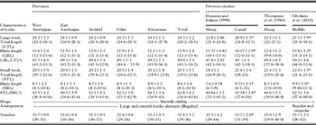
Based on measurements of the hooks of the protoscoleces, the total number and the different parameters related to hook measurements were determined. The morphology of the rostellar characteristics of protoscoleces is summarized in Table 1. The characteristics of the protoscoleces from different parts of Iran were nearly similar. They showed 2 rows of alternating large and small hooks and their shapes were smooth in outline. The numbers of worms recovered from the dog on days 35, 35 and 41 p.i were 223 212 and 285 respectively. The results showed that a small proportion of protoscoleces had developed into adult worms (2·4%). More than 60% of E. granulosus occupied the middle part of the small intestine of infected dogs.
A comparison of the morphological characteristics of protoscoleces derived from buffaloes in the present study with those of previous studies.

* Statistically different from Total column (P<0·05).
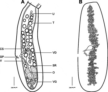
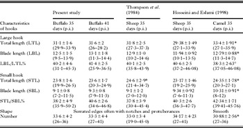
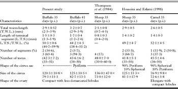
The morphological characteristics of the rostellar hooks of worms (days 35 and 41 p.i.) were studied and the data are summarized in Table 2. In contrast to protoscoleces, the shape of the adult rostellar hooks was rough and had protuberances. The morphological characteristics of adult and gravid (35d.p.i. and 41d.p.i.) strobila were analysed and are summarized in Table 3. The morphology of the strobilla showed that the position of the genital pore in the terminal segment was posterior to the middle of the segment and the testes were distributed throughout the adult segments. The results also showed that the shape of the ovaries was compact with less demarcated lobules (Fig. 1).
Terminal segment of Echinococcus granulosus derived from buffalo. (A) The mature segment of a 35-day-old. (B) The gravid segment of a 41-day-old with prominent thin-shelled eggs (drawings by camera lucida). Abbreviations: CS, cirrus sac; GP, genital pore; O, ovary; SR, seminal receptacle; T, testes; U, uterus; VD, vitelline duct; VG, vitelline glands.

Comparison of the 35- and 41-day-old adult Echinococcus granulosus rostellar hook morphology in the present study (mean±S.D.) with those of Thompson et al. (Reference Thompson, Kumaratilake and Eckert1984) and Hosseini and Eslami (Reference Hosseini and Eslami1998)

* Statistically different from buffalo 35 days post-infection (P<0·05).
Comparison of the 35- and 41-day-old Echinococcus granulosus strobilar morphology in the present study (mean±S.D.) with that of Thompson et al. (Reference Thompson, Kumaratilake and Eckert1984) and Hosseini and Eslami (Reference Hosseini and Eslami1998)

The E. granulosus from buffalo exhibited a relatively rapid rate of growth, reaching a mean total length of 2·9±0·52 mm by day 35 p.i. However, by day 41 p.i., the total length was 3·2±0·7 mm. Maturation was also advanced. At day 35 p.i. 65% of the worms had testes, full growth cirrus sac ovary, uterine streak, vitelline glands and vitello duct. In a further 35% of the worms, a dilated uterus containing developing eggs in the terminal segment was detected. By day 41 p.i. all the worms were gravid and 25% of the worms had thick-shelled embryonated eggs in a fully dilated uterus (Fig. 1). The remaining worms had thin-shelled eggs and none had more than 3 segments.
DISCUSSION
Several criteria have been used to characterize isolates of E. granulosus (Thompson and McManus, Reference Thompson and McManus2002). The morphology of the rostellar hook and adult worms is still believed to be a valid criterion for differentiation of the E. granulosus isolates/strains (Ponce Gordo and Cuesta Bandera, Reference Ponce Gordo and Cuesta Bandera1997; Fasihi Harandi et al. Reference Fasihi Harandi, Hobbs, Adams, Mobedi, Morgan-Ryan and Thompson2002; Tashani et al. Reference Tashani, Zhang, Boufana, Jegi and McManus2002; Ahmadi and Dalimi, Reference Ahmadi and Dalimi2006; Thompson et al. Reference Thompson, Boxell, Ralston, Constantine, Hobbs, Shury and Olson2006; Almeida et al. Reference Almeida, Silva, Neves, Romanii and Sliva2007). It is a comparatively quick and inexpensive method of strain identification, particularly for epidemiological studies. In this regard, the total number, total length and blade length of rostellar hooks have been suggested as one important criterion for differentiation of strains (Ponce Gordo and Cuesta Bandera, Reference Ponce Gordo and Cuesta Bandera1997; Lymbery, Reference Lymbery1998). However, some investigators believe that the use of the morphological characters alone for the determination of the E. granulosus strain could be questionable (Hobbs et al. Reference Hobbs, Lymbery and Thompson1990) and strain identification should include other criteria (Mariaux, Reference Mariaux1996).
Hosseini and Eslami (Reference Hosseini and Eslami1998) and Zhang et al. (Reference Zhang, Eslami, Hosseini and McManus1998) characterized the E. granulosus based on morphology and molecular criteria such as a comparative analysis of the mitochondrial gene. They suggested that 2 separate strains of E. granulosus exist in Iran (sheep strain and camel strain) (Hosseini and Eslami, Reference Hosseini and Eslami1998; Zhang et al. Reference Zhang, Eslami, Hosseini and McManus1998). Further studies by other investigators confirmed the presence of the 2 strains (Fasihi Harandi et al. Reference Fasihi Harandi, Hobbs, Adams, Mobedi, Morgan-Ryan and Thompson2002; Ahmadi, Reference Ahmadi2004; Ahmadi and Dalimi, Reference Ahmadi and Dalimi2006; Rostami Nejad et al. Reference Rostami Nejad, Nazemalhosseini Mojarad, Nochi, Fasihi Harandi, Cheraghipour, Mowlavi and Zali2008; Sharbatkhori et al. Reference Sharbatkhori, Mirhendi, Fasihi Harandi, Rezaeian, Mohebali, Eshraghian, Rahimi and Beigom Kia2010). Recently, another strain, namely G3, was also reported to be present in buffalo and camel (Sharbatkhori et al. Reference Sharbatkhori, Mirhendi, Fasihi Harandi, Rezaeian, Mohebali, Eshraghian, Rahimi and Beigom Kia2010; Amin Pour et al. Reference Amin Pour, Hosseini and Shayan2011a).
Limited surveys have been done on the morphology of E. granulosus originating from buffaloes around the world and no results have been reported from Iran. The rostellar hook characteristics of the protoscoleces isolated from buffaloes did not differ significantly in studied areas. The results showed that the total length, the blade lengths of large and small hooks and the number of the hooks from buffalo isolates show slight similarities to those isolated from sheep but are significantly different from those of camel isolates (Hosseini and Eslami, Reference Hosseini and Eslami1998). The results also showed that the morphological characteristics of the rostellar hooks of adult worms from buffaloes are not significantly different from those of the sheep strain. Interestingly, Gholami et al. (Reference Gholami, Irshadullah and Mobedi2010) reported that the morphology of protoscoleces from Indian buffaloes differed from that observed in Iranian sheep, cattle and buffaloes and may therefore represent a different strain, which is adapted to buffaloes in India.
The growth rate of adult E. granulosus (total worm length, segmentation and maturation) from buffaloes on days 35 and 41 p.i resembles that of the common sheep strain. The form of the strobila and the morphology of the reproductive system are also similar to those from the sheep strain. Gill and Rao (Reference Gill and Rao1967) investigated the morphology of adult worms from buffaloes/dog origin and suggested that the isolates from buffaloes did not correspond to any previously described subspecies, and may be considered as mutants.
The results of morphological studies support the finding of the earlier molecular studies on isolates from buffaloes in Iran which demonstrated that the E. granulosus of buffalo origin is genetically almost similar to the G1 strain. According to comparative genotyping of E. granulosus from buffaloes, it was observed that 92% were infected with the sheep strain (G1) and just 8% of isolates were the buffalo strain (G3) in Iran (Amin Pour et al. Reference Amin Pour, Hosseini and Shayan2011a).
Furthermore, the results showed that the protoscoleces of buffalo origin could not be easily developed into adult worms in dogs. The poor infectivity of protoscoleces from buffaloes is similar to that for those of cattle origin and contrasts with those of sheep and camel origin which can readily infect dogs with a high percentage developing into adult worms (Hosseini and Eslami, Reference Hosseini and Eslami1998). According to the current epidemiological study of hydatid cysts in buffaloes in Iran, despite the relatively high rate of infection (9%), the fertility rate of cysts (7·3%) was low (Amin Pour et al. Reference Amin Pour, Hosseini and Shayan2011b). The G1 genotype seems to become fertilized predominantly in sheep whereas in other hosts it remains mostly infertile (Mwambete et al. Reference Mwambete, Ponce-Gordo and Cuesta-Bandera2004). The low cyst fertility of the sheep strain infecting cattle has been previously reported (Hosseini and Eslami, Reference Hosseini and Eslami1998; Euzeby, Reference Euzeby1991). It seems that most of the buffaloes infected with CE are due to sheep origin (G1) in Iran and the G3 strain has limited distribution in buffaloes. The low host-specificity of the buffaloes is in agreement with findings in Pakistan and India suggesting that isolates of buffalo origin are phenotypically and genetically similar to the sheep strain (G1) and the Tasmanian sheep strain (G2) of Echinococcus (Pedenkar et al. Reference Pedenkar, Gatne, Thompson and Trub2009; Latif et al. Reference Latif, Tanveer, Magbool, Siddiqi, KyawTanner and Traub2010).
In conclusion, these data showed the transmission of the G1 genotype between sheep and buffalo as well as in cattle in Iran. It may be speculated that the sheep strain is the predominant E. granulosus strain in Iran thus the sheep-dog cycle should be targeted in control programmes.
ACKNOWLEGMENTS
We thank Dr Nurolahi, Dr Razbani, Dr Behruzi and Dr Alavi from the veterinary organization of Iran for their help for collecting the samples from different slaughter houses.