The bidirectional superior cavopulmonary anastomosis is a part of staged palliation in patients with single-ventricle physiology.Reference Pridjian, Mendelsohn and Lupinetti1 Performing this anastomosis in patients with bilateral superior caval veins is associated with higher complications. It is further complicated by atresia of one pulmonary artery. This subset poses technical challenges as innovation is needed to direct venous return from both superior caval veins into the single pulmonary artery. We present one such patient with bilateral superior caval veins and atretic left pulmonary artery, in whom the superior cavopulmonary anastomosis was successfully accomplished.
Case report
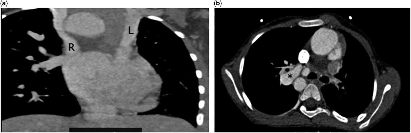
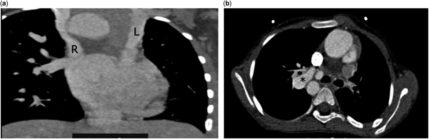
Informed consent was obtained from the parents of the patient to publish report; formal institutional review board approval was waived. A 12-year old girl presented with progressive cyanosis since birth. The parents was aware since birth that she had heart disease, but they had elected for medical follow-up. Now the cyanosis had deepened, with room air saturation being 58%. Transthoracic echocardiography revealed situs inversus, levocardia, normal pulmonary venous drainage, and the inferior caval vein draining to the right atrium. Atrio-ventricular and ventriculo-arterial connections were discordant. A large non-committed ventricular septal defect was present with a hypoplastic morphologic right ventricle. The pulmonary valve was atretic with no antegrade flow across it. The aortic arch was right-sided arch with a large patent arterial duct inserted into an 8 mm-sized right pulmonary artery with stenosis at the site of insertion of the duct. The left pulmonary artery and the area of the pulmonary artery confluence was not visualised. CT angiogram (Fig 1) that was performed showed pulmonary atresia of main and left pulmonary arteries and a large vertical patent arterial duct has been formed in the right pulmonary artery that measured 10 mm. Descending thoracic aorta at the level of the diaphragm measured 14 mm. Similar-sized bilateral superior caval veins were present; the right one drained into the left-sided right atrium via coronary sinus and the left one drained directly into the right atrium. The inferior caval was present on the left side and drained into the right atrium. Multiple small aortopulmonary collaterals arising from the descending aorta were seen. Cardiac catheterisation and cineangiography were confirmatory, with mean pressure in the right pulmonary artery being 14 mmHg.
(a) Coronal section showing bilateral superior caval veins with the right superior caval vein (R) draining to right atrium via the coronary sinus and left superior caval vein (L) directly draining to right atrium. (b) Axial thin section showing right ventricle outflow tract with absent pulmonary arteries and its confluence and then subsequent formation of right pulmonary artery (R) from patent ductus arteriosus and atretic left pulmonary artery. P = right pulmonary artery. Ao = Aorta.
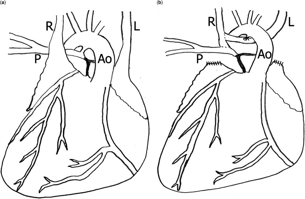
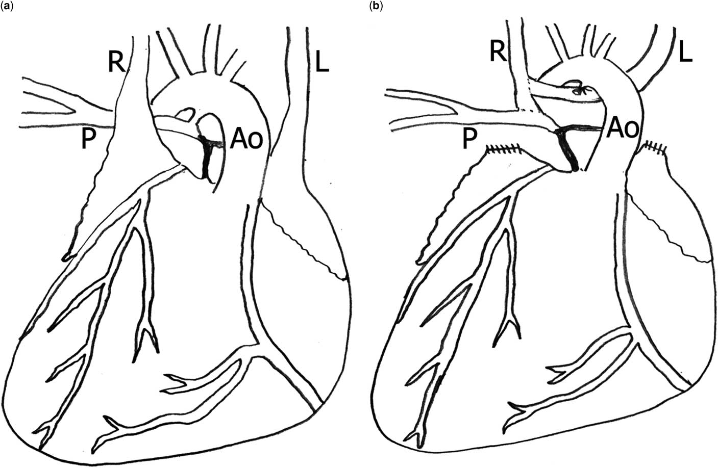
Normothermic cardiopulmonary bypass was established after cannulating the aorta and the right atrium. The individual superior caval were not cannulated because these were bilateral and thought to decompress the cerebral circulation brain even when one of them was clamped for the superior cavopulmonary anastomosis. With the heart beating, a standard right-sided superior cavopulmonary anastomosis was performed after dividing the patent arterial duct. The left superior caval vein was completely mobilised and after division between two clamps, the cardiac end was closed. Its cephalad portion was passed behind the aorta and was anastomosed end-to-side of the right superior caval vein using continuous 6-0 polypropylene (Fig 2). Following this, the patient was uneventfully weaned off cardiopulmonary bypass after which the pressure in the right superior caval vein was 16 mmHg without any across the superior cavopulmonary anastomosis and across the anastomosis between the right and right superior caval veins. The patient was uneventfully extubated after 2 hours. Room air saturation was 88%. Echocardiography on fourth post-operative day showed laminar blood flow across the superior cavopulmonary circuit. At 6 months follow-up, her saturation on room air is in excess of 85% and a completion Fontan is planned after 1 year.
Line drawing of the anatomy and the surgical procedure. (a) Anatomy. Note that there is no left pulmonary artery. (b) The left superior caval vein (L) is anastomosed end-to-side to the right superior caval vein (R). P = right pulmonary artery. Ao = Aorta.
Discussion
The bidirectional superior cavopulmonary anastomosis is a part of staged palliation for patients with a univentricular heart to reduce the volume overload on the single ventricle and improve its natural history.Reference Pridjian, Mendelsohn and Lupinetti1 In patients with bilateral superior caval veins, a bilateral operation is needed. When compared to patients with a single cavopulmonary anastomosis, a bilateral anastomosis is associated with technical difficulties, increased risk of thrombus formation, and a lower rate of conversion to the Fontan circulation.Reference Mayer, Helgason and Jonas2–Reference Forbes, Rosenthal, Reul, Ott and Feltes4 Our patient had bilateral superior caval veins with atresia of the left pulmonary artery. Innovation was therefore needed to direct the blood from both superior caval veins into the right pulmonary artery. We accomplished this by an end-to-side anastomosis of the left superior caval vein and the right superior caval vein. Previously described methods of accomplishing this objective are an interposition graft (prosthetic or biological) between the right and left superior caval veins. In a previous report from Guatemala, Vida and Castaneda have described a Gore-Tex interposition graft between the two superior caval veins creating “a Gore-Tex innominate vein”.Reference Vida, Leon-Wyss, Garcia and Castaneda5 Their technique is easy to perform and can be accomplished irrespective of the distance between the two superior caval veins. However, this graft has no growth potential and there is at risk of thrombosis requiring oral anticoagulant prophylaxis, with progressive occlusion of the graft in up to 30% patients within 3 yearsReference Shintani, Ohta and Minami6 and hence oral anticoagulants are needed.
Autologous saphenous vein and pericardial tube grafts have been used for pulmonary artery reconstruction and also for constructing an innominate vein in such patients, but the unavailability of adequate size grafts is a major drawback.Reference Moreno-Cabral, McNamara, Reddy and Caldwell7, Reference Kosaka, Kurosavwa, Hoshino, Shin’oka, Isomatsu and Tsuji8 We have previously described homograft saphenous veins, homograft common iliac artery, and even an aortic homograft without the valve obtained from our own valve bank to help accomplish the superior cavopulmonary anastomosis.Reference Talwar, Gupta, Choudhary and Airan9
To the best of our knowledge, a direct anastomosis between the two superior caval veins during a superior cavopulmonary anastomosis has not been described. The advantages of this technique are possible growth potential, no need of prosthetic material, minimal risk of thrombosis, and no need of oral anticoagulation. However, this procedure can be difficult in patients with a small and distant left superior caval vein, with risk of kinking of the anastomosis. We preferred to bring the left superior caval vein behind the aorta, because it follows a shorter course and also, it prevents its undue stretching by a dilated aorta, should it be brought in front of the aorta.
Acknowledgements
None.
Financial Support
This research received no specific grant from any funding agency, or from commercial or not-for-profit sectors.
Conflicts of Interest
None.
Ethical Standards
The authors assert that all procedures contributing to this work comply with the ethical standards of the relevant Indian guidelines on human experimentation and with the Helsinki Declaration of 1975, as revised in October 2008, and a need for formal approval was waived off by the Ethics Committee of All India Institute of Medical Sciences, New Delhi, India.