Unilateral discontinuous pulmonary artery is a rare form of CHD that more commonly involves the right pulmonary artery. It is due to the malformation of the sixth aortic arch on the involved side during embryogenesis. Reference Kanno, Maehara, Yamamoto, Nemoto, Kigoshi and Masuda1,Reference Steiropoulos, Archontogeorgis, Tzouvelekis, Ntolios, Chatzistefanou and Bouros2 Restoration of physiologic circulation is essential to prevent long-term complications, which may include pulmonary hypertension, haemoptysis, and recurrent respiratory infections. However, surgical repair can be challenging in cases of long discontinuity. We present a case of semi-autologous reconstruction of discontinuous right pulmonary artery using a main pulmonary artery flap in a 2-month-old infant.
Case presentation
The patient was born at 36 weeks gestation at another institution. He was intubated for almost 2 weeks and was diagnosed with persistent pulmonary hypertension. An echocardiogram showed a patent foramen ovale and a patent ductus arteriosus, but no other evidence of CHD. He continued to have an oxygen requirement and was sent home on oxygen therapy to maintain saturations above 90%. During a follow-up visit, a heart murmur was noted and the mother was told that the ductus arteriosus might still be open, which prompted them to present to our facility for a second opinion.
At presentation to our facility, he was clinically stable with 100% oxygen saturations on 2 L of low-flow nasal cannula. There was no audible murmur. Echocardiogram confirmed that the ductus arteriosus was closed. There were an unusual number of bronchial collaterals seen. The right pulmonary artery was not clearly visualised. A CT angiogram was obtained that showed absence of a central right pulmonary artery with the right lung supplied by multiple systemic collaterals. There was concern at this point that the patient could have discontinuous pulmonary arteries and a catheterisation with pulmonary wedge injection was performed.
Cardiac catheterisation demonstrated a distal right pulmonary hilar stump located 1.5 cm from the main pulmonary artery. The right stump communicated with the collateral vessels. There was no evidence of pulmonary vascular disease in the left lung. The main pulmonary artery mean pressure was 16 mmHg and the left pulmonary artery mean pressure was 12 mmHg. The decision was made to proceed with surgical reconstruction of the right pulmonary artery utilising a flap of the main pulmonary artery.
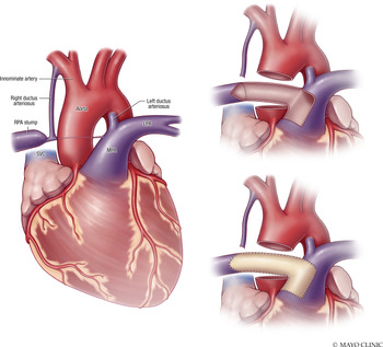
At the time of surgical intervention, the innominate artery was dissected out. A right-sided ductus arteriosus was found on the inferior aspect and was followed to the hilar stump of the right pulmonary artery laterally, and a small band of fibrous tissue connected this to the main pulmonary artery medially. A left ductus was also noted in the usual position, both ducti were ligated. Cardiopulmonary bypass was commenced, and the aorta was cross clamped. The aorta was transected for exposure. The distance from the medial edge of the main pulmonary artery to the hilar stump was just over 1.5 cm. An appropriately sized flap was created from the main pulmonary artery and turned down to serve as the back wall for the right pulmonary artery reconstruction. The distal end of this flap was sutured to the back wall of the diminutive hilar stump (about 4 mm in diameter). A patch of Matracell™ was then cut to size to form the front wall and reconstruct the defect in the main pulmonary artery. This created a well-formed right pulmonary artery with distal diameter of 4 mm, similar to the diameter of the hilar stump (Fig 1).
Left panel demonstrates the anatomy, namely the bi-lateral ductus arteriosus, with the right ductus connected to the right pulmonary artery (RPA) stump. LPA = left pulmonary artery, MPA = main pulmonary artery, SVC = superior vena cava. The right upper panel demonstrates the autologous flap taken from the patient’s main pulmonary artery that has been folded back and sutured to the back wall of the RPA stump to form the posterior of the reconstructed right pulmonary artery. The right lower panel shows the anterior aspect of the reconstructed right pulmonary formed by a patch.

Postoperative CT showed intact proximal and distal anastomoses. Prior to hospital discharge, echocardiography showed a mean gradient across the right pulmonary artery of 8 mmHg. The patient was weaned off oxygen and discharged home on postoperative day 10. The patient was seen 1 month later and had angiography with balloon angioplasty of the residual narrowing. The right ventricular systolic pressure was 33 mmHg. The plan is to perform repeat CT imaging of the right pulmonary artery in 6 months.
Discussion
The prevalence of discontinuous pulmonary artery is estimated to be 1 in 200,000 with only 12% of the cases diagnosed in infancy. Reference Devulapally, Bhatt and Allen3 Resorption of the proximal sixth aortic arch during development is the suggested pathogenic process. Reference Turner, Vincent and Epstein4 The right pulmonary artery is more commonly involved than the left, and in that case it is usually an isolated anomaly. Congenital absence of the left pulmonary artery tends to be associated with more severe cardiovascular malformations. Reference Devulapally, Bhatt and Allen3 Limited literature, especially in infancy, is available in describing surgical techniques for repair. When possible, a simple anastomosis can be made between the main pulmonary artery and distal right pulmonary. This technique is not applicable when longer gaps of discontinuity exist, in part, due to the need for a tensionless repair to prevent subsequent stenosis.
An alternative surgical approach utilises a modified Blalock–Taussig–Thomas shunt or a ductal stent as a staged approach allowing growth of the intrapulmonary arteries on the affected side before connecting the distal pulmonary artery to the main pulmonary artery. Reference Apostolopoulou, Kelekis, Brountzos, Rammos and Kelekis5,Reference Marchau, Boshoff, Gewillig and Mertens6 The clear disadvantage is that the two-staged approach exposes the patient to potential increased morbidity and mortality.
This technique was previously described by El-hattab et al Reference El-Hattab, Calcaterra, Parekh, Rossi, Davis and Turek7 in a 3-month-old infant with a discontinuous right pulmonary artery. A Matracell™ patch was used in our case, whereas a thawed pulmonary homograft was used in their case. ElTayeb, et al Reference Eltayeb, Mongé, Popescu, Sarwark, Harris and Backer8 modified the technique by transecting the aorta to allow for better exposure in a 5-month-old that was showing signs and symptoms of pulmonary hypertension. At 3-month follow-up, the pulmonary artery pressure had normalised, which perhaps suggests that earlier repair is associated with favourable outcome.
Regardless of surgical technique employed, restoration of physiologic blood supply is necessary to allow for appropriate growth and development of the lung and avoid long-term complications including pulmonary hypertension.
Conclusion
Discontinuous pulmonary artery is a rare anomaly with a variable time of presentation and symptoms. In cases of long segment discontinuity, utilisation of an autologous pulmonary artery flap can be considered as an appropriate surgical approach.
Supplementary material
To view supplementary material for this article, please visit https://doi.org/10.1017/S1047951121002018
Acknowledgements
Special thanks to Dr. Joseph A. Dearani and Dr. Jason H. Anderson for their important contribution to the care of this patient.
Financial support
This research received no specific grant from any funding agency, commercial, or not-for-profit sectors.
Conflicts of interest
None.