1. Introduction
Rather than a conventional review, this is a personal account of the development of our understanding of the structural biology of RNA over a 20-year period from 1954 to 1974. In 1954, I started research on RNA structure, and it was in 1974 that we finished the full structure of transfer RNA (tRNA). To the modern, mostly young reader, it is nearly impossible to understand the extent to which in 1954 we were ignorant of the many aspects of RNA chemistry, structure, and biology. The Watson–Crick formulation of the DNA double helix (Watson & Crick, Reference Watson and Crick1953) was published a year earlier, but its recognition and acceptance by the biological community came very slowly (Olby, Reference Olby2003). Understanding RNA's structural and functional aspects was very primitive, almost nonexistent. One can get a good picture of the state of knowledge in 1954 by reading the thorough review of the nucleic acids, published in the Annual Reviews of Biochemistry (Allen, Reference Allen1954).
In the 1930s, it had been proposed that the nucleic acids existed as tetranucleotides, but by 1954, as Allen points out, ‘a flood of evidence has shown that the tetranucleotide theory and all of its correlations are untenable.’ At that time, there was an awareness that the size of nucleic acids was extremely diverse, especially for RNA, but there was no clear demonstration of function. From the structural point of view, the central issue of the day was the nature of the internucleotide linkage in RNA molecules. Although there was evidence that a 3′–5′ linkage was common (Brown & Todd, Reference Brown, Todd, Chargaff and Davidson1955), some evidence suggested that alternative linkages were present. RNA, unlike DNA, had the possibility of forming branch structures using the 2′ hydroxyl group. Evidence was also presented that such structures existed. A variety of different analytical methods were described in Allen's review, often producing results that were contradictory to other methods. Thus, the field of RNA research was characterized by a lack of clarity, both in structure and in function.
2. Fiber X-ray diffraction analysis
Working as a Linus Pauling postdoctoral fellow at Cal Tech in December 1952, I was asked by Pauling to take some X-ray photographs of fibers of DNA. He had already published an erroneous three-stranded DNA structure, and he needed more structural information about the molecule. After a few months, I began to get reasonable fiber X-ray diffraction patterns, but in the spring of 1953, the Watson–Crick paper came out, and it was clear to me that the structure was correct. I then began to wonder about the structure of RNA. Could it form a double helix? The 1953 Watson–Crick paper pointed out that van der Waals crowding by the 2′OH of ribose would prevent it from forming a double helix. Pauling organized a conference on the Structure of Proteins in Pasadena in September 1953, and several people from the nucleic acid field attended, including Jim Watson and Francis Crick, as well as Maurice Wilkins and J. T. Randall. I told Crick that I was planning to study the RNA structure. He said that if I ever got good fiber diffraction from RNA and wished to improve it, I should come to his laboratory in England where they had developed a powerful rotating anode X-ray generator that was useful for small fibers. Watson, who by then had returned to Cal Tech to do a postdoctoral fellowship with Max Delbrück, expressed great interest in the RNA project, and I suggested that he join me in the effort.
By early 1954, we were soliciting RNA samples from many different laboratories. I had developed the technique of drawing oriented fibers from them and carried out X-ray diffraction analyses, but the X-ray diffraction patterns were not encouraging. They looked similar to each other, somewhat suggestive of a double helix but without much definition. Leslie Orgel, another Pauling postdoctoral fellow, was interested in the RNA work and, together with Jim Watson, the three of us discussed these problems extensively. It was thought that RNA might be important in directing the synthesis of proteins. Just how that occurred was mysterious and unknown.
George Gamow, a distinguished theoretical physicist and a friend of Max Delbrück, arrived at Caltech, very much taken by the problem of how proteins are made under the direction of the nucleic acids. He had proposed the first code for determining the sequence of amino acids directly from the DNA double helix (Gamow, Reference Gamow1954). His code was incorrect, but it stimulated a good deal of research. Gamow was a lively personality and had the idea of setting up a club of people interested in the coding problem. The group would be defined by a tie and was called the RNA Tie Club. The club had 20 members, one for each amino acid. Many of the members were friends of Gamow, but it also included many of the principal workers in the then small field of nucleic acid research as well as a number of physicists such as Dick Feynman, Edward Teller, and others. The main purpose of the club was to circulate manuscripts dealing with the code. It was in one of these RNA Tie Club manuscripts that Francis Crick first developed the adapter hypothesis, suggesting that a small RNA molecule might act as an intermediate between the amino acids and the nucleic acid molecule that codes for protein. Later, it was realized that these adapters had been discovered by Mahlon Hoagland and Paul Zamecnik (Hoagland et al. Reference Hoagland, Zamecnik and Stephenson1957), and this led to understanding the role of tRNA in protein synthesis.
But in 1954, our thoughts were much more primitive. We wondered whether it was possible for the RNA molecule to have a form that would bind specific amino acids in some kind of a linear array. Such models seem quite naïve today, but that was our initial thinking. In the X-ray work on RNA fibers, we were using RNA preparations in which the base ratios (A to U, G to C) varied widely. Some were nearly complementary as found in DNA, and others were far removed. Nonetheless, the patterns remained about the same. At the time, we did not understand the variety of different types of RNA in the preparations and there was also a question about whether RNA was a linear polymer, as was the case for DNA. Two papers were published describing these results, one in Nature (Rich & Watson, Reference Rich and Watson1954a) and another sent to PNAS (Rich & Watson, Reference Rich and Watson1954b) by Pauling. The latter wondered about the role of RNA in protein synthesis. A diffraction pattern was included but with no clear interpretation.
In mid-1954, I moved to the National Institutes of Health (NIH), determined to pursue the RNA problem. I wrote to David Davies, another Pauling postdoctoral fellow, urging him join my research laboratory at NIH. I also invited former Pauling graduate student Gary Felsenfeld to join the laboratory.
With somewhat better X-ray cameras than those available at Caltech, I continued to study the diffraction patterns from oriented RNA preparations. This research effort soon took an abrupt change in direction. In 1955, Severo Ochoa, Marianne Grunberg-Manago and others published a paper describing the enzyme polynucleotide phosphorylase (Grunberg-Manago et al. Reference Grunberg-Manago, Ortiz and Ochoa1955). It cleaved a phosphate from nucleoside diphosphates, forming long RNA chains. Soon, we were able to make the enzyme and polymers. The random copolymer containing equal amounts of adenine and uracil residues produced a diffraction pattern very similar to that found in naturally occurring RNA. These synthetic chains were linear with a 3′–5′ linkage, and that made me think it was likely that there were few branched RNA molecules in the natural RNAs. The preparation of polyadenylic acid (poly rA) began to produce an interesting X-ray pattern, highly suggestive of a helical complex. After working on that for some time, it became apparent that more data could be obtained with a more powerful X-ray source and better cameras. Accordingly, I wrote to Francis Crick, and he invited me to Cambridge, England, to use the powerful rotating anode developed at the Medical Research Council unit in the Cavendish Laboratory.
In July 1955, I arrived in Cambridge, England, at roughly the same time as Jim Watson and Leslie Orgel. Thus, we were able to resume the intensive discussions on RNA structure that we had begun at Caltech, with the significant addition of Francis Crick. Improved diffraction patterns of polyadenylic acid were obtained by me and by Jim Watson. Eventually, we solved its parallel, double-stranded, helical structure working with Francis Crick and David Davies (Rich et al. Reference Rich, Davies, Crick and Watson1961).
My visit to Cambridge, England, was unexpectedly extended by work that Crick and I carried out on the structure of polyglycine II (Crick & Rich, Reference Crick and Rich1955), which eventually led to our discovery of the structure of collagen (Rich & Crick, Reference Rich and Crick1955). Staying there for 6 months provided extensive opportunities to discuss RNA structure. Could it form a double helix? In their DNA structure proposal, Watson and Crick pointed out that RNA could not form the same double-helix structure as DNA, but it did not rule out other structures. The four of us – Crick, Watson, Orgel, and I – spent many hours working with molecular models, trying to find stable RNA conformations, but without success. It is striking that none of us suggested that it would be interesting to mix polyriboadenylic acid (poly rA) with polyribouridylic acid (poly rU) to see if that could form a duplex. This is a good illustration of how easy it is to develop unknown biases or blinders concerning plausible experiments.
3. Discovery of hybridization and the RNA double helix
On returning to NIH from England, work on RNA structure continued with David Davies. In early 1956, for some forgotten reason, I mixed a sample of poly rA with poly rU and discovered a remarkable transformation. The sticky droplets with the two polymers quickly became more viscous, and the fibers pulled from it revealed an oriented helical X-ray diffraction pattern. To my astonishment, these two molecules had actually self-assembled with each other to produce a double helix! This was a new type of polymer chemical reaction, one that involved thousands of units specifically binding to each other in an extended array. No one had ever seen a chemical reaction between two different macromolecular polymers in which the monomers from each specifically bound together. A brief note was sent to the Journal of the American Chemical Society in June of that year (Rich & Davies, Reference Rich and Davies1956), describing the work with a preliminary interpretation of the diffraction pattern. The X-ray patterns clearly indicated the formation of a helical complex that was not present in either of the two individual polymers, and most important, the pattern was significantly different from that produced from moist DNA fibers.
This result, which seems so obvious today, was met with some skepticism. While walking down a long corridor at NIH, I met Herman Kalckar, an eminent Danish biochemist. I mentioned that we discovered that poly rA and poly rU formed a double helix. Kalckar was incredulous, ‘you mean without an enzyme?’ he asked. The only double helix known at that time was one made with the DNA polymerase enzyme that Arthur Kornberg had purified (Kornberg et al. Reference Kornberg, Lehman, Bessman and Simms1956). There were many other critics. Some thought it was highly unlikely that macromolecular polymers containing thousands of nucleotides would be able to disentangle themselves and form a regular structure. At a Solvay conference, a distinguished polymer chemist told me that my interpretation was likely to be wrong since both polymer chains were highly negatively charged and that they would never combine. Several physical chemists said it was unlikely that such a reaction would occur because of entropic factors. They did not realize the extent to which the high entropic contribution of solvent binding dominates the thermodynamics of such processes. The great novelty of the reaction sparked the criticism.
Two weeks after sending off the 1956 JACS note, I wrote a letter to Linus Pauling, describing these results. The letter, available in the Pauling archives at Oregon State University, reveals a sense of incredulity on my part that this reaction could happen and that it was ‘completely reproducible.’ This was the first demonstration that RNA molecules could form a double helix. In addition, although no one used the phrase at the time, it was the first hybridization reaction. It represented a paradigm shift in the way chemists and biochemists thought about macromolecular nucleic acids. As pointed out in the JACS letter, we anticipated that ‘this method of forming a two-stranded helical molecule by simply mixing two substances can be used for a variety of studies’ (Rich & Davies, Reference Rich and Davies1956).
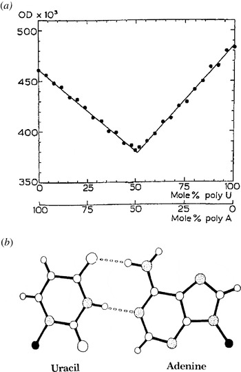
An important method for studying the nucleic acids was measuring their absorbance in the ultraviolet. It had been known for some time that polymerization of nucleotides resulted in a decrease in absorbance at 260 nm. Robert Warner, in Ochoa's department, had described ultraviolet hypochromism on mixing poly rA with poly rU (Warner, Reference Warner1956). Although the mechanism of hypochromicity was not understood at the time, it was a useful tool for analysis. Insight into the reaction of poly rA and poly rU was obtained by carefully measuring hypochromicity in mixtures of varying composition. The very sharp minimum at a 1:1 mole ratio (Figs 1a and 1b) made it possible to estimate the equilibrium constant (Felsenfeld & Rich, Reference Felsenfeld and Rich1957).
(a) Spectroscopic evidence for an RNA duplex. The optical density (OD) at 259 nm of mixtures of poly rA and poly rU. The solutions are in 0·1 m NaCl at neutral pH. The hypochromicity minimum indicates a 1:1 complex (Felsenfeld & Rich, Reference Felsenfeld and Rich1957). (b) Interpretation of the 1:1 complex; a Watson–Crick base-pairing of adenine and uracil. Solid circle, C1′ atom of ribose; speckled circle, nitrogen; large open circle, oxygen.

4. Triple-stranded RNA
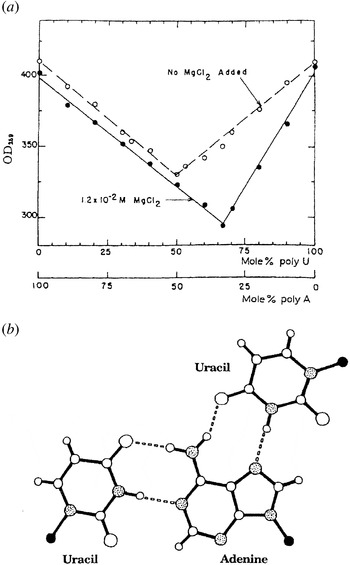
While studying the sedimentation of poly rA, poly rU mixtures in the ultracentrifuge, we detected a more rapidly migrating species. Anticipating that this might be due to additional cations, we found that addition of divalent cations such as magnesium would change the minimum of the poly rA and poly rU reaction (Felsenfeld et al. Reference Felsenfeld, Davies and Rich1957), leading to the formation of a three-stranded molecule (poly rA+2 poly rU) (Fig. 2a). With X-ray and sedimentation studies, we could show that a second strand of poly rU bound in the major groove of the poly rA–poly rU duplex. Addition of this strand did not increase the diameter of the molecule and accounted for the 50% increase in sedimentation constant. We proposed that the second poly rU strand bound through its uracil N3 and O4 by hydrogen bonding to the N7 and N6 of adenine (Fig. 2b). This interpretation was strengthened 2 years later by Hoogsteen's X-ray analysis of the co-crystal containing 9-methyl adenine and 1-methyl thymine (Hoogsteen, Reference Hoogsteen1959), which had the same hydrogen bonding. The 1957 discovery of the RNA triplex was the first indication of the structural complexity inherent in RNA molecules. Over the next several years, a variety of additional polynucleotide interactions were studied (Rich, Reference Rich, McElroy and Glass1957, Reference Rich1958a, Reference Rich1958b; Davies & Rich, Reference Davies and Rich1958), leading to the formation of other two- and three-stranded molecules.
(a) Triplex formation. Optical density (OD) of mixtures of poly rA and poly rU in 0·1 m NaCl at neutral pH with or without 0·012 m MgCl2. The solid line with MgCl2 has a minimum at a 2:1 complex of poly U and poly A (Felsenfeld et al. Reference Felsenfeld, Davies and Rich1957). (b) Interpretation of the hydrogen bonding in the 2:1 complex. The lower Watson–Crick uracil–adenine pair has added to it a uracil residue forming two hydrogen bonds to adenine N6 and N7 (Felsenfeld et al. Reference Felsenfeld, Davies and Rich1957). This type of pairing was later found in a co-crystal structure of adenine and thymine derivatives (Hoogsteen, Reference Hoogsteen1959).

All of these studies revealed the ‘driving power’ accompanying the association of the base π electron systems, which, in effect, assembled into a hydrophobic domain at the center of duplex or triplex RNA. Appreciation of that interaction made it possible to understand the self-assembly of even more complex structures, such as tRNA, ribozymes, pseudoknots, and ribosomes in the future.
Study of the RNA double-helix X-ray diffraction pattern revealed that it was different from the B-DNA double-helix pattern. The diameter of the RNA double helix was 6 Å larger than the B-DNA double helix (Rich, Reference Rich, McElroy and Glass1957). Analysis of the diffraction pattern revealed that the first layer line of the RNA–RNA duplex was stronger than the second – a reversal from that seen in the diffraction pattern of the DNA–DNA duplex (Rich, Reference Rich, McElroy and Glass1957). Another significant difference was that alterations in relative humidity changed the conformation of the DNA double helix, producing both the A and B forms (Franklin & Gosling, Reference Franklin and Gosling1953), while the RNA double helix was unchanged, irrespective of the relative humidity. Only several years later was it realized that the RNA duplex structure was close to the dehydrated A conformation of the DNA duplex (Spencer et al. Reference Spencer, Fuller, Wilkins and Brown1962).
5. Discovery of DNA–RNA hybridization
More than 50 years ago, it was realized that the interaction between DNA and RNA was at the core of molecular biology. The problem was chemical in nature: how could two different types of molecules interact and serve in the transmission of biological information? In the 1950s, it was widely assumed that ‘DNA makes RNA, RNA makes protein.’ This was not based on experimental evidence that DNA and RNA could combine but was more in the nature of an intuitive belief. Two years after moving to MIT in 1958, I was able to carry out a direct experiment: the first DNA–RNA hybridization.
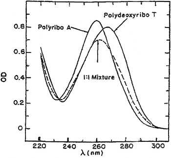
Several investigators at that time had shown that crude preparations of a DNA-dependent RNA polymerase activity would incorporate ribonucleotides into RNA, but the mechanism was not clear. It was widely believed that information transfer went from DNA to RNA, but how did that happen? Could a DNA–RNA hybrid helix form and have the requisite stability to be used for information transfer, in view of the different physical properties and geometries of the RNA and the DNA duplex as outlined above? It became possible to address this problem due to the chemical synthesis of short polymers of deoxythymidylic acid (poly dT) by G. Khorana and colleagues (Tener et al. Reference Tener, Khorana, Markham and Pol1958). Khorana kindly gave me a reaction mixture that I fractionated and took the longer molecules to see if they would react with poly rA. The experiment (Rich, Reference Rich1960) worked. The discovery in 1960 that poly rA and poly dT could form a stable hybrid duplex represented the first experimental demonstration that a hybrid helix could be the method for the transfer of information from DNA to RNA. The evidence was based on measurements of the hypochromism, which occurs when helical molecules are formed (Fig. 3) and the changes in sedimentation rate associated with complex formation. These results indicated that RNA could be made using a single-stranded DNA template to make a complementary RNA strand. As such, it was a model for RNA polymerase activity. When this work was published, the discovery of messenger RNA was still 1 year in the future.
Spectroscopic data showing hybrid helix formation. The optical density (OD) at 259 nm of poly rA, poly dT, and a 1:1 mixture of the two in a dilute salt solution at neutral pH. The optical density of the mixture falls to a minimum in the 1:1 ratio, similar to Figure 1a (Rich, Reference Rich1960).

One year later, in 1961, J. Hurwitz worked with a more highly purified preparation of RNA polymerase (Furth et al. Reference Furth, Hurwitz and Goldmann1961). He used as a template the same short poly dT synthesized by Khorana and was able to synthesize poly rA. This proved that a single stranded template was adequate for RNA polymerase.
The publication in the summer of 1960 (Rich, Reference Rich1960) was the first demonstration of DNA–RNA hybridization, although that term had not yet been invented. That particular hybridization is still widely used today in that immobilized poly dT molecules are used to isolate eukaryotic messenger RNA through their poly rA tails. At the same time in 1960, J. Marmur, P. Doty, and colleagues found they could take denatured DNA molecules and hold them for a prolonged period at an intermediate temperature, called the annealing temperature, thereby allowing the DNA molecules to find each other and re-form a double helix (Doty et al. Reference Doty, Marmur, Eigner and Schildkraut1960; Marmur & Lane, Reference Marmur and Lane1960). One year later, Hall and Spiegelman combined my hybridization work with the annealing technique of Marmur and Doty and found that a newly synthesized RNA strand from T2 virus infection could be annealed with DNA from the virus to make a hybrid helix (Hall & Spiegelman, Reference Hall and Spiegelman1961). Thus, by 1961, both DNA–DNA and RNA–DNA hybridizations were available for many different biological studies.
In 1962 we showed that E. coli transfer RNA (then called soluble RNA) could form a hybrid with E. coli DNA (Goodman & Rich, 1962), thereby demonstrating its origin in the genome. Looking at hybrids of E. coli tRNA with other prokaryotic DNAs made it possible to trace evolutionary changes.
The significance of the discovery of the DNA–RNA hybrid helix is relevant not only to understanding the activity of RNA polymerase but it also explains how reverse transcriptase works, as well as telomerase, retro-transposons, and a variety of other interactions in which DNA and RNA strands combine. Today, fluorescence in situ hybridization is used to form DNA–RNA hybrids to identify specific areas of the genome. Similarly, microarray gene expression profiling studies use DNA–RNA hybrid formation and are dependent on this 1960 discovery.
The full structural analysis of how these RNA and DNA chains accommodated each other was not revealed until much later. In 1982, we solved the first DNA–RNA single-crystal hybrid molecule (Wang et al. Reference Wang, Fujii, van Boom, van der Marel, van Boeckel and Rich1982). It showed that a DNA decamer containing three ribonucleotides formed a hybrid segment that stabilized the entire molecule in the RNA A conformation. In 1992, we carried out a single-crystal X-ray analysis of an Okazaki fragment in which the nucleating ribonucleotides that initiate DNA synthesis in DNA replication bound to DNA (Egli et al. Reference Egli, Usman, Zhang and Rich1992). The presence of a few ribonucleotides was sufficient to convert the entire fragment into the A conformation. This demonstrated the relative conformational flexibility of the DNA backbone, compared to the less flexible RNA strand.
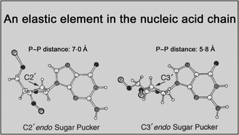
The central role of the ribose ring pucker that differs in DNA and RNA is widely understood today (Rich, Reference Rich2003). The van der Waals crowding, due to the ribose C2′ oxygen, stabilizes ribose into the C3′ endo pucker in the RNA double-helical A conformation (Fig. 4). This interaction provides a sufficient energy barrier to prevent a change in pucker in double-helical RNA, in contrast to the facile manner with which the C2′ endo deoxyribose pucker in DNA changes to C3′ endo on lowering the water content to form A-DNA or to accommodate the presence of RNA segments in a hybrid double helix.
Diagram of deoxyguanosine attached to two phosphates in a polynucleotide chain. Two major types of sugar pucker are illustrated with either C2′ or C3′ above the plane formed by C1′-0-C4′ (endo). For deoxyribonucleotides, the energy barrier between the two puckers is low, while for ribonucleotides, it is considerably higher, giving rise to a more rigid RNA duplex and a more flexible DNA duplex. The differences in the P–P distances results in different conformations for the two duplexes (Rich, Reference Rich2003).

The hybrid DNA–RNA helix remains the bedrock of information transfer in biological systems. The existence of a hybrid helix seems so obvious today that young researchers simply take it for granted. There is little realization of the extent to which scientists once wrestled with the question of how helical polynucleotides with different conformations could react together to make a stable hybrid duplex structure.
6. RNA and the origin of life
In 1961, when asked to contribute an article to a volume dedicated to Albert Szent-Gyorgyi, I decided to write about evolution and the origin of life. This was prompted by the then current ideas largely espoused by the Russian biochemist Oparin, who believed that life began through the creation of primitive protein molecules that provided the necessary environment for developing cells. The greatest weakness in a theory of this type was it did not really explain the evolution of nucleic acid controlled protein synthesis. It seemed more reasonable to suggest that life began with nucleic acids.
It was suggested that primitive polynucleotide chains could act as follows: ‘We postulate that the primitive polynucleotide chains are able to act as a template or a somewhat inefficient catalyst for promoting the polymerization of the complementary nucleotide residues to build up an initial two-stranded molecule. … It may be reasonable to speculate that the hypothetical stem or parent polynucleotide molecule was initially an RNA-like polymer … .’ (Rich, Reference Rich, Kasha and Pullman1962). It was apparent by that time that RNA molecules could contain genetic information, as in the RNA viruses, and forming double-stranded RNA provided a method of replication. They were also believed to play a central role in the synthesis of proteins. DNA was thus regarded as a specialized derivative molecule that evolved later in a form that contained nucleotide sequence information and carried out the molecular replication inherent in propagating living systems.
This article, published in 1962, is probably the oldest statement regarding the role of RNA in the origin of life. Only after many years did the concept of ‘the RNA world’ develop, together with the recognition and enormous growth of ribozyme activities.
7. Anticipation of an RNA regulatory role as in microRNAs
In another symposium volume published in 1961, I described a possible regulatory role for RNA. Long before the role of transcription factors was understood and before messenger RNA was discovered, I suggested that formation of double-stranded RNA on the coding RNA strand would block translation and could be part of a control system for regulating protein synthesis. In a paper discussing the transfer of information in biological systems (Rich, Reference Rich, Davies, Crick and Watson1961), I considered the manner in which RNA polymerase might act. RNA polymerase had not been sufficiently purified at that time to know how it acted, but I discussed the possibility that both strands of the DNA molecule might be transcribed into RNA. What might be the function of these strands? I continue –
There is, however, an interesting possibility in that this may be part of the control apparatus for turning on and off the synthesis of a given class of proteins. One might imagine that one RNA strand is continually used in protein synthesis, while the other strand is used to control the rate at which the operating strand is made available. The synthesis of a given protein would be impeded by the formation of a specific two-stranded RNA molecule which then inactivates the template for protein synthesis. Alternatively, the hydrolysis of the complementary or control strand would then release the operational strand and enable it to participate in the synthesis of proteins (Rich, Reference Rich, Davies, Crick and Watson1961).
Thirty-two years later, G. Ruvkun and V. Ambros (Lee et al. Reference Lee, Feinbaum and Ambros1993; Wightman et al. Reference Wightman, Ha and Ruvkun1993) described the first micro RNA. Micro RNAs control gene expression by forming a duplex with mRNA, somewhat similar to that anticipated in the above excerpt.
8. Purine–pyrimidine co-crystals
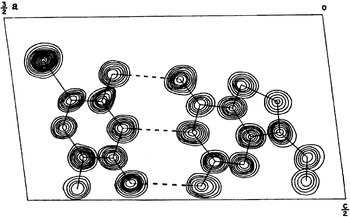
Starting in the early 1960s, there were many single-crystal X-ray diffraction studies involving co-crystals of purine and pyrimidine derivatives similar to Hoogsteen's crystallization of adenine and thymine derivatives cited above (Hoogsteen, Reference Hoogsteen1959). These were useful in obtaining structural information about the association of these nucleic acid components. In 1963, we solved a co-crystal of cytosine and guanine derivatives showing that they were held together by three hydrogen bonds (Sobell et al. Reference Sobell, Tomita and Rich1963) (Fig. 5), not two, as initially suggested by Watson and Crick in Reference Rosenberg, Seeman, Kim, Suddath, Nicholas and Rich1953. This was the first experimental demonstration of the manner in which guanine pairs with cytosine. Pauling had already emphasized the likelihood of three hydrogen bonds based on general structural considerations (Pauling & Corey, Reference Pauling and Corey1956).
First experimental evidence of three hydrogen bonds in the guanine–cytosine Watson–Crick base-pairing. The electron density map shows the structure of the 9-ethyl guanine·1-methyl-5-bromocytosine co-crystal. The dashed lines show the three hydrogen bonds (Sobell et al. Reference Sobell, Tomita and Rich1963).

Several co-crystals were solved of other derivatives of adenine and uracil or adenine and thymine in our laboratory (Mathews & Rich, Reference Mathews and Rich1964; Katz et al. Reference Katz, Tomita and Rich1965; Tomita et al. Reference Tomita, Katz and Rich1967) and elsewhere, as reviewed by Voet & Rich (Reference Voet and Rich1970). At the same time, we were able to demonstrate selective base-pairing in solution. The center of the double helix is a hydrophobic environment due to base stacking. Thus, we studied hydrogen bonding of purine and pyrimidine derivatives in chloroform solution. We measured the infrared spectra and determined association constants. This study showed that adenine paired selectively and most energetically with thymine or uracil derivatives, while guanine paired best with cytosine derivatives. This specificity was called ‘electronic complementarity’ (Kyogoku et al. Reference Kyogoku, Lord and Rich1966).
However, a puzzling trend emerged from the purine–pyrimidine co-crystallographic studies. While guanine and cytosine derivatives consistently showed Watson–Crick pairing, Hoogsteen base-pairing was found in all co-crystals of adenine and thymine/uracil derivatives. This led some investigators to suggest that the double helix itself might be held together entirely by Hoogsteen pairing. The calculated diffraction pattern of such a Hoogsteen-paired DNA duplex had many similarities to that predicted for a double helix held together by Watson–Crick base pairs, even though the fit was not as good (Arnott et al. Reference Arnott, Wilkins, Hamilton and Langridge1965). However, the question remained: What is the real structure of the double helix?
9. The double helix at atomic resolution
X-ray fiber diffraction studies were carried out in several laboratories with both DNA and RNA in the 1950s and 1960s. However, the limitations of such studies were enormous. In fiber X-ray diffraction, a small number of reflections are registered; however, the number of variables needed to define the structure (at least 3N, where N is the number of atoms) is so great that it was clear that fiber diffraction could not ‘prove’ a structure. It could only say that a particular conformation was compatible with the limited diffraction data from fibers.
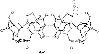
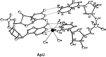
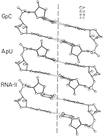
The first single-crystal structures of a double helix were solved in 1973 in my laboratory. This was many years before it was possible to synthesize and obtain oligonucleotides in significant quantities suitable for crystallographic experiments. We succeeded in crystallizing two dinucleoside phosphates, the RNA oligomers guanylyl-3′,5′-cytidine (GpC; Day et al. Reference Day, Seeman, Rosenberg and Rich1973) (Fig. 6) and ApU (Rosenberg et al. Reference Rosenberg, Seeman, Kim, Suddath, Nicholas and Rich1973) (Fig. 7), both of which formed a small fragment of the RNA double helix very similar to the RNA-11 model (Arnott et al. Reference Arnott, Hukins and Dover1972), which had been constructed (Fig. 8). The significant point in these analyses was that the resolution of the diffraction pattern was 0·8 Å. Atomic resolution allowed us to visualize not only the fine details of the sugar phosphate backbone in the form of a double helix but also the positions of ions and water molecules. Further, it could be shown that extending the structure using the symmetry of the two base pairs made it possible to generate long RNA double helices that were quite similar to the structures that had been inferred from fiber X-ray diffraction studies (Rosenberg et al. Reference Rosenberg, Seeman, Day and Rich1976). The bond angles and distances from these structures provided the library of acceptable angles and distances.
Crystal structure of the dinucleoside phosphate, GpC. The structure viewed approximately normal to the bases shows 2 C–G base pairs with a helical stacking. The solid bonds are closer to the reader. The structure was solved at 0·8-Å resolution (Day et al. Reference Day, Seeman, Rosenberg and Rich1973).

The crystal structure of adenylyl-3′,5′-uridine (ApU), solved at 0·8-Å resolution, is shown in perspective together with a sodium ion found in the minor groove that coordinates two bases and four water molecules. This was the first time that this Watson–Crick pairing was seen in a crystal structure, and it appeared on the cover of Nature magazine (Rosenberg et al. Reference Rosenberg, Seeman, Kim, Suddath, Nicholas and Rich1973).

View of the ApU and GpC structures and the RNA-11 model shown approximately normal to the vertical helix axis. The similarities between the structures and the model are quite visible, as is the overall right-handed helical relationship between the successive residues on each of the chains (Day et al. Reference Day, Seeman, Rosenberg and Rich1973).

The GpC structure had the anticipated G·C base pairs connected by three hydrogen bonds. However, the ApU structure showed for the first time that Watson–Crick base pairs formed (Fig. 7) when the molecule was constrained in a double helix, as opposed to the Hoogsteen base pairs that were favored in the single-crystal complexes of adenine with uracil derivatives. I sent preprints to several people, including Jim Watson. He phoned me, saying that after reading the ApU manuscript, he had his first good night's sleep in 20 years! The conclusion of this study was that the uncertainty about the organization of the double helix was resolved. The significance of the double helix at atomic resolution was recognized by the editors of Nature who, in their ‘News and Views’ commentary, called it the ‘missing link’ of nucleic acid structure and pointed out that ‘the many pearls offered’ helped resolve many of the big uncertainties in nucleic acid structure (Nature, 1973, vol. 243: p. 114).
These structures capped the RNA structural effort that I had initiated almost 20 years earlier that started to make progress in 1956 with the recognition that poly rA and poly rU would form a double helix. At atomic resolution, these structures revealed the details of the RNA double helix. High-resolution crystallographic analysis of larger fragments of the double helix (DNA or RNA) did not emerge until almost a decade later with the availability of chemically synthesized and purified oligonucleotides in large enough quantities to permit single-crystal diffraction analysis.
10. The crystal structure of tRNA
By the mid-1960s, a good deal was known about the role of tRNA in protein synthesis. In 1962, we discovered polysomes active in protein synthesis, held together by a messenger RNA strand (Warner et al. Reference Warner, Rich and Hall1962, 1963). In 1964, we found that ribosomes active in protein synthesis contained two tightly bound tRNA molecules and postulated that they occupied adjacent codons on mRNA (naming them the A and P sites, respectively) and discussed how they simultaneously carried out peptidyl transferase activity (Warner & Rich, Reference Warner and Rich1964). However, the structural basis for such tRNA activity was unknown. Holley and co-workers purified and sequenced the first tRNA and suggested it formed a cloverleaf fold (Holley et al. Reference Holley, Apgar, Everett, Madison, Marguisse, Merrill, Penwick and Zamir1965).
Methods for purifying tRNA improved during the 1960s, and many investigators were attempting to form single crystals. This effort was very frustrating, and most people failed repeatedly. In 1968, we were able to obtain single crystals of Escherichia coli tRNAphe (Kim & Rich, Reference Kim and Rich1968). Other groups also obtained single crystals of various tRNAs in that same year, but all of the crystals were poor, somewhat disordered, and with limited resolution. Our earliest crystals diffracted to ~20 Å. By the next year, we were able to get crystals that diffracted to 6 or 7-Å resolution, and a study of the three-dimensional Patterson function using the 12-Å data from the crystals of E. coli tRNAFmet yielded approximate molecular dimensions of 80×25×35 Å (Kim & Rich, Reference Kim and Rich1969). These crystals represented progress of a sort, but the frustration was great because they were not suitable for solving the structure. We spent the next 2 years investigating many different tRNA preparations and crystallization protocols.
By 1971, we reached an exciting goal: Yeast tRNAphe could be crystallized in a simple orthorhombic unit cell with a resolution of 2·3 Å (Kim et al. Reference Kim, Quigley, Suddath and Rich1971) – the first crystals of tRNA suitable for analysis. The key event in making these crystals was the incorporation of spermine, a naturally occurring polyamine. The spermine bound specifically to yeast tRNAphe and stabilized it so that it could form a well-organized crystal. The stabilizating effect of spermine on yeast tRNAphe made it possible to form good crystals in several other lattices as well (Kim et al. Reference Kim, Quigley, Suddath, McPherson, Sneden, Kim, Weinzierl and Rich1973). Analysis of the orthorhombic crystal diffraction pattern showed that it had a characteristic helical distribution of diffracting intensities when viewed in one direction (Kim et al. Reference Kim, Quigley, Suddath and Rich1971). This was taken as a suggestion that short helical segments containing 4–7 base pairs were found in the molecule, a result consistent with the cloverleaf folding of tRNA molecules suggested by Holley (Holley et al. Reference Holley, Apgar, Everett, Madison, Marguisse, Merrill, Penwick and Zamir1965).
10.1 Tracing the backbone of yeast tRNAphe at 4-Å resolution
Myoglobin was the first protein whose three-dimensional structure was solved. The structure was revealed at various levels of resolution. An important milestone was the tracing of the polypeptide chain, which showed how the myoglobin molecule is organized as a series of α-helical and single-stranded regions folded together. This was our first glimpse of how a protein molecule is folded, even though the high-resolution structure had not yet been completed.
The structure of yeast tRNAphe was revealed in a similar, gradual way. Crystallographic research moved more slowly in the early 1970s than today. Computers were primitive; area detectors, cryo-crystallography, and synchrotron beams would be developed in the future. We assembled a cold room around our primitive diffractometer to stabilize the crystals. Heavy-atom derivatives had to be discovered that could be used for phasing the diffraction pattern of a crystalline nucleic acid molecule. This had never been done before, and it took considerable time to discover appropriate derivatives. Three different types of heavy atoms were developed containing platinum, osmium, or samarium ions. The osmium residue was very important since it was known to form complexes with ribonucleotides containing both 2′ and 3′ hydroxyl groups. Only one pair of cis hydroxyl groups was found at the 3′ CCA end of the tRNA molecule. In order to understand its geometry, the structure of an osmium–adenosine complex was solved, which enabled us to visualize the interaction (Conn et al. Reference Conn, Kim, Suddath, Blattman and Rich1974). The position of the single osmium derivative in the tRNA crystal made it possible to identify the 3′ end of the tRNA chain (Kim et al. Reference Kim, Quigley, Suddath, McPherson, Sneden, Kim, Weinzierl and Rich1973). The samarium ions were very useful and occupied more than one site. The platinum residue was somewhat less valuable since it was only useful for 5·5-Å data. An interim electron density map at 5·5 Å made it possible to uncover the external shape of portions of the molecule. However, the true shape and fold of the molecule was not revealed until a map at 4 Å was produced in 1973 (Kim et al. Reference Kim, Quigley, Suddath, McPherson, Sneden, Kim, Weinzierl and Rich1973).
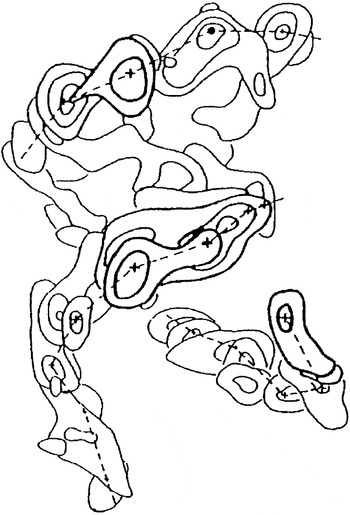
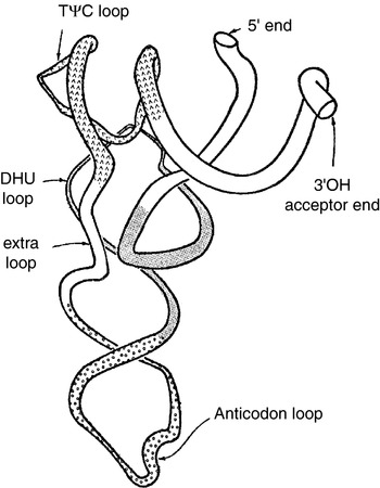
At 4-Å resolution, peaks due to the electron-dense phosphate groups were seen throughout the electron density map. We knew about the distance constraints between adjacent phosphate groups in a polynucleotide chain, and this made it possible to look for peaks between 5 and 7 Å apart (Fig. 9). Tracing the chain led to the discovery that the tRNA molecule had an unusual L shape (Fig. 10). The CCA acceptor helix was collinear with the TΨC helix, and it was almost at right angles to the anticodon stem, which was collinear with the dihydro U stem. The base-paired stems were consistent with Holley's cloverleaf formulation (Holley et al. Reference Holley, Apgar, Everett, Madison, Marguisse, Merrill, Penwick and Zamir1965). The molecule had the shape of an ‘L,’ with the amino acid acceptor 3′ hydroxyl group at one end of the L and the anticodon loop at the other end, over 70 Å away. At the corner of the L, there was a complex folding of the TΨC and dihydro U loops. A perspective diagram of the L-shaped fold was published (Fig. 10).
Superposition of segments of the electron density map of yeast tRNAphe at 4-Å resolution. The + signs indicate positions of electron dense phosphate groups. The dashed lines show segments of three chains. The region between the two upper chains was interpreted as the base pairs of a duplex (Kim et al. Reference Kim, Quigley, Suddath, McPherson, Sneden, Kim, Weinzierl and Rich1973).

Tracing of the entire chain of yeast tRNAphe at 4-Å resolution. This perspective diagram uses a continuous coiled tube for the tracing, and it shows collinearity of the acceptor and TΨC stems, and at right angles, collinear dihydro U and anticodon stems. Shading indicates the different stem-loop segments. The molecule is L-shaped (Kim et al. Reference Kim, Quigley, Suddath, McPherson, Sneden, Kim, Weinzierl and Rich1973). This diagram also appeared on p. 1 of The New York Times on January 13, 1973.

The L-shaped folding of the tRNA polynucleotide chain was a dramatic and surprising discovery, especially the long separation between the acceptor site and the anticodon. The backbone tracing (Fig. 10) was published in Science and in the same week on the front page of The New York Times on January 13, 1973, together with a discussion of its role in protein synthesis. No one had anticipated that the molecule would organize in this fashion.
The L-shaped tRNA molecule is now a standard feature of molecular biology, having been found in virtually all tRNA molecules, even when they are complexed to aminoacyl synthetase enzymes. The significance of the folding is twofold. First, it revealed that the 3′ acceptor end is over 70 Å away from the anticodon loop, which has implications for understanding the interaction between tRNA molecules and some tRNA aminoacyl synthetases. Second and most important, it suggested that the interaction of the tRNA molecules in the ribosome with mRNA occurs at one end of the L, whereas the segment responsible for forming the peptide bond is considerably removed from the site. This separation makes it possible to have great specificity with many interactions at both ends of the molecule.
At that time, I made a proposal regarding the movement of tRNA molecules in the ribosome (Rich, Reference Rich, Nomura, Tissieres and Lengyel1974), as the tRNA structure presented a dilemma. A codon occupies ~10 Å (3×3·4 Å) when the bases are stacked. Thus, the center-to-center distance between the A- and P-site codon is 10 Å. The chain tracing revealed an anticodon stem ~20 Å in diameter. Thus, the paradox: How can two tRNAs simultaneously occupy the A and P sites? The answer, I suggested, was that the mRNA ‘bends or turns a corner’ between A and P site, allowing both tRNAs to make contact. In the crystal structure of the ribosome solved 27 years later, tracing the path of mRNA (Yusupova et al. Reference Yusupova, Yusupova, Cate and Noller2001) revealed a kink of ~45° between the A- and P-site codons. The tRNA chain tracing was thus valuable in anticipating some features of the ribosome machinery.
10.2 The full tRNA structure at 3-Å resolution
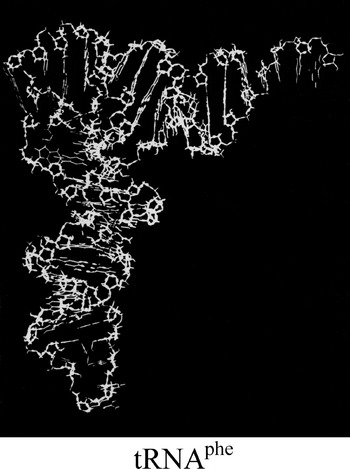
This tracing was visualized in more detail a year later in our 3-Å analysis of the folding of yeast tRNAphe in the orthorhombic lattice (Fig. 11) (Kim et al. Reference Kim, Suddath, Quigley, McPherson, Sussman, Wang, Seeman and Rich1974a). Simultaneously, Klug and colleagues published the 3-Å structure of the same spermine-stabilized yeast tRNAphe in the monoclinic lattice (Robertus et al. Reference Robertus, Ladner, Finch, Rhodes, Brown, Clark and Klug1974). Both papers confirmed the L-shaped folding of the polynucleotide chain, even though the lattices were different. The 3-Å structures were very similar and revealed in great detail the manner in which base-pairing of nucleotides in the double-helical regions stabilizes the molecule (Fig. 11). The folding was also held together by a variety of hydrogen-bonding interactions, including many in the nonhelical, single-stranded regions of the molecule. These additional hydrogen-bonding interactions included only one Watson–Crick pairing; many other triplexes and other single hydrogen bonding interactions were found. The importance of such alternative non-Watson–Crick hydrogen bonds explained why the model builders of that period, trying to predict the structure of tRNA, were all incorrect. They relied excessively on Watson–Crick base pair interactions and did not recognize the stabilizing effect of many other types of hydrogen bonds. The L-shaped folding was predicted to be a general conformation found in all tRNA molecules (Kim et al. Reference Kim, Sussman, Suddath, Quigley, McPherson, Wang, Seeman and Rich1974b). Subsequent work amply verified the relative constancy of the hydrogen-bonding networks (Quigley et al. Reference Quigley, Wang, Seeman, Suddath, Rich, Sussman and Kim1975; Quigley & Rich, Reference Quigley and Rich1976).
The complete structure of yeast tRNAphe appeared on the cover of Science magazine. The view is perpendicular to the plane of the L-shaped molecule with the anticodon on the bottom (Kim et al. Reference Kim, Suddath, Quigley, McPherson, Sussman, Wang, Seeman and Rich1974a).

We have known of the L-shaped folding of tRNA molecules since 1973. The full understanding of why this particular folding is so robust and the manner in which modified nucleotides are important, both to its folding and its function, is still a work in progress. However, the L-shaped folding provides the essential information regarding the interactions and movements of tRNA in protein synthesis.
tRNA was the first macromolecular nucleic acid structure to be solved, and it was the only one available for study for the next 20 years. In 1994, the hammerhead ribozyme structure was solved (Pley et al. Reference Pley, Flaherty and McKay1994). At its core was a special ‘U-turn’ hydrogen bonding conformation that was first described stabilizing the tRNA anticodon and TΨC loops (Quigley & Rich, Reference Quigley and Rich1976).
11. Epilogue
The period from 1954 to 1974 represented the era of RNA awakening. Virtually nothing was known about RNA structure or function at the beginning. In 1954, there was no three-dimensional structural information available and some uncertainty about internucleotide linkages. Functional knowledge was limited, mostly anecdotal and usually incorrect. Twenty years later, the role of different RNA species in protein synthesis was understood, and the broad outlines of its structural biology had been determined. Since 1974, there has been a further flowering and diversification of knowledge about RNA function and structure as well as the introduction of many new species of RNA, including ribozymes, splicesomes, pseudoknots, RNAi, microRNA as well as a dizzying number of newly discovered small RNA species. That process is likely to continue without abatement for many years in the future.