Introduction
The population of primordial follicles present in the ovarian cortex can be a source of thousands of oocytes for maturation and fertilization in vitro, which can revolutionize current assisted reproduction technologies. However, primordial follicles isolated from ovarian tissue and cultured in vitro do not initiate growth (McLaughlin et al., Reference McLaughlin, Albertini, Wallace, Anderson and Telfer2018; Telfer and Andersen, Reference Telfer and Andersen2021). Thus, a consensus has been reached that the activation of primordial follicles can only be achieved by culturing the ovarian cortex containing follicles and stromal cells (Yang et al., Reference Yang, Zhu and Jin2020; Barbato et al., Reference Barbato, Genovese, De Gregorio, Di Nardo, Travaglione, De Napoli, Fragomeni, Zanetti, Adiga, Mondrone, D’Hooghe, Zheng, Longobardi, Catapano, Gualtieri and Talevi2023). Although the contribution of the ovarian stroma to regulating follicle growth is not completely known, preserving the structural integrity of the tissue and the physical interactions between follicles and their surrounding environment is essential for in vitro follicle activation (Grosbois et al., Reference Grosbois, Bailie, Kelsey, Anderson and Telfer2023). Therefore, an efficient in vitro culture system needs to maintain healthy follicles and stromal cells to preserve extracellular matrix components (Tang, Reference Tang2020; Barbato et al., Reference Barbato, Genovese, De Gregorio, Di Nardo, Travaglione, De Napoli, Fragomeni, Zanetti, Adiga, Mondrone, D’Hooghe, Zheng, Longobardi, Catapano, Gualtieri and Talevi2023; Grosbois et al., Reference Grosbois, Bailie, Kelsey, Anderson and Telfer2023).
Over the years, several studies have been proposed to analyse the nutritional requirements of bovine ovarian tissues in vitro with the purpose of establishing an ideal culture medium (Telfer et al., Reference Telfer, McLaughlin, Ding and Thong2008; McLaughlin et al., Reference McLaughlin, Albertini, Wallace, Anderson and Telfer2018). However, some barriers have not yet been sufficiently overcome. For example, the occurrence of oxidative stress during the culture period remains a recurrent limitation. Indeed, oxidative stress has been strongly associated with decreased ability of follicles and somatic cells to neutralize reactive oxygen species, which are among the most prominent physiological inducers of cell damage and apoptosis (Cacciottola et al., Reference Cacciottola, Manavella, Amorim, Donnez and Dolmans2018; Soto-Heras and Paramio, Reference Soto-Heras and Paramio2020). Thus, the success of in vitro culture of preantral follicles strongly depends on the supplementation of culture medium with antioxidant substances. In this sense, Vasconcelos et al. (Reference Vasconcelos, Costa, Azevedo, Paulino, Soares, Souza, Carneiro, Brito, Catunda Júnior and Silva2022) investigated the use of essential oils to improve in vitro ovarian tissue culture systems.
Some studies have investigated a range of biological activities of Lippia sidoides essential oil (LSEO), with emphasis on its antioxidant activity (Da Silva et al., Reference Da Silva, Silva, Andrade, Gratieri, Setzer, Maia and Da Silva2017; Nascimento et al., Reference Nascimento, Silva, Cascaes, Costa, Figueiredo, Costa, Andrade and De Faria2021; Ziyatdinova and Kalmykova, Reference Ziyatdinova and Kalmykova2023). Monteiro et al. (Reference Monteiro, De Melo Leite, Bertini, De Morais and Nunes-Pinheiro2007) showed that LSEO (7.27 µg/mL) inhibited 50% of the free radical (DPPH). In addition, studies conducted on several species of the genus Lippia have identified thymol as the main active component (Mota et al., Reference Mota, Lobo, Costa, Costa, Rocha, Rocha e Silva, Pohlit and Neto2012; Aquino et al., Reference Aquino, Sales, Soares, Martins and Costa2014; Parente et al., Reference Parente, Custódio, Cardoso, Lima, Melo, Linhares, Siqueira, Nascimento, Catunda Júnior and Melo2018) with concentrations ranging from 34% to 95%. The interaction of thymol and the other constituents of LSEO has been frequently associated with the biological activities of these oils (Ruberto and Baratta, Reference Ruberto and Baratta2000). Thymol has already increased glutathione concentrations (Saccol et al., Reference Saccol, Uczay, Pês, Finamor, Ourique, Riffel, Schmidt, Caron, Heinzmann, Llesuy, Lazzari, Baldisserotto and Pavanato2013; Reyes-Becerril et al., Reference Reyes-Becerril, Gijón, Angulo, Vázquez-Martínez, López, Junco, Armenta, Guerra and Angulo2021) and reduced cell apoptosis rates during in vitro maturation of oocytes (Pereira, Reference Pereira2015), as well as improved the development and quality of buffalo blastocysts produced in vitro (Sollecito et al., Reference Sollecito, Pereira, Grázia, Neves, Couto, Andrade, Miranda, Silva and Borges2019). It is hypothesized that LSEO improves follicular survival and growth, as well as the expression of mRNA and activity of antioxidant enzymes, such as superoxide dismutase (SOD1), catalase (CAT), peroxiredoxin-6 (PRDX6) and glutathione peroxidase 1 (GPX1) in bovine ovarian tissue cultured in vitro for 6 days.
The present study aims to investigate the chemical composition of LSEO and the influence of different concentrations (400, 800, 1600 or 3200 µg/mL) of this oil on primordial follicles activation and survival, ovarian stromal cell density and collagen distribution in ECM in bovine ovarian tissue after 6 days of in vitro culture. In addition, its effects on the levels of thiol and mRNA for CAT, SOD1, GPX1 or PRDX6, and on the activity of antioxidant enzymes CAT, SOD1 and GPX1 were also investigated.
Material and methods
Chemicals
The culture media and other substances used in this study were purchased from Sigma Chemical Co. (St Louis, MO, USA), unless otherwise stated. The essential oil of Lippia sidoides, with 100% purity, was purchased from the Brazilian aromatherapy company Laszlo (Belo Horizonte, MG, Brazil).
Essential oil analysis
The chemical analysis of the essential oil constituents was performed with a Shimadzu QP-2010 Ultra instrument employing the following conditions: Column: Rtx-5 MS (Crossbond, 5% diphenyl/95% dimethylpolysiloxane) measuring 30 m × 0.25 mm × 0.25 μm df; carrier gas: He (24.2 mL/min, in constant linear velocity mode); injector temperature of 250°C, in split mode (1:100); and detector temperature of 250°C. The column temperature was programmed for 35–180°C at 4°C/min, then 180–280°C at 17°C/min, and 280°C for 10 min. Mass spectra were obtained at electron impact of 70 eV. The volume of essential oil injected was 1 μL and the components were identified from its gas chromatography (GC) retention times, calculated by linear interpolation relative to retention times of main compounds and by comparison with mass spectra those present in the computer data bank (NIST) and published literature (Adams, Reference Adams2007)
Ovarian sources and in vitro culture of cortical tissue
Bovine ovaries (n = 16) from mixed breed cows were collected at a local slaughterhouse. Immediately after slaughter, each pair of ovaries was washed in ethanol (70%) for approximately 10 s, followed by two washes in 0.9% saline solution supplemented with penicillin (100 µg/ml) and streptomycin (100 µg/ml). After washing, each pair of ovaries was individually transported to the laboratory in falcon tubes containing α-MEM supplemented with penicillin (100 µg /mL) and streptomycin (100 µg/mL) at 4°C, within 1 h. This study was approved by the Committee of Ethics and Animal Welfare of the Federal University of Ceará (N° 09/2021).
In the laboratory, ovarian cortical tissue from the same pair of ovaries was cut into sixty-eight slices (3 mm × 3 mm × 1 mm) in dissection medium composed of α-MEM supplemented with penicillin (100 µg/ml) and streptomycin (100 µg/ml). For each of the eight animals, two cortical slices were fixed in paraformaldehyde (4%) for 24 h at 4°C for histological analysis (uncultured control) as well as for analysis of the extracellular matrix and stromal cells. The remaining fragments were cultured in 24-well culture dishes for 6 days (Passos et al., Reference Passos, Costa, Da Cunha, Silva, Ribeiro, De Souza, Barroso, Dau, Saraiva, Gonçalves, Van den Hurk and Silva2016). The in vitro culture was performed with 5% CO2 in a humidified incubator at 38.5°C. The basic culture medium consisted of α-MEM (pH 7.2–7.4) supplemented with ITS (10 μg/ml insulin, 5.5 μg/ml transferrin, and 5 ng/ml selenium), 2 mM glutamine, 2 mM hypoxanthine, antibiotics (100 µg/ml penicillin and 100 µg /ml streptomycin) and 1.25 mg/ml of bovine serum albumin (α-MEM+). The fragments were cultured in 0.5 mL of control medium (α-MEM+) alone or in this control medium (α-MEM+) supplemented with different concentrations of LSEO (400, 800, 1600 or 3200 μg/mL). The LSEO was diluted in dimethyl sulphoxide to ensure its bioavailability and its concentrations were chosen according to Costa et al. (Reference Costa, Arruda, Barroso, Melo, De Carvalho and Catunda2018). Using the DPPH assay, these authors demonstrated that LSEO antioxidant activity increased significantly as its concentration increased from 400 to 800, 1600, and 3200 μg/mL. Every 2 days, 60% of the culture medium was replaced with fresh medium. Each treatment was repeated eight times, with one cow in each repetition. After the culture period, the fragments were processed for morphological evaluation, real-time quantitative polymerase chain reaction, and biochemical analyses.
Morphological analysis and investigation of follicular development in vitro
Histological analysis was performed according to the methodology of Bizarro-Silva et al. (Reference Bizarro-Silva, Santos, Gerez, González, Lisboa and Seneda2018). Uncultured tissues (D0) and tissues cultured in vitro were fixed in paraformaldehyde (4%) for 24 h, dehydrated in increasing concentrations of ethanol, cleared with xylene and embedded in paraffin. Serial sections (7 µm thick) were made and, every 5 sections, stained with haematoxylin and eosin. Only preantral follicles with an oocyte nucleus in the section were analysed to avoid double counting. The slides were examined under a microscope (Nikon, Tokyo, Japan) at magnifications of 100 and 400 times. The developmental stages of preantral follicles were classified as primordial (one layer of flattened or flattened and cuboidal granulosa cells surrounding the oocyte) or growing follicles (primary: one layer of cuboidal granulosa cells and secondary: two or more layers of cuboidal granulosa cells surrounding the oocyte) (Figueiredo and Lima, Reference Figueiredo and Lima2017). These follicles were classified as morphologically normal when they had intact oocyte surrounded by well-organized granulosa cells without pyknotic nucleus. Degenerated follicles had a retracted oocyte and/or is surrounded by disorganized granulosa cells, which are separated from the basement membrane (Telfer and Andersen, Reference Telfer and Andersen2021). Previous studies have shown that the main indicators used to classify the follicles as morphologically normal are the absence of pyknosis in the nuclei of oocytes and granulosa cells, as well as the absence of cytoplasmic retraction. Morphological analysis is a widely recognized method for determining healthy follicles in cultured bovine ovarian tissues (Bizarro-Silva et al., Reference Bizarro-Silva, Santos, Gerez, González, Lisboa and Seneda2018; Telfer and Andersen, Reference Telfer and Andersen2021; Bergamo et al., Reference Bergamo, Bonato, Bizarro-Silva, Bonato, González, Rossaneis, Verri, Morotti and Seneda2022; Deligiannis et al., Reference Deligiannis, Kask, Modhukur, Boskovic, Ivask, Jaakma, Damdimopoulou, Tuuri, Velthut-Meikas and Salumets2024). Overall, 100–160 follicles were evaluated for each treatment. The percentages of healthy primordial and developing follicles were calculated before (uncultured control) and after culture in a given medium
Stromal cell density and extracellular matrix analysis
To assess the density of ovarian stromal cells, tissue sections were stained with haematoxylin and eosin and the number of stromal cells was counted in a 10 mm2 area as previously described Cavalcante et al. (Reference Cavalcante, Matos-Brito, Paulino, Silva, Aguiar, De Almeida, Souza, Vasconcelos, De Assis, Silva and Silva2019) For each treatment, ten fields from sections of five different animals were evaluated.
The collagen analysis was performed by using Picrosirius Red (Abcam Kit) according to the methodology described by Rittié (Reference Rittié2017). To this end, ovarian sections were deparaffinized in xylene and incubated in Sirius Red solution (0.1%) for 1 h at room temperature. Then, the excess dye was removed with an acetic acid solution (0.5%) and the sections were dehydrated and analysed in an optical microscope (Nikon, Eclipse, TS 100, Japan) at 400x magnification. For each treatment, the percentage of area occupied by collagen fibres in ten different fields was measured using a camera coupled to a microscope (Nikon, Eclipse, TS 100, Japan) and the images were evaluated by Image J software (Version 1.51p, 2017). The collagen fibres were stained in red by picrosirius, while follicles remained unstained. The quantification of percentage of collagen fibres in uncultured and cultured tissues was performed by Image J software. The intensity of collagen fibre staining was determined by evaluating the average pixel intensity of the total photographed area after subtraction of the background.
Expression of mRNA for SOD1, CAT, PRDX6 and GPX1 in ovarian tissues
For analysis of gene expression, ovarian tissues were stored at −80°C immediately after end of the culture period until the extraction of total RNA for further analysis of expression of SOD1, CAT, PRDX6 and GPX1 mRNAs. Tissues cultured in control medium (α-MEM+) and those cultured in medium supplemented with 400 or 800 μg/mL were selected to investigate mRNA expression. The ovarian tissues were macerated under sterile conditions and then subjected to extraction of total RNA, by using a TRIzol® purification kit (Invitrogen, São Paulo, Brazil), and quantification of mRNA was performed using SYBR Green. The PCR reactions were composed of 1 μL cDNA as a template in 7.5 μL of SYBR Green Master Mix (PE Applied Biosystems, Foster City, CA, USA), 5.5 μL of ultra-pure water, and 0.5 μM of each primer. The primers were designed to amplify SOD1, CAT, PRDX6, GPX1 and GAPDH (Table 1). The melting curve analysis of PCR products was used to evaluated specificity of each primer pairs. The thermal cycling profile for the first round of PCR was initial denaturation and activation of the polymerase for 10 min at 95°C, followed by 40 cycles of 15 s at 95°C, 30 s at 58°C, and 30 s at 72°C. The final extension was for 10 min at 72°C. All reactions were performed in a Step One Plus instrument (Applied Biosystems, Foster City, CA, USA). The 2^ΔΔCt method was used to transform the Ct values into normalized relative expression levels (Livak and Schmittgen, Reference Livak and Schmittgen2001).
Primer pairs used in real-time PCR

Table 1. Long description
A table with five rows and four columns detailing primer sequences for real-time PCR analysis. The columns are labeled ‘Target gene’, ‘Primer sequence (5’ -> 3’)’, ‘Sense (S), anti-sense (As)’, and ‘GenBank accession no.’. The rows list the following information: Row 1: Target gene: GAPDH; Primer sequence: TGTTTGTGATGGGCGTGAACCA ATGGGCGCGTGGACAGTGGTCATAA; Sense (S), anti-sense (As): SAs; GenBank accession no.: GI: 402744670. Row 2: Target gene: PRDX6; Primer sequence: GCACCTCCTCTTACTTCCCGGATGCGGCCGATGGTAGTAT; Sense (S), anti-sense (As): SAs; GenBank accession no.: GI: 59858298. Row 3: Target gene: GPX1; Primer sequence: AACGTAGCATCGCTCTGAGGGATGCCCAACTGTTGTGCAG; Sense (S), anti-sense (As): SAs; GenBank accession no.: GI: 156602645. Row 4: Target gene: SOD1; Primer sequence: GTGAACAACCTCAACGTCGCGGGTTCCTCCACCACCGTTAG; Sense (S), anti-sense (As): SAs; GenBank accession no.: GI: 31341527. Row 5: Target gene: CAT; Primer sequence: AAGTTCTGCATCGCCACTCAGGGGCCCTACTGTCAGACTA; Sense (S), anti-sense (As): SAs; GenBank accession no.: GI: 402693375.
Biochemical analysis
To produce the homogenate for biochemical analyses, each 100 mg of ovarian tissue was homogenized 3 to 4 times, for 10 s at 4°C, in 900 μL of buffer (10 mM Tris-HCl, 0.9% NaCl (w/v), pH 7.4), containing inhibitors of protease (5 mg/mL aprotinin and 34.8 mg/mL PMSF) using an Ultra-Turrax homogenizer T25. The samples were centrifuged at 4°C and 720 G for 10 min. The supernatant was used to test the activity of the enzymes and thiol content, after determining the protein content of the sample using the Bradford method (Bannister and Calabrese, Reference Bannister and Calabrese1987).
Total proteins (Bradford method)
The protein concentration was determined using the Bradford method. This method uses Coomassie blue (Quick start/Bradford; Catalogue No. 500-0205; Bio-Rad) to determine the total concentration of proteins in each extract sample. In contact with proteins, the Coomassie blue stain forms a complex and emits a blue luminescence. The absorbance is directly related to the protein concentration of the sample and evaluated spectrophotometrically at a wavelength of 595 nm. The total protein concentration in samples was determined using a standard curve of bovine albumin (0, 2.5, 5, 10, 15, 25, 35 and 50 mg/mL), which was used to standardize the levels of pro-oxidants (thiol) and antioxidants (SOD1, CAT, GPX1), as described below.
Determination of pro-oxidant based on the thiol content
The total thiol content was determined in the supernatant of the tissue homogenate using 5,5`-dithiobis 2-nitrobenzoic acid (DTNB; D8130; Sigma–Aldrich Chem. Co, St Louis, USA) as an index of reduced thiol molecules. Thiol residues react with DTNB (10 mM), cleaving the disulphide bond to form 2-nitro-5-thiobenzoate anion (NTB2–) at a neutral pH. The NTB2– is quantified in a spectrophotometer by measuring absorbance at 412 nm, with results expressed as nMol of reduced DTNB per milligram of protein (Ellman, Reference Ellman1959)
Activity of SOD1, CAT and GPX1 enzymes
The SOD1 activity was measured as the inhibition of adrenaline auto-oxidation (Bannister and Calabrese, Reference Bannister and Calabrese1987). Adrenaline oxidation, in the presence of CAT in basic medium, leads to the formation of O2– radical, which SOD1 reacts with, thus inhibiting the oxidation of adrenaline. The CAT solution (0.048 mg/mL; c9322; Sigma–Aldrich Chem. Co St Louis, USA) was performed adding (7:3) to glycine buffer, pH 10.2 (Dinâmica Química, São Paulo, Brazil). Three different volumes (10, 20 or 40 μL) of ovary homogenate were then added to the solution and then adrenaline (0.218 mg/mL; E4260; Sigma–Aldrich Chem. Co, St Louis, USA) was added to start oxidation. Oxidation was evaluated at 480 nm every 10 s for 180 s.
The CAT activity was measured as the consumption of H2O2 as a substrate at 240 nm (Flohé and Günzler, Reference Flohé and Günzler1984). A solution of H2O2 (152 µL/mL; PH09717RA; Êxodo Científica, Sumaré, Brazil) and phosphate-buffered saline (PBS; pH7.4) was mixed in a quartz cuvette at room temperature, and then 50 μL of the ovary homogenate were added. Every 30 s, the consumption of H2O2 was measured twice.
The GPX1 activity was measured by the oxidation of NADPH. NADPH is consumed by glutathione reductase (GR; G3664; Sigma–Aldrich) to convert oxidized glutathione (GSSG) to reduced glutathione (GSH). In the presence of H2O2, GPX oxidizes GSH to GSSG and reduces the peroxides to alcohols and water. The consumption of NADPH is directly proportional to the consumption of H2O2 and, consequently, to the activity of GPX (Furtado et al., Reference Furtado, Martins, Oliveira, Guerreiro, De Sá, Ferraz, Ceccatto, Rodrigues and Araújo2021). The reactions were prepared by mixing 500 µL potassium phosphate buffer (100 mM), which is composed of 13.6 g/L monobasic potassium phosphate (P0662; Sigma–Aldrich) added to 1.86 g/L EDTA (5 mM; E4884; Sigma–Aldrich; pH 7.4) and 38 µg/mL GR, 3 µg/mL GSH, and 100 µL homogenate of the ovarian tissue samples for 10 min at room temperature so that GPX cysteine can contact GR and GSH. Finally, 100 µL NADPH and 120 s later, 100 µL of H2O2 were added to the mixture, and NADPH oxidation was measured as the reduction in NAPDH with absorbance at 340 nm, being evaluated every 10 s for 300 s (Furtado et al., Reference Furtado, Martins, Oliveira, Guerreiro, De Sá, Ferraz, Ceccatto, Rodrigues and Araújo2021)
Statistical analysis
Statistical analysis was performed using GraphPad Prism software (version 9.0). Initially, data were assessed for normality and homogeneity of variances using the Shapiro–Wilk and Brown-Forsythe tests, respectively. Data sets that simultaneously met these criteria were classified as parametric and analysed by one-way ANOVA, followed by Tukey’s post hoc test for multiple comparisons between experimental groups, including variables related to morphologically normal follicles, primordial and developing follicles, collagen content, and thiol levels, as well as for activity of SOD1, CAT, and GPX1. For data that did not meet the assumptions of normality and/or homogeneity, the nonparametric Kruskal–Wallis test was adopted, followed by Dunn’s multiple comparison test, applied to the analyses of stromal cell density and mRNA levels of PRDX6, GPX1, SOD1, and CAT. All values were expressed as mean ± standard error of the mean (SEM), and differences between groups were considered statistically significant when P < 0.05.
Results
Chemical composition of LSEO
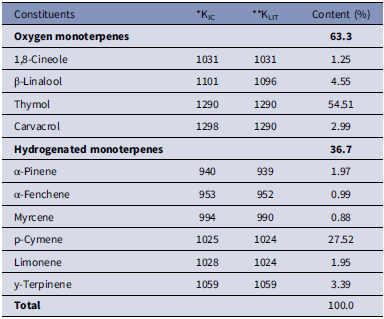
The components of LSEO, their percentages and retention index (RI) are listed in Table 2. The constituents were identified by gas chromatography-mass spectrometry (GC/MS) and represented as 100.0% LSEO. The oxygen monoterpenes represented 63.3%, while hydrogenated monoterpenes were found in lower percentages (36.7%). Thymol (54.5%) and para-cymene (27.5%) were the major components identified in LSEO.
Chemical constituents of Lippia sidoides essential oil identified by GC-MS and their relative contents

Table 2. Long description
A table comparing chemical constituents of Lippia sidoides essential oil identified by GC-MS and their relative contents. The table has 12 rows and 4 columns. Column headers are Constituents, KIC, KLIT, and Content (percent). Row labels include Oxygen monoterpenes and Hydrogenated monoterpenes. Oxygen monoterpenes include 1,8-Cineole, beta-Linalool, Thymol, and Carvacrol. Hydrogenated monoterpenes include alpha-Pinene, alpha-Fenchene, Myrcene, p-Cymene, Limonene, and gamma-Terpinene. Thymol has the highest content at 54.51 percent, followed by p-Cymene at 27.52 percent. The total content sums to 100 percent.
*KIC – calculated Kovats index; ** *KLIT – Kovats index literature.
Effects of LSEO on follicular survival after in vitro culture
For the follicular survival analysis, the primordial and developing follicles (both primary and secondary) were classified collectively as either normal or degenerated. After six days of culture, regardless of treatment, a significant reduction in the percentage of morphologically normal follicles was observed compared with non-cultured tissues (P < 0.05). However, the presence of 400 or 800 μg/mL of LSEO in the culture medium promoted significant improvement in follicular morphology preservation compared with tissues cultured in the control medium (α-MEM+). Conversely, tissues cultured with the highest LSEO concentrations (1600 and 3200 μg/mL) exhibited no statistical differences compared with those cultured in the control medium (Figure 1A).
Morphological features of primordial (A, B), primary (C, D) and secondary follicles (E, F) classified as normal (A, C, E) or degenerated (B, D, F) after 6 days of in vitro culture of bovine ovarian cortex in control medium alone or supplemented with Lippia sidoides essential oil (LSEO). The percentages (mean ± SEM) of morphologically normal follicles (primordial, primary, and secondary follicles together) in uncultured tissues and tissues cultured in control medium (α-MEM+) alone or supplemented with 400, 800, 1600, or 3200 µg/mL of LSEO for 6 days (n = 8 biological replicates). Different letters indicate significant differences between the groups (P < 0.05; one-way ANOVA followed by Tukey’s post-hoc test).

Figure 1. Long description
Morphological features of primordial (A, B), primary (C, D) and secondary follicles (E, F) classified as normal (A, C, E) or degenerated (B, D, F) after 6 days of in vitro culture of bo-vine ovarian cortex in control medium alone or supplemented with Lippia sidoides essential oil (LSEO). The percentages (mean ± SEM) of morphologically normal follicles (primordi-al, primary, and secondary follicles together) in uncultured tissues and tissues cultured in control medium (α-MEM+) alone or supplemented with 400, 800, 1600, or 3200 μg/mL of LSEO for 6 days (n = 8 biological replicates). Different letters indicate significant differ-ences between the groups (P < 0.05; one-way ANOVA followed by Tukey’s post-hoc test).
Effects of LSEO on activation and development of primordial follicles
Ovarian tissues cultured in all treatments showed a reduction in the percentage of primordial follicles and an increase of developing follicles at primary and secondary stages, when compared with uncultured tissues. For this analysis, only morphologically normal follicles were considered. However, the presence of 400 µg/mL LSEO in the culture medium significantly (P < 0.05) increased the percentage of developing follicles when compared with tissues cultured in the control medium alone or supplemented with 800, 1600 and 3200 µg/mL LSEO. Additionally, tissues cultured with 800, 1600 and 3200 µg/mL LSEO also showed a higher percentage of developing follicles (P < 0.05) when compared with those cultured in control medium (Figure 2A).
Percentages (mean ± SEM) of morphologically normal primordial and developing follicles in fresh (non-cultured) bovine ovarian tissues and after 6 days of in vitro culture in control medium or in medium supplemented with 400, 800, 1600, or 3200 µg/mL Lippia sidoides essential oil (LSEO). Data are expressed as percentages of each follicle category (n = 8 biological replicates). Different letters indicate significant differences (P < 0.05; one-way ANOVA followed by Tukey’s post hoc test).

Figure 2. Long description
Percentages (mean ± SEM) of morphologically normal primordial and developing follicles in fresh (non-cultured) bovine ovarian tissues and after 6 days of in vitro culture in control medium or in medium supplemented with 400, 800, 1,600, or 3,200 μg/mL Lippia sidoides essential oil (LSEO). Data are expressed as percentages of each follicle category (n = 8 bio-logical replicates). Different letters indicate significant differences (P < 0.05; one-way ANOVA followed by Tukey’s post hoc test).
Effects of LSEO on stromal cell density after in vitro culture
After 6 days of culture, regardless of the treatment, a significant reduction in the percentage of stromal cell density was observed when compared with non-cultured tissues (P < 0.05, Figure 3A). The presence of 400 µg/mL LSEO in cultured medium, however, increased the percentage of stromal cells when compared with those cultured in control medium (P < 0.05). In addition, 800 or 1600 µg/mL LSEO did not influence stromal cell density, while a reduction in the number of cells was seen in tissues cultured with 3200 µg/mL LSEO (Figure 3B).
Stromal cell number (cells/100 µm2, mean ± SEM) in uncultured tissues (A) and in tissues cultured for 6 days in control medium (B) or in medium supplemented with 400, 800, 1600, or 3200 µg/mL Lippia sidoides essential oil (LSEO) (C–F).. Different letters indicate significant differences (P < 0.05; Kruskal–Wallis test followed by Dunn’s multiple comparison test). Representative histological sections stained with haematoxylin and eosin are provided to illustrate tissue structure. Scale bars: 100 µm. n = 8 biological replicates.

Figure 3. Long description
Stromal cell number (cells/100 μm2, mean ± SEM) in uncultured tissues (A) and in tissues cultured for 6 days in control medium (B) or in medium supplemented with 400, 800, 1,600, or 3,200 μg/mL Lippia sidoides essential oil (LSEO) (C–F).. Different letters indicate sig-nificant differences (P < 0.05; Kruskal–Wallis test followed by Dunn’s multiple comparison test). Representative histological sections stained with hematoxylin and eosin are provided to illustrate tissue structure. Scale bars: 100 μm. n = 8 biological replicates.
Evaluation of collagen in ovarian extracellular matrix
Figure 4 shows a significant reduction in collagen fibres in tissues cultured in all treatments (P < 0.05) when compared with non-cultured tissues. In contrast, ovarian cortex cultured with 400 µg/mL LSEO had higher rates of collagen fibres than those cultured in control medium (P < 0.05). In addition, 800, 1600 or 3200 µg/mL LSEO did not influence collagen fibres (Figure 4).
Analysis of extracellular matrix density in bovine ovarian tissues after 6 days of in vitro culture in control medium alone or supplemented with Lippia sidoides essential oil (LSEO). (A–F) Representative histological sections stained with Picrosirius Red illustrating the distribution of collagen fibres and matrix organization in: (A) non-cultured tissue and tissues cultured in (B) control medium (α-MEM+) alone or supplemented with 400, 800, 1600, or 3200 µg/mL LSEO (C–F). (G) Quantification of high-density matrix (HDM) area (%). Data are presented as mean ± SEM (n = 8 biological replicates). Different letters indicate significant differences (P < 0.05; one-way ANOVA followed by Tukey’s post hoc test). The asterisk (*) denotes a significant difference between non-cultured control and cultured tissues.

Figure 4. Long description
Analysis of extracellular matrix density in bovine ovarian tissues after 6 days of in vitro culture in control medium alone or supplemented with Lippia sidoides essential oil (LSEO). (A–F) Representative histological sections stained with Picrosirius Red illustrating the distribution of collagen fibers and matrix organization in: (A) non-cultured tissue and tissues cultured in (B) control medium (α-MEM+) alone or supplemented with 400, 800, 1,600, or 3,200 μg/mL LSEO (C–F). (G) Quantification of high-density matrix (HDM) area (%). Data are presented as mean ± SEM (n = 8 biological replicates). Different letters indicate significant differences (P < 0.05; one-way ANOVA followed by Tukey’s post hoc test). The asterisk (*) denotes a significant difference between non-cultured control and cultured tissues.mosic. A trident is placed diagonally behind it. A small bird sits atop the rudder. Either side of the rudder are two fish, possibly dolphins, swimming downwards. In the top right-hand corner, a creature with the body and head of a horse and the tail of a sea dragon swims upwards. The mosaic appears to have been partially restored and there is a small patch missing on the right-hand side.
Quantification of mRNA for SOD1, CAT, PRDX6 and GPX1 in ovarian tissues
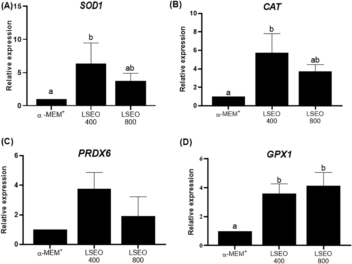
Figure 5 shows the mRNA levels for SOD1, CAT, PRDX6 and GPX1 in bovine ovarian tissues after 6 days of culture. The presence of 400 µg/mL LSEO in the culture medium increased the mRNA levels for SOD1, CAT and GPX1, while 800 µg/mL LSEO increased RNA expression for GPX1 in ovarian tissues, when compared with those cultured in control medium (P < 0.05). On the other hand, 800 µg/mL LSEO did not alter the expression of mRNA for SOD1 and CAT. The expression of mRNA for PRDX6 was not influenced by LSEO.
mRNA levels (mean ± SEM) of SOD1 (A), CAT (B), PRDX6 (C), and GPX1 (D) in bovine ovarian tissues cultured in vitro for 6 days in control medium alone or supplemented with 400 or 800 µg/mL of Lippia sidoides essential oil (LSEO); n = 8 biological replicates. Different letters indicate significant differences (P < 0.05; Kruskal–Wallis test followed by Dunn’s multiple comparison test).

Figure 5. Long description
mRNA levels (mean ± SEM) of SOD1 (A), CAT (B), PRDX6 (C), and GPX1 (D) in bovine ovarian tissues cultured in vitro for 6 days in control medium alone or supplemented with 400 or 800 μg/mL of Lippia sidoides essential oil (LSEO); n = 8 biological replicates. Different letters indicate significant differences (P < 0.05; Kruskal–Wallis test followed by Dunn’s multiple comparison test).
Activity of SOD1, CAT and GPX1 enzymes
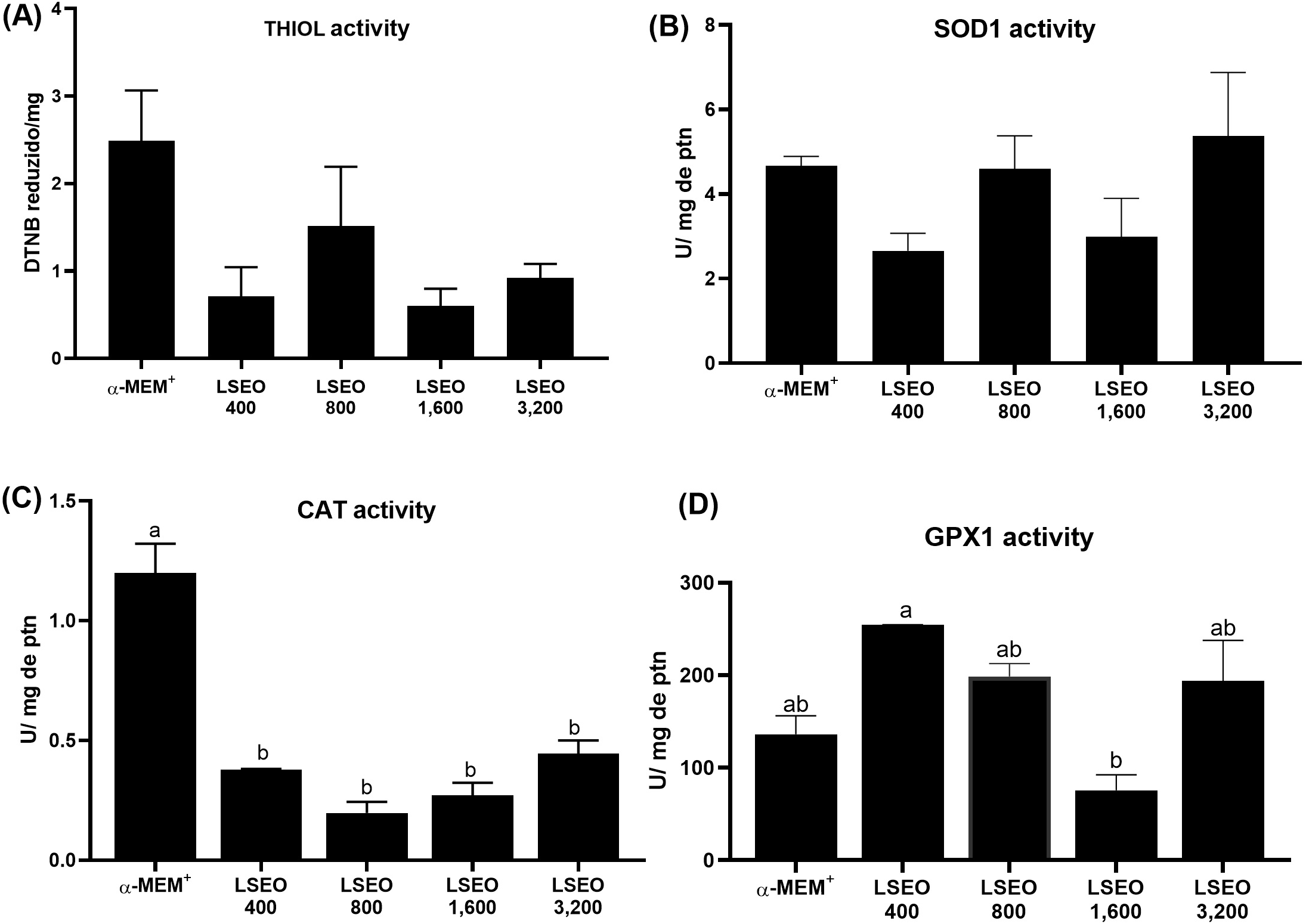
The LSEO at all tested concentrations reduced CAT activity in cultured bovine ovarian tissues (P < 0.05), but did not influence the levels of thiol and activity of SOD1 and GPX1 (P > 0.05), when compared with tissues cultured in control medium (Figure 6). The activity of GPX1 in ovarian tissues cultured with 400 µg/mL LSEO was, however, higher than that seen in tissues cultured with 1600 µg/mL LSEO (P < 0.05).
Thiol levels (A) and enzymatic activity of CAT (B), SOD1 (C), and GPX1 (D) in bovine ovarian tissues cultured in vitro for 6 days in control medium alone or supplemented with 400, 800, 1600, or 3200 µg/mL of Lippia sidoides essential oil (LSEO); n = 8 biological replicates. Different letters indicate significant differences (P < 0.05; one-way ANOVA followed by Tukey’s post hoc test).

Figure 6. Long description
Thiol levels (A) and enzymatic activity of CAT (B), SOD1 (C), and GPX1 (D) in bovine ovarian tissues cultured in vitro for 6 days in control medium alone or supplemented with 400, 800, 1,600, or 3,200 μg/mL of Lippia sidoides essential oil (LSEO); n = 8 biological replicates. Different letters indicate significant differences (P < 0.05; one-way ANOVA followed by Tukey’s post hoc test).
Discussion
Essential oils are important biotechnological products composed of a complex mixture of organic compounds that can be used to supplement culture media and, consequently, improve in vitro cell culture systems (Vasconcelos et al., Reference Vasconcelos, Costa, Azevedo, Paulino, Soares, Souza, Carneiro, Brito, Catunda Júnior and Silva2022). In this context, identifying the chemical constituents of essential oils is an important step in evaluating a given biological response under in vivo or in vitro conditions (Tak and Isman, Reference Tak and Isman2017). In our research, the composition of LSEO indicated the presence of 63.3% oxygenated compounds. This complex mixture of organic compounds can act synergistically or antagonistically, modulating biological activity (Huang et al., Reference Huang, Lao, Pan, Chen, Zhao, Gong, Xie and Mo2021). We demonstrated for the first time that, during in vitro culture of bovine ovarian tissue, LSEO (400 and 800 µg/mL) increases follicle activation, development, and survival. These results may be related to the maintenance of redox balance (Sá et al., Reference Sá, Bruno, Guerreiro, Cadenas, Alves, Cibin, Leal-Cardoso, Gastal and Figueiredo2018). The lower percentage of morphologically normal follicles in tissues cultured in control medium may be associated with increased oxidative stress. Previous studies have demonstrated the positive effect of extracts and essential oils of the genus Lippia at low concentrations on the in vitro viability of bovine germ cells (Pereira, Reference Pereira2015; Sollecito et al., Reference Sollecito, Pereira, Grázia, Neves, Couto, Andrade, Miranda, Silva and Borges2019). In addition, it has been shown that the development of primordial follicles to the secondary stage in mammals can only be achieved through the culture of intact ovarian cortex (Yang et al., Reference Yang, Zhu and Jin2020), which requires the optimization of in vitro culture conditions (Sá et al., Reference Sá, Bruno, Guerreiro, Cadenas, Alves, Cibin, Leal-Cardoso, Gastal and Figueiredo2018). Therefore, the protective activity of LSEO may be strongly related to the presence of phenols, such as thymol (54.5%), which has been shown to be responsible for the antioxidant activity of many essential oils in which this molecule appears as the main constituent (Ruberto and Baratta, Reference Ruberto and Baratta2000).
This study shows that 400 µg/mL of LSEO increases the density of stromal cells in cultured ovarian tissues, which may have contributed to maintaining a higher percentage of morphologically normal follicles. This finding is relevant, considering that healthy stromal cells secrete factors that directly influence follicular survival and development in cattle (Barbato et al., Reference Barbato, Genovese, De Gregorio, Di Nardo, Travaglione, De Napoli, Fragomeni, Zanetti, Adiga, Mondrone, D’Hooghe, Zheng, Longobardi, Catapano, Gualtieri and Talevi2023). Furthermore, it is already known that follicular growth is strongly regulated by ovarian stromal cells, which support the tissue and perform several important functions, for example, in cell-tissue communication (De Assis et al., Reference De Assis, Azevedo, De Lima Neto, Costa, Paulino and Barroso2022). Corroborating our results, Mbemya et al. (Reference Mbemya, Guerreiro, Donfack, Silva, Vieira and Sousa2017) had already demonstrated the relationship between follicular morphology and stromal cell density during goat ovarian tissue culture, reinforcing the importance of the stromal microenvironment for in vitro follicular viability.
Interestingly, 400 µg/mL of LSEO also increases collagen fibres in cultured tissues. The ECM is composed of a network of collagen-rich proteins, polysaccharides, and water (Alamgeer et al., Reference Alamgeer, Uttra, Qasim, Ikram, Saleem and Niazi2020) that together perform a variety of cellular functions in the ovary, regulating follicular development through signalling cascades strongly influenced by biochemical and biomechanical properties (Zhao et al., Reference Zhao, Zhang, Gao, Chang, Leung and Tan2023). In mammals, maintenance of the ECM microarchitecture provides a permissive microenvironment that regulates cell development, steroidogenesis, cell aggregation, morphology, and cell-cell and cell-tissue communication (Theocharis et al., Reference Theocharis, Skandalis, Gialeli and Karamanos2016; Grosbois et al., Reference Grosbois, Bailie, Kelsey, Anderson and Telfer2023). Thus, our results show that LSEO improves the relationship between stromal density and ECM remodelling, which is consistent with previous studies that analysed the antioxidant potential of other substances in the follicular dynamics of in vitro cultured bovine ovarian tissue (Costa et al., Reference Costa, Vasconcelos, Azevedo, Teixeira de Assis, Paulino, Silva, Silva and Batista2021; Silva et al., Reference Silva, Lima, Ferreira, Silva, Alves, Alves, Oliveira, Morais, Rodrigues, Santos and Figueiredo2022; Caetano Filho et al., Reference Caetano Filho, Paulino, Bezerra, Azevedo, Barroso, Costa, Amorim and Silva2024; Costa et al., Reference Costa, Silva, Filho, Bezerra, Azevedo, Silva and Silva2025).
The LSEO reduced CAT activity in cultured bovine ovarian tissues, which may be due to a limitation of its substrate, hydrogen peroxide (H2O2). The CAT breaks down H2O2 to control its intracellular concentration (Nandi et al., Reference Nandi, Yan, Jana and Das2019; Belo and Sousa, Reference Belo and Souza2016). In the ovary, xanthine oxidase is involved in the formation of superoxide radicals and H2O2 after the hydroxylation of hypoxanthine to xanthine and finally to uric acid (Margolin and Bearman, Reference Margolin and Behrman1992). Abbasi et al. (Reference Abbasi, Gharaghani, Benvidi, Rezaeinasab and Saboury2018) reported that thymol, the main component of LSEO, is able to enter into the catalytic centres of xanthine oxidase and to inhibit its activity through the direct binding to flavin adenine dinucleotides centre, which consequently reduce the formation of free radicals. Thus, an optimized redox environment caused by the presence of LSEO can be associated with reduced levels of H2O2, and consequently, reduced CAT activity due to substrate reduction. Additionally, when compared with tissues cultured in control medium, the GPX1 activity was not influenced by LSEO. This finding is biologically relevant, considering that GPX1 uses reduced glutathione to neutralize H2O2 and thus contribute to the control of oxidative stress (Kurutas Reference Kurutas2016; Wang et al., Reference Wang, Tang, Wang, Tan, Song, Zhou and Li2021; Jamova et al., Reference Jamova, Alomar, Alwasel, Nepovimova, Kuca and Valko2024).
In terms of mRNA expression, ovarian tissues cultured in the presence of 400 µg/mL of LSEO exhibited elevated levels of CAT, GPX1 and SOD1 transcripts. However, this transcriptional upregulation was not associated with increased enzyme activity. These findings are consistent with previous studies on the use of essential oils in bovine ovarian tissue culture (Azevedo et al., Reference Azevedo, De Assis, Silva, Costa, Souza and Silva2024; Silva et al., Reference Silva, Costa, De Lima Neto, Caetano Filho, De Assis, Aguiar, Silva, Martins, Araújo, Matos, Costa and Silva2024). According to Maier et al. (Reference Maier, Güell and Serrano2009), the correlation between mRNA and protein abundance and activity depends on various biological factors, such as translation efficiency and protein half-life.
In conclusion, the presence of 400 μg/mL LSEO in culture medium increases follicular growth, survival, stromal cell density and collagen fibres, as well as mRNA levels for SOD1, CAT and GPX1 in cultured bovine ovarian tissues. Additionally, the LSEO at all tested concentrations reduced CAT activity in cultured bovine ovarian tissues.
Supplementary material
The supplementary material for this article can be found at https://doi.org/10.1017/S0967199426100616.
Author contributions
Francisco Freire Caetano Filho: Formal analysis – Equal, Investigation – Equal, Methodology – Equal, Writing – original draft-Equal; Vitória Santos Bezerra: Formal analysis – Equal, Investigation – Equal, Methodology – Equal; Laís R. Melo Paulino: Formal analysis – Equal, Investigation – Equal, Methodology – Equal; Geovany Amorim Gomes: Conceptualization – Equal, Investigation – Equal, Methodology – Equal, Writing – review & editing – Equal; Francisco Flavio da Silva Lopes: Formal analysis – Equal, Investigation – Equal, Methodology – Equal; Selene Maia Morais: Investigation – Equal, Methodology – Equal, Writing – review & editing – Equal; Valdevane Rocha Araújo: Formal analysis – Equal, Investigation – Equal, Methodology – Equal; Solano Dantas Martins: Formal analysis – Equal, Investigation – Equal, Methodology – Equal; Francisco das Chagas Costa: Formal analysis – Equal, Investigation – Equal, Methodology – Equal; Jose Roberto V. Silva: Conceptualization – Equal, Funding acquisition – Equal, Supervision – Equal, Writing – review & editing – Equal.
Funding
This work was supported by the National Council for Scientific and Technological Development (CNPq, Brazil, Grant No. 407992/2021-9) and Coordination for the Improvement of Higher Education Personnel (CAPES).
Competing interests
The authors declare that there is no conflict of interest that would prejudice the impartiality of this scientific work.
Ethical standards
The authors affirm that all procedures contributing to this work comply with the ethical standards of the relevant national and institutional committees on human experimentation and with the Helsinki Declaration of 1975, as revised in 2008.
The authors affirm that all procedures contributing to this work comply with the ethical standards of the relevant national and institutional guides on the care and use of laboratory animals.