Introduction
The COVID-19 pandemic is a disease that leads to acute respiratory infection, threatening the lives of many. Having affected more than 450 000 000 individuals and 6 000 000 associated deaths worldwide, urgent initiatives into antiviral therapy are the need of the hour (Ref. 1). The aetiological agent for COVID-19, the SARS Coronavirus-2 (SARS-CoV-2) belongs to the same beta-coronavirus family consisting of SARS Coronavirus 2003 (SARS-CoV) and Middle East Respiratory Syndrome Coronavirus (MERS-CoV), from the past decade. SARS-CoV-2 is believed to have originated from the natural bat reservoir, akin to the previous 2002 SARS-CoV outbreak (Ref. Reference Ye2). Albeit scoring a lower estimated fatality rate at 3.4% compared to the 9.6 and 40% fatality rate for SARS-CoV and MERS-CoV, respectively, the SARS-CoV-2 and its variants are currently evolving quickly at a global scale causing large-scale human infections at a rapid pace (Refs Reference Zumla, Hui and Perlman3–5).
Coronaviruses are enveloped, spherical-shaped, non-segmented positive-sense single-stranded RNA (+ssRNA) viruses with a diameter of 65–125 nm (Ref. Reference Shereen6) that fall under the family of Coronaviridae and can be subdivided into four different genera: alpha (α), beta (β), gamma (γ) and delta (δ) (Ref. Reference Wu7). Typically, coronaviruses have club-shaped spike projections on the virion surface, resembling the solar corona, hence, the name Coronavirus (Refs Reference Low8, Reference Fehr and Perlman9). Being in the same genus as the SARS-CoV and MERS-CoV, the SARS-CoV-2 closely resembles the general structure and genomic configuration of SARS-CoV (Ref. Reference Chen, Liu and Guo10) with 94.4% similarity in the amino acid sequence for open reading frame (ORF) 1a and 1b (Ref. Reference Low8). The SARS-CoV-2 genome is approximately 29.9 kilobases (kb) in size with a typical 5’ cap and a poly (A)-3'tail to mimic an mRNA for translation (Ref. Reference Naqvi11). The ORF 1a and 1b replicase genes that encode various non-structural proteins (NSPs) for viral replication take up two-thirds of the viral RNA genome at the 5’ end (Ref. Reference Astuti and Ysrafil12). The remaining one-third of the genome encodes for the structural proteins at the 3’ end, namely the spike protein (S), envelope protein (E), membrane protein (M) and nucleocapsid protein (N) (Ref. Reference Astuti and Ysrafil12). The general genomic layout for the SARS-CoV-2 can be denoted as follows [5′-leader-UTR-replicase-S-E-M-N-3′-UTR-poly (A) tail], with the accessory genes placed in between the structural genes at the 3′ end (Ref. Reference Kumar and Saxena13).
The RNA-dependent RNA polymerase (RdRp) enzyme plays a vital role in the viral genome replication and mRNAs synthesis. Several non-structural genes encoding the RdRp have been well described in an array of +ssRNA viruses in the past; the nsp12-RdRp in SARS-CoV and MERS-CoV, nsp5-RdRp in hepatitis C virus (HCV), nsp5-RdRp in dengue virus, nsp5-RdRp in West Nile virus and nsp5-RdRp in Zika virus (ZIKV) (Refs Reference Afdhal14, Reference Min15). Given the highly conserved RdRp domains in +ssRNA viruses and also within the Coronaviridae family, the RdRp becomes a good target for viral inhibition and drug repositioning studies from other viruses described above (Ref. Reference Amin and Jha16). Akin to the SARS-CoV, the SARS-CoV-2 RdRp comprises three nsps, the central nsp12 component with nsp7 and nsp8 as a co-factor for efficient processing. A structural study of RdRp revealed high homology of RdRp domains between SARS-CoV and SARS-CoV-2 with 96.35, 98.8 and 97.5% similarity in nsp12, nsp7 and nsp8, respectively (Ref. Reference Wang17). Thus, RdRp might be a pivotal key of SARS-CoV-2 for antiviral drug treatment, especially concerning drug repositioning of RdRp inhibitors, such as the Ribavirin, Remdesivir, Sofosbuvir, Galidesivir and Tenofovir (Ref. Reference Low, Farouk and Lal18).
In the conquest for the RdRp inhibitor for SARS-CoV-2, it is essential to consider several FDA-approved drugs for drug repositioning, especially for urgent times of the COVID-19 pandemic. To date, there are only two approved RdRp inhibitors for the COVID-19, namely the Remdesivir and Molnupiravir. Despite so, the efficacy of both drugs remains debatable due to the lack of significant clinical outcomes reported. WHO Solidarity trial results revealed that Remdesivir treatment had no significant impact on mortality, in which 69% of Remdesivir-treated remained hospitalised compared to 59% of the control group after 7 days. Moreover, the lack of difference between the mortality rate of control and treated groups (10.9 versus 11.1%) reaffirms our point (Ref. 19). It is to note that the intravenous administration, along with the high cost (approximately $2600 per course), limits the usage of Remdesivir as COVID-19 treatment (Ref. Reference Dyer20). In contrast, the newly discovered Molnupiravir had demonstrated significant efficacy and safety against COVID-19 (Ref. 21). However, there is a lack of published findings and clinical trials due to its recent discovery; thereby further findings are required to conclude the efficacy of Molnupiravir. Taken together, the uncertainty of existing treatments calls for the search for more drugs, such as RdRp inhibitors.
Interestingly, it has been postulated that the viral RdRp is closely related to the activity of human telomerase reverse transcriptase (hTERT), a major contributor to various cancers. Hence, the potential of RdRp inhibitors for COVID-19 treatment may also act on cancer cells via hTERT which cannot be overlooked. In light of the aforementioned, this manuscript aims to investigate the molecular structure and function of RdRp in SARS-CoV-2 and its corresponding similarity to the hTERT, thereby allowing us to begin to apply the gains made in cancer research towards SARS-CoV-2 RdRp antiviral therapy.
The significance and molecular structure of RdRp
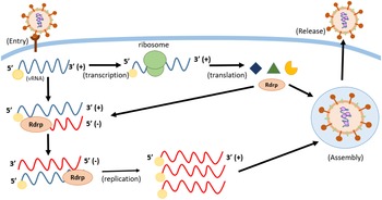
The functional role of the RdRp enzyme in a virus lies within its transcription and replication activity. RdRp or RNA replicase catalyses the replication of viral RNA from the original RNA template. During replication of SARS-CoV-2, the RdRp will synthesise complementary negative-sense RNA copies from the positive-strand template. The negative strand then serves as the new template for the replications of positive-sense RNA genomes to facilitate virus replication. Notably, there have been reports on RdRp activities in the hTERT, an RNA-dependent polymerase counterpart that reverse transcribes the telomere repeat, lengthening telomeres in DNA strands, thereby allowing replication of both embryonic stem cells and adult stem cells via protection from enzymatic end-degradation, maintaining the chromosomal and genomic stability (Refs Reference Maida, Yasukawa and Masutomi22, Reference Machitani23). However, dysregulation or overexpression of hTERT is a prominent trait in cancer cells, a response from the lengthening of telomeres that causes a cell to become immortal (Refs Reference Maida, Yasukawa and Masutomi22, Reference Machitani23). In light of the aforementioned, it is essential to elucidate the similarities and significance of viral RdRp and hTERT in response to repositioning available anticancer drugs for COVID-19 (Fig. 1).
The life cycle of positive-sense single-stranded RNA (+ssRNA) virus. The positive-sense viral RNA genome can directly translate to viral proteins, such as the viral RdRp protein. Conversely, the +ssRNA acts as a template for synthesising complementary negative-sense RNA that subsequently synthesises many new +ssRNA copies for replication. The process mentioned above requires viral RdRp to replicate the viral genome.

The viral RdRp in SARS-CoV-2
The virus genome of many RNA viruses is single-stranded, such as influenza A virus (IAV), flaviviruses, SARS-CoV, MERS-CoV and the current ongoing SARS-CoV-2. Regardless of positive (i.e., SARS-CoV-2) or negative sense (i.e., IAV) RNA viruses, the viral RdRp is essential for viral transcription and replication. For instance, the +ssRNA genome in SARS-CoV can function as an mRNA for direct protein translation, such as the RdRp protein or serve as a template for the production of the negative-strand via RdRp (Ref. Reference Machitani23). The SARS-CoV-2 RdRp structure comprises viral nsp12, nsp7 and nsp8, akin to the aforementioned SARS-CoV. The nsp12 catalytic subunit contains an N-terminal nidovirus RdRp-associated nucleotidyltransferase domain, an interface domain followed by a C-terminal RdRp domain that entails a right-handed domain comprising the fingers, palm and thumb subdomains (Ref. Reference Hillen24). On the other hand, the accessory co-factors, nsp7 and nsp8 are present to further stabilise the conformation of nsp12 and increase the RdRp template binding and processivity (Ref. Reference Gao25). The overall structure of the RdRp complex goes by nsp8 pair binding to nsp12 (nsp8-1) and nsp7 (nsp8-2), forming an nsp12-nsp7-nsp8 complex (Ref. Reference Gao25). The general replication process by the RdRp initiates nucleotide triphosphate (NTP) binding, following which a conformational change in the active site happens, phosphatidyl transfer and subsequent formation of the phosphodiester bond with the existing nucleotide chain with the aid of Mg2+ ions followed by translocation of the newly bound NTP and chain elongation (Ref. Reference Wu and Gong26) (Fig. 2).
The general domain structure of SARS-CoV-2 RdRp comprised the nidovirus RdRP-associated nucleotidyltransferase (NiRAN), interface and RdRP domains. The NiRAN domain entails three conserved sequence motifs: AN, BN and CN (within residues 51–249) connected to the interface domain (residues 250–365). Within the RdRp domain, it is composed of three right-handed structures, namely finger (residues 366–581 and 621–679), palm (residues 582–620 and 680–815) and the thumb (residues 816–920) subdomains with polymerase motifs A–G spanning across the RdRp domain.

The hTERT in mammalian cells
Telomere, a repetitive sequence of non-coding DNA situated at the chromosome ends, functions to protect the chromosomes from damage (Ref. Reference Maida and Masutomi27). Telomeres shortened progressively in cells through rounds of cell division, leading to cell senescence. The regulation of telomeres is maintained by the telomerase reverse transcriptase enzyme, a close counterpart to RdRp (Ref. Reference Gilson and Ségal-Bendirdjian28). The catalytic component of the telomerase is denoted as the hTERT or telomerase reverse transcriptase (TERT). The structure of hTERT comprises a telomerase essential N-terminal domain, the telomerase RNA-binding domain, the reverse transcriptase catalytic (RT) domain and the C-terminal extension (CTE) domain. Notably, the RT and CTE domain arrangement has the right-handed structure comprising the fingers, palm and thumb subdomains, akin to the aforementioned viral RdRp (Ref. Reference Wyatt, West and Beattie29). It has been postulated that hTERT and RNA component of mitochondrial RNA processing endoribonuclease together generate double-stranded RNAs that can be further processed into small interfering RNA in a Dicer-dependent manner, a feature of RdRp (Ref. Reference Maida30). Apart from this, the upregulation of hTERT during mitotic cell division of germline cells and cancer cells further implores the RdRp activity in hTERT (Ref. Reference Roake and Artandi31). The ribonucleoprotein complex responsible for telomere lengthening entails the telomerase-encoding hTERT and a functional RNA, namely the human telomerase RNA component (Ref. Reference Jafri32). The human telomeric DNA contains a variable number of G-rich, non-coding tandem repeats of double-stranded DNA sequence, 5′-TTAGGG-3′, followed by a terminal 3′ G-rich single-stranded overhang (Ref. Reference Somanathan and Baysdorfer33). The synthesis of telomeres begins with the appropriate positioning of primer at the 3′ end, followed by the availability of deoxyribose nucleotide triphosphate (dNTP) and template at the active site of the telomerase RNA. Here, the stem IV distal loop is associated with TERT for the stimulation of nucleotide addition to the template towards the 5′-end. Finally, the stem III pseudoknot undergoes conformational changes to dissociate the newly formed strand from the template, freeing the active site from successive binding (Ref. Reference Lai, Miller and Collins34). Akin to the aforementioned viral RdRp, the hTERT uses Mg2+ ions required for the catalysis of dNTP addition at the active site (Ref. Reference Lai, Miller and Collins34). In light of the similar structure and function of hTERT and RdRp (summarised in Table 1), it is essential to study currently approved cancer inhibitors for repositioning as antiviral drugs against the RdRp of SARS-CoV-2 (Fig. 3).
The general domain structure of hTERT comprises TEN (telomerase essential N-terminal domain), telomerase RBD (RNA-binding domain), central RT (reverse transcriptase) and CTE (C-terminal extension) domains. Akin to the SARS-CoV-2 RdRp, the hTERT consists of a right-handed structure, namely finger, palm and thumb subdomains.

The functional properties that confer significant similarities in role between human telomerase reverse transcriptase (hTERT) and viral RNA-dependent RNA-polymerase (RdRp)

The potential of anticancer drugs for the inhibition of viral RdRp
The participation of hTERT in germline cells and somatic stem cells to prevent concurrent telomere shortening in pluripotent stem cells upon successive rounds of cell division is well documented (Ref. Reference Takahashi and Yamanaka35). However, the transcriptional regulation by hTERT can contribute towards telomerase activation to cause cancer (Ref. Reference Kyo36). Several factors have been postulated to the cancer occurrence, by means of direct or indirect activation of hTERT promoter from cellular transcriptional activators, such as c-Myc, Sp1, HIF-1, AP2, or repressors, such as p53, WT1 and Menin (Ref. Reference Rafnar37). These studies have revealed that upregulation of hTERT from single-nucleotide polymorphisms (SNP) in the hTERT gene and mutation in the hTERT promoter occurs in some tumours, such as melanoma, malignant glioma, hepatocellular carcinoma and urothelial carcinoma (Ref. Reference Rafnar37). In addition, a study has shown that the interference of RdRp activity in hTERT leads to inhibitory effects on cancer cell growth, paving ways for the RdRp inhibitors in hTERT for anticancer regimen (Ref. Reference Yasukawa38). Given that the aforementioned viral RdRp closely resembles the structure and RdRp activity in hTERT, it brings massive potential to the current anticancer drugs or hTERT inhibitors to inhibit RdRp in SARS-CoV-2 thereby exterminating viral infection (Fig. 4).
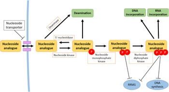
An overview of the mechanism of action of nucleoside analogues. The cellular uptake of nucleoside analogues is carried out via an active process followed by phosphorylation step by nucleoside kinases (nucleoside kinase, nucleoside monophosphate kinase and nucleoside diphosphate kinase). The production of nucleoside analogue phosphates can inhibit nucleic acid synthesis by inhibiting essential DNA/RNA polymerases. Nucleoside analogue phosphates also inhibit ribonucleotide reductase M1 (RRM1), and DNA synthesis, terminating the growing chain in cancer.

Nucleoside or nucleotide analogues are widely used to treat viral diseases and cancers due to the foundation of RdRp and hTERT activity requiring new nucleoside and/or nucleotide incorporation. The transport of nucleoside and nucleotide analogues into cells involves nucleoside transporters (Ref. Reference Cano-Soldado and Pastor-Anglada39). These nucleoside and nucleotide analogues get phosphorylated by multiple nucleoside kinases. The cellular uptake of these nucleoside analogues begins with the phosphorylation of nucleoside kinase, followed by the nucleoside monophosphate kinase and nucleoside diphosphate kinase (Ref. Reference Jordheim40). This cascade of reactions leads to the accumulation of tri-phosphorylated nucleoside, inhibiting essential RdRp/hTERT polymerase enzymes for DNA/RNA synthesis in viruses and cancer. Moreover, the accumulation of both di- and tri-phosphorylated nucleosides has been shown to inhibit the key enzyme ribonucleotide reductase M1, an enzyme that is essential for the production of deoxyribonucleotides for DNA synthesis via conversion of ribonucleoside diphosphates to deoxyribonucleoside diphosphates (Ref. Reference Ahmad and Dealwis41). Apart from the aforementioned mode of action, the nucleoside and nucleotide analogues can induce chain termination in the growing viral DNA or RNA chain (Ref. Reference Tárkányi and Aradi42). This is contributed by the absence of a 3′ hydroxyl group in the analogues, preventing the formation of the 3′-5′ phosphodiester bonds between the analogue and the new 5′ nucleoside triphosphates (NTPs), thereby terminating the growing chain during the RNA/DNA-dependent DNA synthesis in viruses and DNA polymerases in cancer (Ref. Reference Richman43).
As aforementioned, the structural and functional resemblance in viral RdRp and hTERT could bring new insights for repositioning existing anticancer or hTERT inhibitors for viral RdRp, especially in urgent times like the current ongoing SARS-CoV-2 global pandemic. On top of that, further studies on inhibitors against the RdRp activity for hTERT might bring serendipitous encounters on the therapeutic effects on both RNA viruses and cancers, paving the way for more uses in both viral RdRp and hTERT inhibitors. With that being said, the antiviral nucleoside and nucleotide analogues have shown better tolerance profiles in mammalian cells compared to the anticancer nucleoside analogues, opening possibilities for a better anticancer drug with an optimal pharmacological safety profile (Ref. Reference Jordheim40).
The potential of anti-cancer drugs to inhibit RdRp in SARS-CoV-2
As described earlier, nucleoside or nucleotide analogues are widely used as antiviral and anticancer agents. For instance, a study has shown that 5-fluoro-2′-deoxyuridine triphosphate or Floxuridine, a nucleoside analogue, can effectively get substituted into the telomere DNA sequence, hTERT, and inhibit the binding of essential telomere end-binding complexes, activating ataxia-telangiectasia mutated and Rad3-related DNA damage response that leads to cancer cell death in hepatic metastases (Ref. Reference Zeng44). Given the close association of nucleoside analogues in viral and cancer polymerases, this drug is now being considered as a potential antiviral candidate against SARS-CoV-2 (Refs Reference Wu45, Reference Tomar, Krugliak and Arkin46).
While many inhibitory activities for virus and cancer revolve around nucleoside analogues, an interesting study illustrated that the VX-222, a non-nucleoside inhibitor (NNI) developed to inhibit the HCV RdRp initially, now sees a possible role in the inhibition of RdRp activity in human hTERT (Refs Reference Machitani23, Reference Yi47, Reference Di Bisceglie48). VX-222 is a polymerase inhibitor that binds to the thumb domain of NS5B in HCV RdRp (Ref. Reference Yi47). With the similar right-handed structure in hTERT, the VX-222 could potentially be used as an anticancer drug upon further assessments and clinical studies (Ref. Reference Yi47). With limited studies on hTERT inhibitors for viral RdRp and vice versa, it is not surprising that dual-mode drugs (anti-hTERT and antiviral RdRp) have yet to be discovered and studied in detail, especially in the context of drug repositioning.
To recall, hTERT, a classical enzyme found only in the cancer cells that reverse telomere shortening in cells that would usually bring cells to senescence, limiting the cycle of one cell can replicate. RdRp is an enzyme produced only by the virus to enhance viral replication machinery upon cell infection. Given the similarity and specificity stated above, the potential for anticancer inhibitors for SARS-CoV-2 RdRp and antiviral inhibitors for hTERT should not be overlooked. Apart from the inhibitor's original mode of action, it might suggest an anti-hTERT or antiviral RdRp effect in future. Discussed below are examples of potential anti-cancer drugs with RdRp inhibitory activity against the SARS-CoV-2 (Fig. 5).
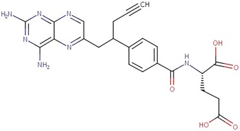
The chemical structure of Pralatrexate (Source: https://www.ncbi.nlm.nih.gov/books/NBK547901/table/Prelatrexate.T1/?report=objectonly).

Pralatrexate
Pralatrexate is an FDA-approved folate analogue inhibitor and antineoplastic chemotherapy drug, marketed as ‘Folotyn’ for refractory peripheral T-cell lymphoma. Folotyn is postulated to show antiviral activity against SARS-CoV-2 (Refs Reference Hong49, 50). The well-studied activity of Pralatrexate against T-cell lymphoma lies within its high affinity for reduced folate carrier-1 (RFC-1) and competitive inhibition of dihydrofolate reductase (DHFR), a crucial enzyme that produces co-factors that are necessary for DNA synthesis in cancer cells. Pralatrexate selectively enters RFC-1 expressing cancer cells, and competes for the folate binding site of DHFR, subsequently hindering tetrahydrofolate synthesis, causing depletion of nucleotide precursors, thereby terminating cell growth (Ref. Reference Serova51).
Among the 1906 approved drugs, pralatrexate has demonstrated a strong association with SARS-CoV-2 RdRp. Pralatrexate can interact with 16 amino acid residues in RdRp, forming a stable complex via polar and charge interactions. In particular, Pralatrexate forms hydrogen bonds with ARG569, ASN496, ASN497, LYS500, GLN573 and GLY590 residues; Pi-Alkyl or Alkyl interaction with LEU576 and LYS577, and salt bridges with ARG569 and LYS500 (Ref. Reference Zhang52). Interestingly, the RdRp shares many similar physio-chemical features with the DHFR. The high number of charged and polar residues may explain why Pralatrexate possesses this antiviral activity (Ref. Reference Zhang52). In addition, it has also been postulated that Pralatrexate binds to the RdRp cavity via non-covalent binding, hindering the interaction between RNA primer and the RdRp cavity, thus halting viral replication (Ref. Reference Bae53). The result was profound at 24 h SARS-CoV-2 post-infection in Vero cells upon Pralatrexate administration. Notably, the inhibitory effects are much lesser at 48 h post-infection in Calu-3 cells, thereby suggesting Pralatrexate administration only for early phase infection (Ref. Reference Bae53). Also, Pralatrexate has a low EC50 value of 0.008 μM post-infection, outperforming the inhibitory activity of Remdesivir (EC50 value of 8.777 μM) and various other tested drugs (Ref. Reference Zhang52) (Fig. 6).
The chemical structure of Corilagin (Source: https://comptox.epa.gov/dashboard/chemical/details/DTXSID90865084).

Corilagin
Aside from synthetic compounds, natural derivatives are also gaining traction in the research community, such as plant extracts. Corilagin, an ellagitannin, is an active component found in a variety of ethnopharmacological plants (e.g., Phyllanthus niruri L. and P. urinaria L. and P. amarus) which were first discovered to suppress the reverse transcriptase activity of avian myeloblastosis virus (AMV), an RNA tumour virus (Refs Reference Li54, Reference Kakiuchi55). The effects of inhibitory activity of ellagitannin on the reverse transcriptase in AMV were measured using polyadenylic acid-oligothymidylic acid as a template-primer, in which, Corilagin showed a comparable inhibitory activity at a concentration of 10−5 M via the incorporation of deoxythymidine monophosphate (Ref. Reference Kakiuchi55). Apart from that, Corilagin has also been reported with antimicrobial, antioxidant, anti-inflammatory, antitumor and antiviral activities. Over the years, Corilagin was reported to attenuate the growth of ovarian cancer through various signalling pathways, namely the AKT/ERK and TGF-β signalling pathways. In addition, Corilagin increases the sensitivity of ovarian cancer towards chemotherapy thus improving therapeutic efficacies (Ref. Reference Jia56). Besides that, Corilagin also shows positive outcomes in hepatocellular carcinoma and breast cancer via induction of G2/M phase arrest and reactive oxygen species-dependent apoptosis, respectively (Refs Reference Zheng57, Reference Tong58). In view of the above, Corilagin has been suggested for its antiviral properties in addition to the solid potential anticancer drug.
The Corilagin extracted from P. amarus has been shown to inhibit HCV RdRp. At 20 μM, Corilagin significantly inhibits the NS5B RdRp activity by interacting with the amino acid residues (β-hairpin) in NS5B RdRp (Ref. Reference Reddy59). This leads to the possibility that Corilagin may also possess antiviral activity against SARS-CoV-2. In an in-silico docking analysis, Corilagin showed a high binding affinity at −9.30 kcal/mol towards the active site residues (ASN705, GLN724, HIS133, LEU207 and TYR129) of RdRp in SARS-CoV-2 via hydrogen bonding (Ref. Reference Hiremath60). A study conducted by Li et al. further demonstrated the strong binding affinity of Corilagin for RdRp in SARS-CoV-2 as an NNI (Ref. Reference Li61). The study agreed with the strong binding affinity of Corilagin to the palm subdomain of RdRp, where G616 to Y619 (motif A), D761 to V763 (motif C), K798 (motif D), E811 to S814 (motif E) and I548 to K551 (motif F) are located (Ref. Reference Li61). This suggests that Corilagin may suppress SARS-CoV-2 replication by preventing conformational changes and nucleotide incorporation by RdRp. The interaction between Corilagin and residues in motif F may block NTP entry to the active site of RdRp, effectively inhibiting viral RNA synthesis.
Distinct from other RdRp inhibitors, Corilagin can circumvent the exoribonuclease (ExoN) proofreading (Nsp10/14) in SARS-CoV-2. Of note, the ExoNs or also known as the exonuclease ribonucleases are enzymes that are responsible for the RNA degradation via removal of nucleotides from the 5’ end or the 3’ end of the RNA structure, a critical feature for the synthesis of multiple RNAs from the RNA template in RNA viruses (Ref. Reference Minskaia62). Recently, the bifunctional role of SARS-CoV-2 nsp14 has been further unravelled, in which, the N-terminus ExoN domain has been implicated in proofreading role by removing misincorporated nucleotides from the 3’ end of the nascent RNA strand and the C-terminus N7-methyltransferase domain methylates the guanine at the N7 position, forming a cap-0 structure (m7GpppA…), a feature that aids for host immune escape (Refs Reference Ogando63–Reference Viswanathan65).
Given its nature of being an NNI, Corilagin exerts antiviral activity by preventing conformational changes in the SARS-CoV-2 RdRp which are vital for RNA transcription, thereby giving a more pronounced inhibitory effect as compared to the nucleoside analogue inhibitors (NI) that can develop resistance in the virus over time since it requires two phosphorylation steps conferred by the viral and host enzymes (Ref. Reference Li61). Owing to its different mechanism, the Corilagin is much more resistant towards the SARS-CoV-2 proofreading activity and much more suited as an antiviral agent for SARS-CoV-2 as compared to other NI drugs, such as Remdesivir (Ref. Reference Li61). Over and above that, the usage of Corilagin warrants an enhanced inhibitory effect at very low EC50 values at 0.13 μmol/l, which is comparable to the Remdesivir which scored 0.06 μmol/l (Ref. Reference Li61). Accompanied with an appealing docking score (−9.30 kcal/mol) in comparison with Remdesivir at –7.6 kcal/mol, Corilagin holds a vast potential for drug repositioning against the SARS-CoV-2 (Ref. Reference Hiremath60).
Lycorine
Akin to the Corilagin, Lycorine is a benzyl phenethylamine alkaloid found in the well-studied medicinal plant, from Amaryllidaceae species – Lycoris radiate, Leucojum aestivum and Hymenocallis littoralis. Owing to its divergent chemical structures, which reflects on the strength of biological properties, Lycorine and its derivatives are rapidly drawing the interest of many. The pharmacological properties of Lycorine have since been discovered, including anti-inflammatory, antimicrobial, antiparasitic, antitumor and antiviral (Ref. Reference Roy66). At 0.5–5 μM, Lycorine was reported to inhibit ZIKV effectively via the direct inhibition of RdRp activity, successfully attenuating viral replication. It was then discovered that Lycorine binds to the finger domain of RdRp preferentially (Ref. Reference Chen67). Notably, Lycorine extracted from L. radiata has demonstrated the ability to inhibit SARS-CoV in the past (Ref. Reference Li68). Also, Lycorine has been reported to suppress other coronaviruses such as the MERS-CoV, HCoV-OC43 and HCoV-NL63 in in-vitro and in-vivo studies (Ref. Reference Shen69) (Fig. 7).
The chemical structure of Lycorine (Source: https://www.biocrick.com/Lycorine-BCN2409.html).

Although the mechanism of action behind Lycorine against the aforementioned coronaviruses is yet to be determined, the close genomic resemblance of SARS-CoV and SARS-CoV-2 makes Lycorine a strong candidate to inhibit RdRp activity in SARS-CoV-2 (Refs Reference Ren70, Reference Zhang71). For instance, Jin et al. has shown that Lycorine acts as a non-nucleoside analogue, successfully inhibiting all three coronaviruses with more pronounced effects against SARS-CoV and SARS-CoV-2, as compared to MERS-CoV. Akin to Remdesivir, Lycorine binds to the same catalytic active site and interacts with similar amino acid residues (Asp623, Asn691 and Ser759) in SARS-CoV-2 RdRp, thereby suggesting RdRp inhibitory activity, subsequently causing RNA chain termination and attenuation in viral replication (Ref. Reference Jin72). This study also showed a greater binding affinity by Lycorine for SARS-CoV-2 RdRp compared to Remdesivir at −6.2 versus −4.7 kcal/mol (Ref. Reference Jin72).
Apart from its antiviral potential, Lycorine is also gaining attention for its anti-cancer properties. For instance, Lycorine can suppress the growth, migration and invasion of breast cancer cells by inducing apoptosis via the blocking of the sarcoma/focal adhesion kinase pathway. Such anticancer activity was also reported in in-vivo studies where breast tumour metastasis was inhibited (Ref. Reference Li73). Interestingly, Lycorine has much lower toxicity than Paclitaxel, a first-line chemotherapy drug, making it a much better candidate (Ref. Reference Ying74). Lycorine also plays a role in inhibiting gastric cancer by downregulating the protein stability of myeloid cell leukaemia-1, a member of an anti-apoptotic BCL-2 family protein that confers drug resistance in tumours and renders gastric cancer cells to apoptosis (Ref. Reference Li73).
Conclusion
As viral RdRp has high conserved motifs across the RNA virus species, especially the coronavirus family, it is essential to study the molecular structure and potential RdRp inhibitors as an antiviral strategy. Notably, the hTERT also shows RdRp activity against high tumourigenicity. Given that, the viral RdRp and hTERT in cancers, both share right-handed structure characteristics with the typical finger, palm and thumb domains, and this makes them important candidates to be evaluated and considered for anticancer as well as antiviral drugs. While cancer progression is different from viruses, they share a common characteristic: utilising RdRp activity for survival, paving the way for more uses in both viral RdRp and anti-cancer inhibitors. For instance, Floxuridine, an hTERT inhibitor, is now being analysed as a potential antiviral candidate against SARS-CoV-2. Also, VX-222, a NNI developed for HCV RdRp initially, now sees a possible role in inhibition of RdRp activity in human hTERT. With limited studies on hTERT inhibitors for viral RdRp and vice versa, it is not surprising that dual-mode drugs (anti-hTERT and antiviral RdRp) have yet to be discovered and studied in detail, especially in the context of drug repositioning. Therefore, the study of potential anticancer inhibitors for SARS-CoV-2 RdRp remains of great interest. Drug inhibitors that act upon RdRp might give a positive outcome for anticancer, and vice versa. As such, Pralatrexate, Corilagin and Lycorine have shown SARS-CoV-2 RdRp inhibitory activity and some anti-cancer properties, this makes the aforementioned drugs potential for anticancer studies which could bring more uses to the drugs. In the conquest for antiviral drug hunting, especially in urgent times of need like the SARS-CoV-2 pandemic, it is vital to study the potential anticancer drug portfolio for possible drug repositioning of antiviral RdRp.
Author contributions
ZYL and AJWY contributed equally to the preparation and drafting of the manuscript. Critical revisions of the article were done by SKL.
Conflict of interest
None.