Introduction
Pathogens of oral cavity play an important role in the development and progression of periodontitis destroying the periodontal tissues. Reference Di Benedetto, Gigante, Colucci and Grano1 Periodontitis is characterized by gingival inflammation, irreversible alveolar bone loss, and loosening and/or loss of teeth. Reference Yucel-Lindberg and Bage2–Reference Page, Eke, Jin, Liu and Lin4 The immune response seems to be crucial, not only to the development of periodontitis, but also contributing to their systemic effects through high inflammatory cells activity. Reference Shah, Thomas and Mehta5–Reference Cardoso, Reis and Manzanares-Céspedes7 It is well established that periodontitis modulates several diseases, including cardiovascular diseases, diabetes, cancer, chronic respiratory diseases, and adverse effects during pregnancy. Reference González-Jaranay, Téllez, Roa-López, Gómez-Moreno and Moreu8
Clinical and experimental studies show an association between maternal periodontitis and adverse outcomes during gestation, including preterm birth, low birth weight as well as alterations in the uterine microenvironment. Reference Corbella, Taschieri, Francetti, De Siena and Del Fabbro9,Reference Srinivas, Sammel and Stamilio10 On the other hand, there were no studies evaluating the impact of periodontitis during pregnancy on the immune system of offspring.
Considering that an adverse uterine microenvironment may lead to a fetal reprogramming contributing to development of many diseases, such as asthma, Alzheimer, hypertension among others, in earlier study, our group showed the putative programming mechanisms induced by adverse intrauterine microenvironment on the immune system of offspring. Reference Ibrahim, Barioni and Heluany11–Reference Maiellaro, Correa-Costa and Vitoretti13 Indeed, we also evaluated in the last study the role of periodontitis in the uterine microenvironment. We found elevated messenger RNA (mRNA) levels of interleukin (IL)-6, IL-10, and interferon (IFN)-γ in the uterus, while reduced expression of IL-6 and IFN-γ in the placenta. Periodontitis did not modify the expression of cycloogenase (COX)-1 and 2 in the uterus, while COX-2 was increased in the placenta.
Thus, it is reasonable to admit that these changes in the uterine microenvironment could cause alterations in the genome of offspring, leading to a risk factor for the onset of inflammatory diseases, including asthma. We investigated this hypothesis evaluating the development of experimental asthma in the offspring from mothers with periodontitis.
Our study prioritized the quantification of cells migrated into the bronchoalveolar lavage (BAL), edema, tracheal responsiveness, levels of cytokines, anaphylactic antibodies, and mucus production, since these parameters are important biomarkers of asthma.
Materials and methods
Animals
Mothers
Female and male 2-month-old Wistar rats were obtained from the University Nove de Julho and maintained in a light- and temperature-controlled room (12/12-h light–dark cycle, 21 ± 2°C), with free access to food and water. The virgin females were caged overnight with male (1:1), and the vaginal smears were carried out to identify the presence of spermatozoids, and therefore, confirm the coitus. Pregnancy was confirmed by maternal weight (weekly). The total number of pregnant rats used in this study was 12 (n = 6 for each group). We also used six male rats for mating.
Pups
Thirty days after the birth (natural delivery), pups’ males and females of each mother, generated by different matrices, were mixed and kept in litters of 10 animals for group. These procedures were adopted in order to eliminate the formation of groups containing only puppies’ brothers and a single mother, excluding the possibility of the results being influenced by individual susceptibility. Indeed, we mixed the results obtained from both sex, because we observed no differences between males and females responses. It is important to mention that only one male pup and one female pup per litter were used at any time point.
The offspring was assigned into four groups:
-
(1) B/B group, identified as basal offspring from basal mothers.
-
(2) B/OVA group, identified as asthmatic offspring from basal mothers.
-
(3) P/B group, identified as basal offspring from mothers with periodontitis.
-
(4) P/OVA group, identified as asthmatic offspring from mothers with periodontitis.
Periodontitis induction in the mothers
Considering that experimental periodontitis in animals is developed within 7–15 days, we induced periodontitis in the female rats 10 days before mating. Thus, we guarantee that the rats would be with periodontitis during all pregnancy, mainly in the first gestational trimester that is the most critical.
Female rats were anesthetized by intraperitoneal route with ketamine (100 mg/kg) and xylazine (10 mg/kg) for the induction of periodontitis according to Candeo et al. Reference Candeo, Rigonato-Oliveira and Brito14 . Briefly, we gently introduced silk ligature into the interproximal area between the first mandibular molar and the second mandibular molar in order to avoid damaging the periodontal area around the first molar. As control group, we used pregnant rats not submitted to periodontitis (basal group).
Experimental asthma in the offspring
Male and female rats (30 days) were sensitized with 10 μg of ovalbumin (OVA) + 10 mg of alumen by subcutaneous route. One week later, the rats received another injection with OVA-alum by same route (booster). At days 12–14, the rats were submitted to an inhaled antigen challenge (aerosolized 1% OVA in phosphate-buffered saline (PBS), 15 min) in order to induce the local inflammatory response that is characteristic of asthma. We used an ultrasonic nebulizer device (Icel®, São Paulo, Brazil) coupled to a plastic inhalation chamber (20 l). The analyses were performed 24 h after the last OVA challenge.
Quantification of leukocytes in the BAL
In order to determine the number of cells recruited into the BAL, the tracheae of mice were cannulated, and the alveolar space was flushed twice with PBS (1.5 ml total volume). The collected BAL fluid was centrifuged (1500 rpm for 15 min at 20°C), and the resulting cell pellet was resuspended in 1 ml of PBS. The cell suspension was stained with crystal violet, and the total cell number was determined microscopically using a Neubauer chamber.
Edema and tracheal responsiveness analysis
Edema was determined by wet:dry weight ratio, while the tracheal responsiveness was quantified by the isometric force. For this, tracheal rings were mounted in a 15-ml organ bath by means of two steel hooks. Force contraction was recorded using a force displacement transducer and a chart recorder (Powerlab®, Labchart; AD Instruments, Dunedin, Nova Zelândia). Tracheal rings were suspended in organ bath filled with continuously aerated (95% O2 and 5% CO2) Krebs–Henseleit solution at 37°C. After 40 min, the tracheal tension was adjusted to 1 g and was added methacholine (MCh, 10−3 M) in order to obtain maximum contractile response.
Quantification of cytokines in the bronchoalveolar fluid
Levels of cytokines (IL-6, IL-4, IL-1β, and IL-10) were determined in the BAL fluid by ELISA, according to manufacturer’s instructions (BD® Biosciences, New Jersey, NJ, USA). The results were expressed as picograms of cytokine produced per milliliter.
Evaluation of gene expression of cytokines
Lung samples were snap-frozen in liquid nitrogen. The total RNA wasted using Trizol Reagent (Invitrogen, Carlsbad, CA, USA) according to the protocol provided by Invitrogen. RNA concentrations were determined by spectrophotometer readings at absorbance 260 nm. First-strand complementary DNAs were synthesized using the Moloney Murine Leukemia Virus reverse transcriptase (Promega, Madison, WI, USA). Reverse transcription polymerase chain reaction was performed using the TaqMan real-time PCR assay (Applied Biosystem, Foster City, CA, USA) for the following molecules: IL-6 (Rn00561420_m1), IL-10 (Rn00563409_m1), IL-4 (Rn01456865_m1), IL-1β (Rn00580432_m1), and hipoxantina-guanina fosforibosiltransferase (HPRT) (Rn01527838_g1).
Cycling conditions were as follows: 10 min at 95°C followed by 45 cycles at 20 s each at 95°C, 20 s at 58°C, and 20 s at 72°C. Analysis was performed using Sequence Detection Software 1.9 (SDS), and mRNA expression was normalized to HPRT expression.
Evaluation of mucus production and cell infiltrate by histology
After anesthesia, lung fragments were removed and fixed in 10% formalin solution for 24 h. After that, the fragments were dehydrated in alcohol followed by diaphanization by xylene. Therefore, the fragments were embedded in paraffin, deparaffinized, and sectioned at 5 μm (microtome HYRAX M60; Zeiss, Germany). After this period, the lungs were submitted to histological analysis. For this, the slides were stained with hematoxylin and eosin for cell infiltrate quantification and with periodic acid-reactive Schiff (PAS) for mucus quantification. The histomorphometric technique was used and all data were analyzed through the Software Axiovision v.3.1 (Zeiss, Germany).
Passive cutaneous anaphylaxis
Passive cutaneous anaphylaxis reaction is an assay used to indirectly measure anaphylactic antibodies in rats. Reference Mota and Wong15 The Immunoglobulin E (IgE)-dependent skin reaction was generated by sensitizing the skin of unmanaged rats with an intradermal injection of sera from serially diluted sensitized rats (1:2 to 1:256). After 24 h of cutaneous administration of the seriously diluted serum, recipient rats received intravenously a solution containing 500 mg OVA + 2.5 mg Evans blue dye dissolved in NaCl (0.9%). Thirty minutes later, the rats were euthanized, the skin was removed, and the dye diameter was determined by the size of the spot caused by the dye measured on the inner surface of the skin. Anaphylaxis titers were represented by the highest serum dilution resulting in a stain greater than 5 mm in diameter.
Statistical analysis
Data were expressed as the means ± SEM, and comparisons among the experimental groups were analyzed by two-way ANOVA followed by the Student’s Newman–Keuls test for multiple comparisons using the GraphPad prism software V.5, San Diego, CA, USA. P values less than 0.05 were considered statistically significant.
Results
Periodontitis during pregnancy did not alter the number of cells recovered in the BAL of offspring
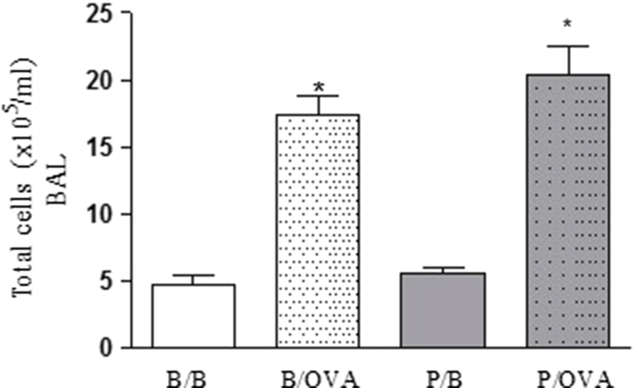
Fig. 1(a) showed increased number of cells recruited into the BAL in both B/OVA and P/OVA groups when compared to respective control groups (B/B and P/B groups). No differences were found between B/B and P/B groups.
Effects of maternal periodontitis in the cell influx into the bronchoalveolar lavage (BAL) of asthmatic pups. Groups of pregnant rats were submitted or not to periodontitis. Thirty days after the birth, the animals were sensitized and challenged with OVA. After 24 h of the last challenge, the number of cells recruited into the BAL was evaluated. Data represent the mean ± SEM of 10 pups.

Periodontitis during pregnancy reduced cellular infiltration in the parenchyma of offspring when submitted to experimental asthma
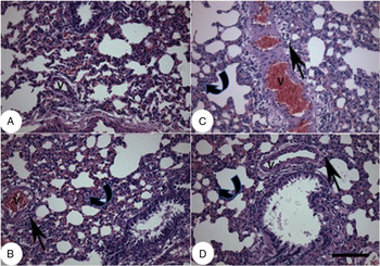
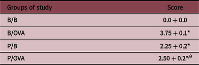
Fig. 2 and Table 1 represent the histopathological analysis. In panel (a), we can observe that B/B group showed lung under normal conditions presenting intact bronchial, vascular (V), and alveolar architecture. Panel (b) represents B/OVA group with parenchymal (curved arrow) and perivascular (V) leukocyte infiltrate (arrow).
Effects of maternal periodontitis in the cell influx into the parenchyma of asthmatic pups. Groups of pregnant rats were submitted or not to periodontitis. Thirty days after the birth, the animals were sensitized and challenged with OVA. After 24 h of the last challenge, the histological analysis was evaluated. Panels (a) and (b) represent B/B and B/OVA groups; panels (c) and (d) represent P/B and P/OVA groups.

Evaluation of inflammatory cells recruited in the parenchyma

Score: 0 = absence of perivascular and parenchymal infiltrated; 1 = presence of some leukocytes infiltrated in the perivascular area; 3 = presence of many leukocytes infiltrated in the perivascular area; 4 = presence of many leukocytes infiltrated in the perivascular and parenchymal areas.
* P < 0.05 in relation B/B group.
θ P < 0.05 in relation B/OVA group.
In panel (c), we can observe that P/B group had intense perivascular (V) leukocyte infiltrate (arrow) but reduced parenchymal infiltration (curved arrow). On the other hand, in panel (d), a significant reduction of leukocyte infiltration (arrow and curved arrow) in the P/OVA group was found. Staining: hematoxylin and eosin. Bar: 100 µm.
Periodontitis during pregnancy reduced maximum contractile response to MCh of offspring when submitted to experimental asthma
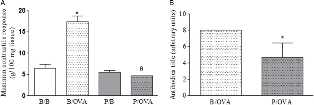
Fig. 3(a) showed increased tracheal responsiveness to MCh in B/OVA group when compared to B/B group. On the other hand, tracheal reactivity of P/OVA group was reduced when compared to B/OVA group.
Effects of maternal periodontitis in the tracheal responsiveness as well as in the antibodies IgE titles of asthmatic pups. Groups of pregnant rats were submitted or not to periodontitis. Thirty days after the birth, the animals were sensitized and challenged with OVA. After 24 h of the last challenge, the tracheal responsiveness and antibodies anti-OVA were quantified. Data represent the mean ± SEM of 10 pups.

In panel (b), we can observe that periodontitis reduced the production of anti-OVA antibody titers.
Periodontitis during pregnancy reduced lung edema of offspring when submitted to experimental asthma
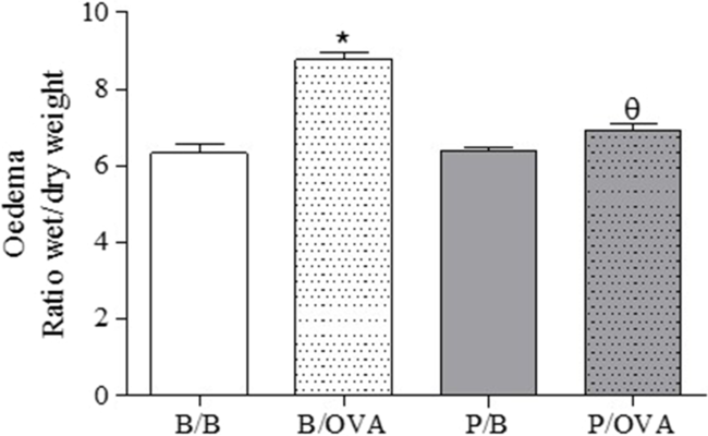
Fig. 4 showed increased lung edema in B/OVA group when compared to B/B group. Moreover, lung edema of P/OVA group was reduced when compared to B/OVA group. No differences were observed between B/B and P/B groups.
Effects of maternal periodontitis in the lung edema of asthmatic pups. Groups of pregnant rats were submitted or not to periodontitis. Thirty days after the birth, the animals were sensitized and challenged with OVA. After 24 h of the last challenge, the lung edema was evaluated. Data represent the mean ± SEM of 10 pups.

Periodontitis during pregnancy did not alter the mucus production in the lung of offspring when submitted to experimental asthma
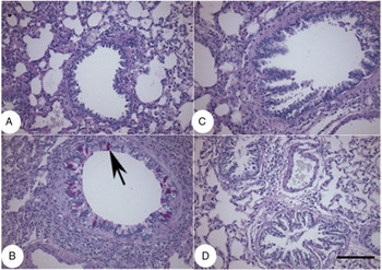
Fig. 5 showed that B/B group presented lung under normal conditions without mucus staining (panel (a)). In panel (b), we can observe that B/OVA group presented excess of mucus production in the lumen and in the goblet cells (arrow). We can also observe that P/B and P/OVA groups (panels (c) and (d)) had no mucus production. Coloring: PAS. Bar: 100 µm.
Effects of maternal periodontitis in the mucus production of asthmatic pups. Groups of pregnant rats were submitted or not to periodontitis. Thirty days after the birth, the animals were sensitized and challenged with OVA. After 24 h of the last challenge, the histological analysis was evaluated. Panels A and B represent B/B and B/OVA groups; panels C and D represent P/B and P/OVA groups.

Periodontitis during pregnancy did not alter the cytokines production in the BAL fluid of offspring when submitted to experimental asthma
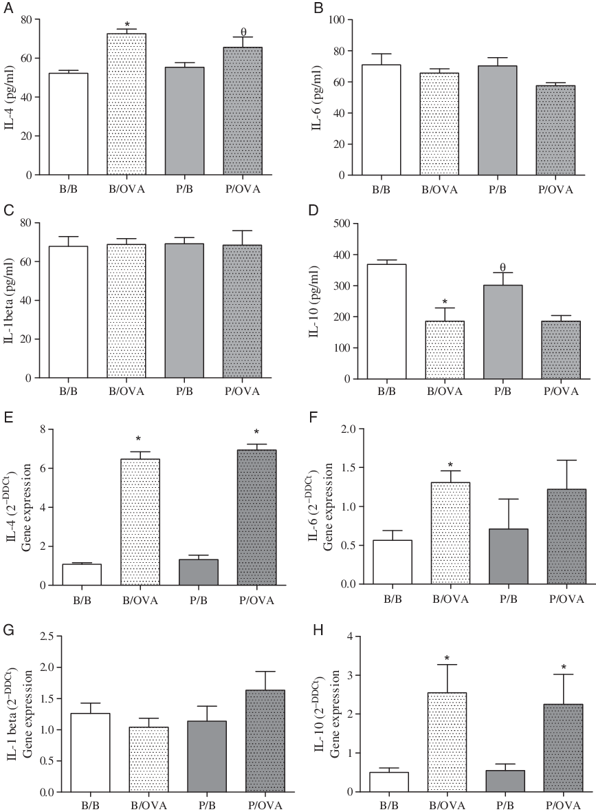
Fig. 6(a) showed that experimental asthma increased the levels of IL-4 in the BAL fluid in all groups of study (B/OVA and P/OVA) when compared to control groups non-asthmatic (B/B and P/B groups). In panels (c) and (e), no differences were observed in the levels of IL-6 and IL-1β among groups of study.
Effects of maternal periodontitis in the level of cytokines as well as the gene expression of asthmatic pups. Groups of pregnant rats were submitted or not to periodontitis. Thirty days after the birth, the animals were sensitized and challenged with OVA. After 24 h of the last challenge, the cytokines were quantified. Data represent the mean ± SEM of 10 pups.

As can be seen in panel (g), asthma reduced the level of IL-10 in all groups when submitted to experimental asthma (B/OVA and P/OVA groups). Panel (b) showed that periodontitis did not alter IL-4 expression in offspring’s lung. We also observed that OVA increased the expression of this cytokine in relation to their respective control groups (B/B and P/B groups)
In panel (d), we observed that only B/OVA group showed elevated gene expression of IL-6 in relation to respective control group (B/B). We did not observe differences between the other groups of study. Similarly, panel (f) showed that there were no differences in IL-1β expression among groups of study. As can be seen in panel (h), OVA increased IL-10 gene expression relative to their respective control groups (B/B and P/B groups).
Discussion
Periodontitis is an infectious inflammatory lesion that surrounds the supporting tissues of the teeth, leading to loss of connective insertion, alveolar bone, and root cementum. Reference Di Benedetto, Gigante, Colucci and Grano1–Reference Warren, Watkins, Cowen, Hand, Levy and Kuthy3 Studies show the influence of periodontitis on the exacerbation and/or modulation of systemic diseases, including effects on pregnancy. Reference Cardoso, Reis and Manzanares-Céspedes7 However, systematic reviews and meta-analyses still claim that the quality of evidence is very poor. Reference Iheozor-Ejiofor, Middleton, Esposito and Glenny16,Reference Ide and Papanou17 Since this relationship between pregnancy and periodontitis is not fully understood, and considering that the impact of these effects on offspring has not been evaluated, in this study, our objective was to investigate the impact of maternal periodontitis on the offspring’s immune system.
For this, we use a well-established experimental model of periodontitis, which mimics the main characteristics of disease in humans, such as high alkaline phosphatase production, increased total bone loss and elevation of inflammatory cytokines such as IL-6, a biomarker of disease. Reference Ren and Du18,Reference Chen, Chang, Huang, Huang and Tsai19 To evaluate the impact of periodontitis on the offspring’s immune system, we induced experimental asthma in pups. The experimental asthma model using OVA, an inducing agent widely used in the literature. Reference Hwang, Jin and Choi20–Reference Siqueira, Evangelista and Dos Santos24
Some hypotheses may be raised to explain the interaction between periodontitis and pregnancy. Among them, epigenetic modifications deserve highlighting. Periodontitis by inducing systemic effects could alter the uterine microenvironment by inducing epigenetic changes, which in turn predispose the onset of young or adult diseases in the offspring, such as asthma. Reference Hannson and Gluckman25 Another hypothesis is that reactive oxygen species may be increased in utero by periodontitis-inducing systemic effects, and such species could generate an adverse intrauterine environment contributing to changes in the fetal gene and thus altering the immune response of offspring. Reference Maiellaro, Correa-Costa and Vitoretti13 Thus, we investigated the impact of maternal periodontitis on the development of asthma in pups.
Our data showed that maternal periodontitis increased the development of lung inflammatory allergic response in the offspring. This was the first indication of an interference of maternal periodontitis in the offspring’s immune system.
Sequentially, we investigated the tracheal contractile response to cholinergic stimulation, an important parameter in asthma, and the bigger responsible for hospitalizations. We observed that periodontitis caused a reduction in tracheal reactivity despite of increased lung inflammation. However, this dissociation between cellular and smooth muscle events is not surprising, since in previous studies we have shown that these events are not necessarily coordinated. Reference Hamasato, de Lima, de Oliveira, dos Santos Franco, de Lima and Palermo-Neto26 Further studies need to be driven in order to understand the mechanisms involved in tracheal hyporresponsiveness periodontitis-induced. Eedema was also reduced by periodontitis. Thus, both results show us that periodontitis modulated the immune system response of offspring. If we consider that the inflammatory response is necessary as defense mechanism, this reduction does not seem to be beneficial.
When we investigated both cytokine production and gene expression by lung, we observed that maternal periodontitis did not alter any of the cytokines evaluated (IL-4, IL-6, IL-1β, and IL-10). Thus, in future studies, we need to evaluate other mediators such as those originating from arachidonic acid metabolism in order to explain the effects of maternal periodontitis on cell recruitment, edema, and tracheal reactivity.
Since asthmatic response is dependent on the antibody production, we investigate the effect of maternal periodontitis on anti-OVA antibody. Our data showed that maternal periodontitis reduced the production of anti-OVA antibodies. Such reduction may explain the reduced cell migration, edema, and contractile response of the trachea. Indeed, at the first time, the influence of maternal periodontitis in the immune system of offspring is showed.
Finally, the histological evaluation showed that maternal periodontitis in non-asthmatic offspring induced intense perivascular leukocyte infiltrate, although it did not trigger cell migration into the alveolar space. Such effects may occur due to a modulation of adhesion molecules reducing the migration of cells from parenchyma to alveolar space. However, maternal periodontitis in asthmatic animals caused a reduction in leukocyte infiltrate, while increased number of cells were found in BAL. Again, this result may involve modulation of adhesion molecules. It will be interesting to investigate in future studies the expression of intercellular cell adhesion molecule-1, vascular cell adhesion molecule-1, and selectins. Regarding mucus production, maternal periodontitis did not cause any changes.
We can conclude that periodontitis has relevant impacts on the offspring’s immune system, blunting the response to allergic and inflammatory stimulus. This study shows the important role of oral health during pregnancy and opens possibilities for future studies in order to explain the effects of periodontitis during pregnancy in the offspring.
Funding
This study was sponsored by Fundação de Amparo à Pesquisa do Estado de São Paulo (FAPESP: 2017/0644-9) and CNPq 305099/2017-5.
Competing interests
The authors declare that there are no conflicts of interest regarding the publication of this paper.
Ethical approval and accordance
The animals were obtained from the University Nove de Julho and the experiments were approved by the Animal Care Committee University Nove de Julho (CoEP-UNINOVE, AN0006/2017). The methods used in the present study were carried out in accordance with the relevant guidelines.