Introduction
Septal deviations are commonly found during examination of the nose. Patients can be asymptomatic but often complain of nasal airway obstruction. A systematic analysis published in 2010 showed an increased risk of rhinosinusitis in patients with septal deviation; however, the clinical relevance is thought to be small.Reference Orlandi1 Septal deviation with intranasal contact points may also cause facial pain, although the evidence is conflicting.Reference Harrison and Jones2 Correcting the deviated septum can relieve nasal airway obstruction, provide access for endoscopic sinus surgery and may treat facial pain due to septal spurs.Reference Chung, Batra, Citardi and Lanza3
The first attempts at septoplasty date back to ancient Egypt, where a broken nose was treated by external fixation and plugging each nostril with grease-coated linen to stabilise the septum.Reference Fettman, Sanford and Sindwani4 Later on, obstructing spurs were removed using full-thickness resection. This technique was abandoned once the complications of septal perforations were discovered. Other techniques, such as applying pressure and creating septal fractures with splinting, were tried but ultimately abandoned when submucous resection was developed.Reference Aaronson and Vining5 KillianReference Killian6 described the importance of accessing the subperichondrial plane and FreerReference Freer7 highlighted the importance of the L-shaped cartilaginous strut to provide nasal support.
In 1991, Lanza et al.Reference Lanza, Kennedy, Zinreich and Lee8 and StammbergerReference Stammberger and Hawke9 first introduced the technique of endoscopic septoplasty. This led to the development of multiple endoscopic techniques, each with its own advantages and limitations. This paper aims to provide a comprehensive literature review of endoscopic septoplasty and to discuss the results of endoscopic spur resection at the University Hospitals Leuven over the past 15 years.
Materials and methods
A comprehensive literature review was carried out using PubMed and Google Scholar databases. A search was performed using the Medical Subject Heading terms ‘nasal septum/surgery’ and ‘endoscopy’. Afterwards, key word searches using the terms ‘endoscopic septoplasty’, and ‘endoscopic’ and ‘septoplasty’, were carried out to find additional articles.
The search results were limited to articles in English. Related articles and citations were reviewed. Only articles published in peer-reviewed journals that were relevant to our study design, and which discussed endoscopic septoplasty techniques, applications and outcomes, were selected. Personal opinions were excluded.
The search resulted in 393 articles, of which 34 were considered relevant; these are discussed below.
Discussion
Advantages and limitations
The most obvious advantage of endoscopic septoplasty is the improved visualisation. The increased magnification, lighting and field of view, compared to the traditional headlight approach, offer the surgeon a better view of the septal deformity, without distortion of the nasal anatomy from speculum retraction. This is particularly useful when dealing with areas that are otherwise difficult to access, such as posterior septal deformities.
Secondly, endoscopic septoplasty provides the option of limited flap dissection for isolated deformities, therefore reducing post-operative oedema and the risk of mucosal tears. Prospective randomised trials by Sathyaki et al.,Reference Sathyaki, Geetha, Munishwara, Mohan and Manjuanth10 and Paradis and Rotenberg,Reference Paradis and Rotenberg11 reported a decrease in mucosal tears, ranging from two to nearly four times less than conventional septoplasty. If a mucosal tear occurs, a more controlled flap elevation can limit the extent of the laceration. The increased visualisation can also aid in the prevention and early recognition of mucosal tears.
In revision septoplasty, adhesions from previous dissection and cartilage resection can interfere with smooth flap elevation. Challenging mucosal dissection can be limited by placing the incision immediately anterior to an isolated septal deflection. This usually means placing the incision more posterior in the nose, which can be challenging using the headlight approach. Similarly, limited flap dissection can reduce the need to elevate flaps without underlying cartilage. This is an important advantage in patients with pre-existing septal perforations.Reference Hwang, McLaughlin, Lanza and Kennedy12
Thirdly, a smooth transition from endoscopic septoplasty to endoscopic sinus surgery is possible, avoiding annoying exchanges between the headlight and the endoscope.Reference Getz and Hwang13 Spur resection only takes 5 minutes to perform, so endoscopic septoplasty can be a more time-efficient procedure in some cases.Reference Vanclooster and Jorissen14 In more extensive septoplasty procedures, operative times are similar to those in traditional septoplasty.Reference Getz and Hwang13
Lastly, the use of endoscopes is helpful for teaching purposes. With the traditional approach, teaching is often limited to verbal descriptions and suboptimal visualisation for the assistant. The use of video monitors provides the assistant and operating theatre staff with valuable learning opportunities. A senior surgeon can in turn observe and correct the assistant when performing the procedure.
Endoscopic septoplasty is not suitable for every deviation. Contraindications for using an endoscopic approach include deformities that are better corrected by means of septorhinoplasty, such as septal deviations associated with major external deviations.Reference Hwang, McLaughlin, Lanza and Kennedy12 As Killian incisions are often used in endoscopic techniques, deviations of the caudal septum are also harder to address. Steadying the weight of the endoscope while operating so anteriorly in the nose, and the inability to use a nasal speculum for exposure, are also major drawbacks.Reference Chung, Batra, Citardi and Lanza3 In order to deal with these limitations, Nakayama et al.Reference Nakayama, Okushi, Yamakawa, Kuboki and Haruna15 proposed an endoscopic technique using batten grafts to correct mild to moderate caudal septal deviations.
Surgical techniques
Many different endoscopic septoplasty techniques have been proposed, depending on the septal deviation characteristics and surgeon preferences. The different septoplasty techniques and their results are discussed below (Table 1).Reference Chung, Batra, Citardi and Lanza3, Reference Nakayama, Okushi, Yamakawa, Kuboki and Haruna15–Reference Wang, Han, Zhang, Zang, Li and Liu25
Endoscopic septoplasty techniques

*In cases of a limited septal deviation or single spur, the incision can be placed immediately anterior to the deviation, and only a limited unilateral flap is elevated. †No further information about these numbers is available. ‡Concomitantly performed inferior turbinate reduction is also thought to have an impact on nasal airway obstruction. **There was failure to correct the caudal septum deviation in two patients. The study was limited by its small size, short follow-up duration and simultaneously performed endoscopic sinus surgery. §The study was limited by its small size and because other septal deviations were simultaneously corrected. 2-OCA = 2-octyl cyanoacrylate adhesive; VAS = visual analogue scale; NOSE = Nasal Obstruction Symptom Evaluation
‘Classic’ endoscopic septoplasty
In this technique, the patient is positioned, prepared and draped as for endoscopic sinus surgery. The procedure is performed under general anaesthesia. Cocaine, oxymetazoline or adrenaline is applied endonasally with the aid of cottonoids, and local anaesthetic (1 per cent lidocaine with 1:200 000 adrenaline) is infiltrated in the submucoperichondrial plane in the incision and flap elevation areas.
Depending on the surgeon's preference, a 0° or 30° endoscope can be used. Standard Killian incisions are used for the majority of septal deviations. The incision is made along the least deviated site; however, some surgeons prefer using a same side approach for each patient.Reference Chung, Batra, Citardi and Lanza3, Reference Hwang, McLaughlin, Lanza and Kennedy12, Reference Pons, Champagne, Genestier and Ballivet de Régloix16 In cases of an isolated posterior deformity, the incision can be placed immediately anterior to the deviation to prevent unnecessary flap dissection.
The mucoperichondrial and mucoperiosteal flap is elevated using a Cottle or Freer elevator. A vertical full-thickness incision through the septal cartilage is made several millimetres posterior to the mucosal incision, and the contralateral flap is raised. The deviated parts of the cartilaginous and bony septum are exposed, and can now be corrected with endoscopic scissors, punches or forceps.Reference Hwang, McLaughlin, Lanza and Kennedy12 Pieces of bone and cartilage can be kept in a saline solution and replaced when needed. The flaps are then repositioned and the nasal cavity is checked bilaterally for any remaining deviations.
The septal incision is closed with a running absorbable suture, often accompanied by a transseptal absorbable quilting stitch.Reference Hwang, McLaughlin, Lanza and Kennedy12, Reference Getz and Hwang13 Septal splints and nasal packing are not consistently required, but may be used in patients with an increased risk of bleeding or synechiae formation.Reference Chung, Batra, Citardi and Lanza3
Back-and-forth endoscopic septoplasty
The use of back-and-forth endoscopic septoplasty has been proposed,Reference Trimarchi, Bellini, Toma and Bussi17 approaching the chondro-vomerine junction with a back-and-forth technique, as described by Mantovani et al.Reference Mantovani, Mazzola and Cioccarelli26 for traditional septoplasty.
Scraping the fused perichondrial-periosteal tissue in a postero-anterior direction facilitates the interruption of the connective tissue fibres. At this site, the inner layer of the perichondrium merges with the inner layer of the vomer periosteum, extending into and through the suture. Trimarchi et al.Reference Trimarchi, Bellini, Toma and Bussi17 claim the back-and-forth technique applies less force to this critical area between the cartilaginous septum and the vomero-premaxillary crest, preventing tears.
In Trimarchi and colleagues’ study of 218 patients who underwent back-and-forth endoscopic septoplasty, 4 (1.85 per cent) developed a septal perforation.Reference Trimarchi, Bellini, Toma and Bussi17 Of these patients, one was symptomatic and needed surgical correction. These results are similar to the findings of other studies, as discussed below.
Pons et al. endoscopic septoplasty
In the technique described by Pons et al.,Reference Pons, Champagne, Genestier and Ballivet de Régloix16 a large arc-shaped incision is placed very anteriorly. This allows the creation of a large operative cavity, facilitating endoscopic navigation. Visualisation is further enhanced by systematically resecting a 2 cm long strip of anterior cartilage.
Powered endoscopic septoplasty
In powered endoscopic septoplasty,Reference de Sousa, Iniciarte and Levine18–Reference De Sousa Fontes, Sandrea Jimenez and Chacaltana Ayerve21 a microdebrider can be used to shave the deviation, even with the remaining septum. This allows simultaneous aspiration and removal of blood and debris, improving visualisation. Alternatively, the ultrasonic bone aspirator can be used to remove bony spurs and correct a hypertrophied maxillary crest.
The ultrasonic bone aspirator uses high frequency vibrations to disrupt hydrogen bonds and cause denaturation of proteins. Compared to the micro drill, the use of ultrasonic bone aspirator is claimed to prevent heat-related injury and damage to the surrounding soft tissue. However, disadvantages include the slow speed, the constant need for saline irrigation and the bulky straight tip.
Batten graft for caudal septum deviation
In septoplasty procedures, a hemitransfixion incision is preferably avoided, to prevent potential disruption of nasal tip support. However, caudal septum deviations are hard to correct using a Killian incision, and so hemitransfixion or open septorhinoplasty approaches are often required. With the help of batten grafts, Nakayama et al.Reference Nakayama, Okushi, Yamakawa, Kuboki and Haruna15 suggest a technique to endoscopically correct the deviated caudal septum without using a modified Killian incision. An important requirement is a straight caudal end of the deviated septum.
In this technique, septal cartilage is harvested with preservation of the L-strut. The harvested cartilage is trimmed to fit the length of the caudal end and inserted at the concave side as a batten graft (Figure 1). The graft and deviated caudal septum are then sutured together with absorbable sutures.
Septal cartilage (a) is taken (b) and used as a batten graft (c). A partial-thickness incision is made in some cases (d). The septal cartilage and batten graft are sutured together (e & f). Reproduced with permission.Reference Nakayama, Okushi, Yamakawa, Kuboki and Haruna15

Open book method
In the open book method,Reference Prepageran and Lingham22 a vertical incision is placed just anterior to the point of maximal deviation on the convex side. The mucoperichondrial flap is elevated, and afterwards cleaved horizontally along the axis of maximal deviation. The flaps are then opened superiorly and inferiorly, providing optimal visualisation. The septal cartilage is incised and the contralateral flap is elevated, without cleaving the flap. After correction of the cartilaginous and bony deviations, the flaps are repositioned. If the flaps oppose well, suturing is not required.
Although this technique requires the mucoperichondrial flap to be cleaved, the authors reported no septal perforations in their study of 34 cases.Reference Prepageran and Lingham22
Full-thickness horizontal mucosal incision
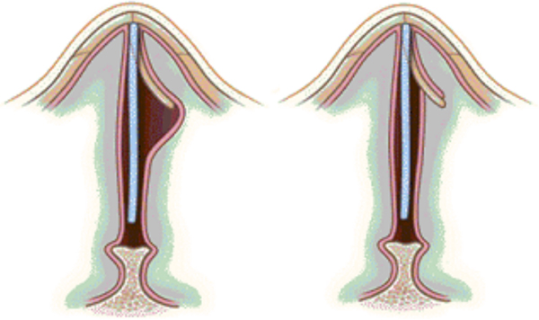
Correction of high bony deviations can be a problem because of the risks of L-strut damage and cerebrospinal fluid leakage. In the full-thickness horizontal mucosal incision technique,Reference Hyun, Kim, Lee, Yoon and Kim23 at least 1 cm of the dorsal bony septum is left uncorrected, keeping the L-strut intact. This may cause a ‘tenting effect’ of the mucosal flap, obstructing the nasal cavity (Figure 2). By making a full-thickness incision in the septal mucosa, just above the cutting edge of the deviated bony septum, this ‘tenting effect’ is resolved. The flap is repositioned to the midline and the cross-sectional area of the nasal cavity is increased.
Full-thickness horizontal mucosal incision to resolve the ‘tenting’ effect. Reproduced with permission.Reference Hyun, Kim, Lee, Yoon and Kim23

Hyun et al.Reference Hyun, Kim, Lee, Yoon and Kim23 observed no septal perforations using this technique. However, their study was limited by its small size (10 patients).
Scoring incisions with 2-octyl cyanoacrylate adhesive
In this technique,Reference Kim, Kim, Park, Kang, Lim and Yu24 partial-thickness incisions are made on the concave cartilaginous surface to affect the interlocked cartilaginous stresses and bend the tissue contralaterally. They are placed on the concave side of the cartilage at 2–4 mm intervals, to approximately half of the cartilage thickness. Because the cartilage frequently reverts to its original deviation once healing occurs, this technique uses a 2-octyl cyanoacrylate adhesive, which acts as a wedge in the incisions to achieve structural stability.
In a study of 24 patients undergoing scoring incisions with 2-octyl cyanoacrylate adhesive, 3 patients (12.5 per cent) developed a temporary foreign body reaction characterised by septal swelling lasting over one month.Reference Samad, Stevens and Maloney27 Further studies are needed to analyse the association between the use of 2-octyl cyanoacrylate adhesive and complications such as foreign body reactions.
Resection of three high tension lines
The septoplasty with three high tension lines resection procedureReference Wang, Han, Zhang, Zang, Li and Liu25 is based on exposure and mobilisation of three high tension lines of the nasal septum, located at the anterior, inferior and posterior attachments of the septal cartilage. The septal cartilage is mobilised by resection of these three attachments. Instead of removing the attachments, the three bony or cartilaginous bars created during this mobilisation are swung in a midline position. The cartilage is then sutured into place at the nasal spine using absorbable sutures.
Outcomes
Chung et al.Reference Chung, Batra, Citardi and Lanza3 reported complete resolution of nasal airway obstruction in 70 per cent of 20 patients who underwent endoscopic septoplasty without concomitant endoscopic sinus surgery. An additional 20 per cent of patients noted improvement. Similar results have been reported with other endoscopic techniques, as shown in Table 1.
These rates are comparable to those described in traditional septoplasty (significant improvement of symptoms in 71 per cent of patients).Reference Siegel, Gliklich, Taghizadeh and Chang28 Other studies reported success rates varying from 63 per cent to 89.5 per cent, depending on the follow-up duration, patient characteristics and assessment methods.Reference Samad, Stevens and Maloney27–Reference Gandomi, Bayat and Kazemei34 However, these retrospective studies are inherently limited by their study design, and caution is therefore needed when comparing these results.
A recent review by Champagne et al.Reference Champagne, Ballivet de Régloix, Genestier, Crambert, Maurin and Pons35 analysed the differences in functional outcome exclusively using results from prospective randomised trials. Both endoscopic and traditional septoplasty improved all symptoms related to septal deformity, but no significant difference could be shown between the two.
Larger prospective randomised trials are needed to further analyse the differences in functional outcome between endoscopic and traditional septoplasty.
Complications
Major complications such as cerebrospinal fluid leak, blindness and saddle nose deformity have all been described with traditional septoplasty techniques.Reference Ketcham and Han36 Injury to the cribriform plate, optic canal or dorsal support is also possible in endoscopic surgery; therefore, careful dissection and maintaining proper orientation remain crucial. However, these complications are rare, and no reports of their occurrence in endoscopic septoplasty can be found in the literature.
Minor complications include perforation, epistaxis, septal haematoma, infection, synechiae formation, and transient dental pain or numbness. Septal perforations are often small and asymptomatic, but some may need further treatment. They occur mostly as a result of bilateral opposing mucosal tears, but may also be caused by a disruption of septal blood supply.Reference Chung, Batra, Citardi and Lanza3, Reference Bloom, Kaplan, Bleier and Goldstein37 Gentle elevation of the mucoperichondrial and mucoperiosteal flaps, and avoidance of the supraperichondrial plane, can help with prevention. Intra-operative tears need to be repaired immediately, and high pressure forces due to septal splints and nasal packing should be avoided to prevent septal necrosis.Reference Bloom, Kaplan, Bleier and Goldstein37
Epistaxis is usually modest, and risks are minimised by avoiding anticoagulants peri-operatively. Patients are also advised to refrain from heavy lifting during the first two weeks after surgery.Reference Sautter and Smith38 A septal haematoma can be easily recognised and drained during the first post-operative visit. Preventive measures such as quilting sutures, and posterior or inferior septal incisions for drainage, may help to reduce the risk.Reference Sautter and Smith38 Dental complaints are caused by injury to the nasopalatine nerve. They usually resolve within several weeks or months, but permanent damage can occur. Avoiding major dissection of the anterior nasal septal and nasal floor mucosa can help minimise the risk.Reference Sautter and Smith38
In a retrospective analysis of 116 patients, Chung et al.Reference Chung, Batra, Citardi and Lanza3 reported transient dental pain or numbness as the most common complication (4.3 per cent), followed by asymptomatic septal perforation (3.4 per cent), synechiae formation (2.6 per cent), epistaxis (0.9 per cent) and septal haematoma (0.9 per cent). Comparable rates for epistaxis, septal perforation and synechiae formation were found in traditional headlight septoplasty.Reference Chung, Batra, Citardi and Lanza3 Similar results were found in a retrospective study by Hwang et al.,Reference Hwang, McLaughlin, Lanza and Kennedy12 who found synechiae formation in 4.9 per cent and septal haematoma in 0.9 per cent, but observed asymptomatic septal perforation in only 0.9 per cent, and found no dental complaints or epistaxis in 111 patients undergoing endoscopic septoplasty.
De Sousa et al.Reference de Sousa, Iniciarte and Levine18 studied the results of powered endoscopic nasal septal surgery in 2730 patients. Septal haematoma was the most common complication (1.8 per cent), followed by septal perforation (0.7 per cent). No other complications were documented, but the follow-up period of three to six weeks can be considered short.
Of course, direct comparison of these retrospective studies remains difficult because of the heterogeneity in patient populations, indications and surgical techniques.
Endoscopic spur resection: our experience
Methods
A retrospective analysis was performed to identify patients undergoing endoscopic spur resection at the University Hospitals Leuven between 2001 and 2015. The medical records were reviewed for: patient characteristics, medical history, operative indication, associated procedures, follow-up duration, symptom outcome and complications. Exclusion criteria were: incomplete medical records and a follow-up period of less than two months. Permission from the ethics committee was obtained prior to commencing the study.
Technique
The procedure is performed under general anaesthesia. The patient is placed in a supine position, and is prepared and draped as for endoscopic sinus surgery. First, cottonoids with a 5 per cent cocaine solution are placed in the nose as a topical decongestant. Unless performed for access, septoplasty is carried out after endoscopic sinus surgery. Lidocaine 1 per cent with 1:200 000 adrenaline is infiltrated in the submucoperichondrial plane, anteriorly above and under the spur. The incision is then made horizontally, along the apex of the spur, and the mucoperichondrial or mucoperiosteal flaps are elevated superiorly and inferiorly using a Cottle elevator. Afterwards, the Cottle elevator is used to fracture the spur, and the spur is removed using forceps. Lastly, the flap edges are approximated and no suturing is necessary. Septal splints and nasal packing are only placed on indication.
Results
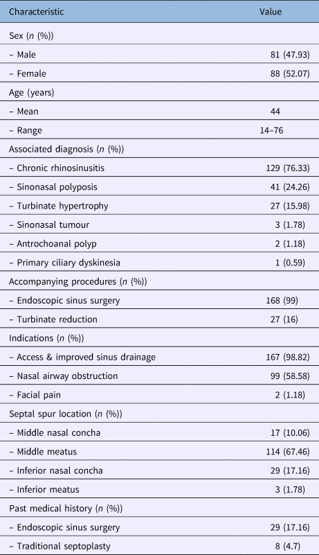
A total of 205 patients who underwent endoscopic spur resection were identified. Thirty-six patients were excluded because of incomplete medical records or loss to follow up. Patient characteristics and demographics are shown in Table 2.

All patients were treated with maximal medical therapy before planning surgery. All procedures were performed by the same surgeon (MJ). Follow-up appointments, which included nasal endoscopy and suction cleaning, were carried out at one, two and three weeks, and at two months post-operatively. Longer follow up was provided on indication.
No intra-operative complications were encountered during endoscopic septoplasty and no cases required conversion to traditional septoplasty. The procedure took 5 minutes on average.
Access was successfully gained in all cases. Correction of the septal deviation was achieved in 167 patients (98.82 per cent). Two patients had a residual septal deviation during follow up, but only one was symptomatic and required revision septoplasty.
Of the 99 patients complaining of nasal airway obstruction, 74 (74.75 per cent) experienced complete resolution of symptoms. An additional 20 patients (20.20 per cent) experienced a marked improvement. Five patients (5.05 per cent) noted persistent nasal airway obstruction. Two patients underwent endoscopic septoplasty for septal spur-related facial pain; neither of these patients experienced an improvement.
No major complications occurred in our series. Four patients (2.37 per cent) developed epistaxis post-operatively and one patient (0.59 per cent) presented with septal haematoma. Synechiae formation was seen in three patients (1.78 per cent), all of whom were asymptomatic. Transient dental hyperesthesia occurred in one patient (0.59 per cent). One patient (0.59 per cent) developed a small (1.5 × 3 mm) and asymptomatic septal perforation. No differences in complication and outcome rates were noted when comparing patients with and without a history of previous nasal surgery.
Discussion
As discussed previously, direct comparison of these results with other series is difficult. Still, our outcome and complication rates are similar to those reported earlier in this essay. Interestingly, we noted no differences in complication and outcome rates when comparing patients with and without a history of previous nasal surgery.
Remarkably, neither of the two patients with spur-related facial pain experienced an improvement. As discussed by Harrison and Jones,Reference Harrison and Jones2 studies report variable outcomes when dealing with septal spur-related facial pain. This may be explained by an inadequate selection of candidates for surgery, or because of neuroplasticity and placebo effects.
Other authors have described largely similar techniques for endoscopic spur resection, although some differences exist. Chung et al.Reference Chung, Batra, Citardi and Lanza3 preferred to remove the spur with a large through-cutting punch. If the spur is too thick, a powered septoplasty burr can be used. As in our technique, the contralateral mucosal flap is always left undisturbed. Giles et al.Reference Giles, Gross, Abram, Greene and Avner39 suggest making a vertical mucosal incision, immediately caudal to the deviation. A limited flap dissection is then made bilaterally, and the spur is removed with straight-biting forceps or a small flat chisel.
Our study is limited by its retrospective design and lack of controls. Simultaneously performed endoscopic sinus surgery is also assumed to have contributed to the improvement of symptoms in our patients. A nine-year follow-up study showed that both subjective and objective changes in nasal airway patency take place over time; hence, our follow-up period of two months can be considered relatively short.Reference Jessen, Ivarsson and Malm40 Ideally, prospective randomised trials, with a longer follow-up period, are needed to further investigate the effects of endoscopic spur resection.
Conclusion
Endoscopic septoplasty provides an alternative way to correct a deviated nasal septum, with outcome and complication rates similar to those seen in traditional approaches. It offers multiple advantages, including improved visualisation, the option of limited flap dissection and teaching benefits. Many different techniques have been proposed, each with its own advantages and limitations. Endoscopic spur resection, as conducted at the University Hospitals Leuven, showed favourable results. It is a quick, safe and efficient way to manage isolated septal spurs, especially when combined with endoscopic sinus surgery.
Competing interests
None declared