Case discussion
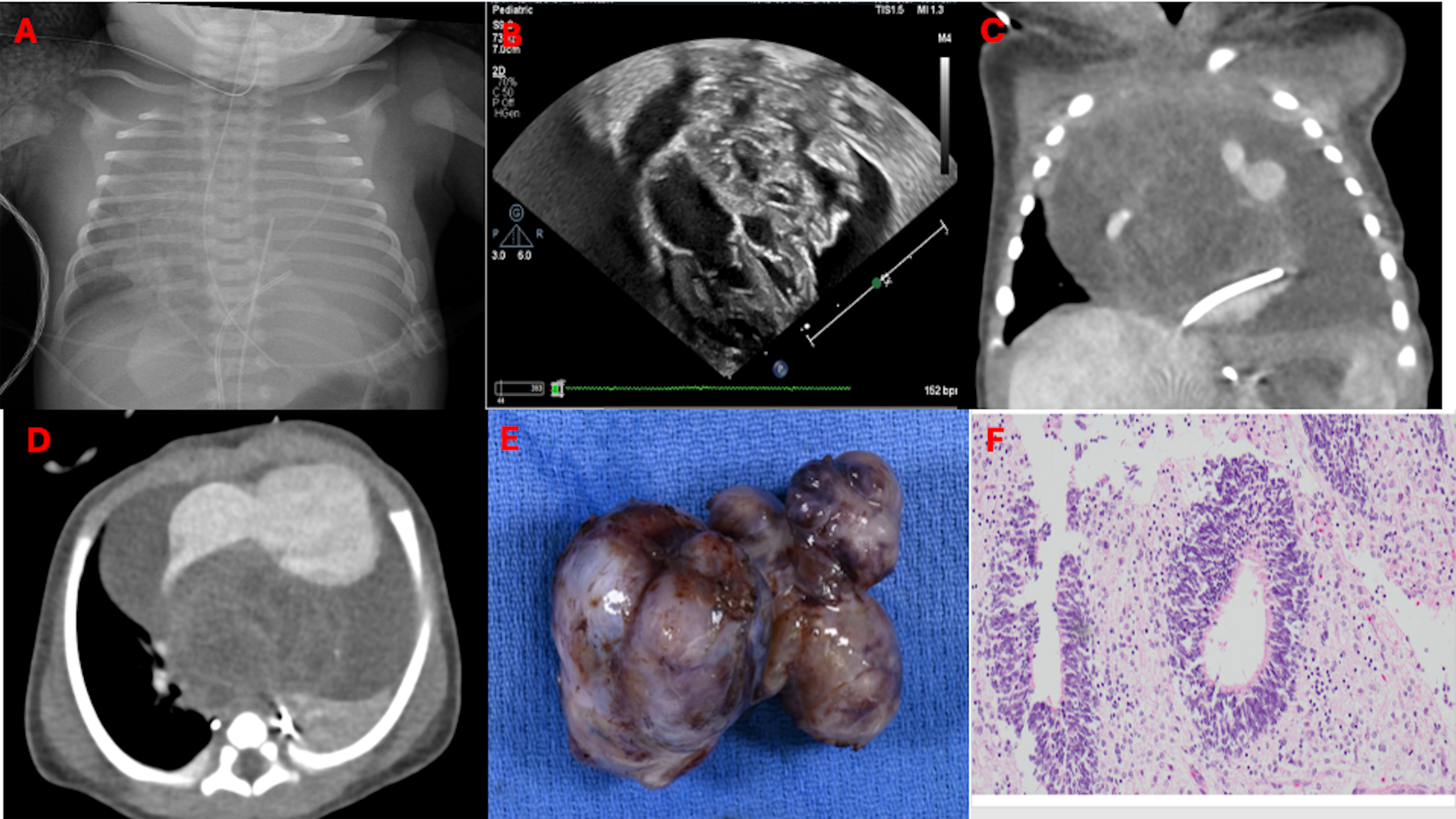
A term female neonate with an unremarkable antenatal history presented at birth with severe respiratory distress requiring non-invasive positive pressure ventilation. Chest radiography showed cardiomegaly (Figure 1A). Echocardiography revealed a well-encapsulated, heterogeneous, multicystic mass (4.5 × 5 cm) extending from the superior heart to the right of the main pulmonary artery, causing marked extrinsic distortion of the left atrium, right pulmonary artery, ascending aorta, and systemic/pulmonary veins, with a large pericardial effusion and a large patent ductus arteriosus (Figure 1B).
Multimodal imaging and pathological findings of anterior mediastinal mass. Panel A: chest radiograph showing large soft tissue density with a small amount of normal aerated lung. Panel B: Two-dimensional (2D) apical four-chamber still-frame echocardiographic image demonstrating a large intrapericardial mass causing extrinsic distortion of cardiac anatomy with compression of systemic and pulmonary veins and the left atrium. Panel C, D: computed tomographic images in coronal and transverse sections demonstrating an extremely large heterogeneous mass in the anterior mediastinum, centred in the region of the intra-arterial septum. Panel E: gross specimen of pericardial mass. Panel F: histological examination demonstrating a primitive neuroepithelial rosette composed of mitotically active, hyperchromatic pseudostratified columnar cells surrounding a central empty lumen, representing immature neural tube-like structures in the neuroectodermal component of an immature teratoma.

Figure 1. Long description
Panel A: A chest radiograph showing a large soft tissue density with a small amount of normal aerated lung. Panel B: A two-dimensional apical four-chamber echocardiographic image demonstrating a large intrapericardial mass causing distortion of cardiac anatomy and compression of systemic and pulmonary veins and the left atrium. Panel C: A computed tomographic image in a coronal section showing an extremely large heterogeneous mass in the anterior mediastinum, centered in the region of the intra-arterial septum. Panel D: A computed tomographic image in a transverse section showing the same mass. Panel E: A gross specimen of the pericardial mass. Panel F: A histological examination showing a primitive neuroepithelial rosette composed of mitotically active, hyperchromatic pseudostratified columnar cells surrounding a central empty lumen, representing immature neural tube-like structures in the neuroectodermal component of an immature teratoma.
Chest tomographic angiography revealed a large heterogeneous anterior mediastinal mass compressing the right atrium and encasing the aortic root, ascending aorta, and proximal arch vessels, causing near-complete left lung collapse and partial collapse of the right upper and middle lobes. (Figure 1C,D). Cardiac magnetic resonance imaging, though superior for tissue characterisation, was deferred considering severe respiratory distress and the need for urgent surgical intervention.
Differential diagnoses included teratomas, rhabdomyoma, fibromas, haemangioma, and myxomas, as well as malignant rhabdomyosarcomas and fibrosarcoma. The extracardiac pericardial location, well-encapsulated multiloculated cystic-solid heterogeneity, large pericardial effusion, great vessel attachment, and coarse calcifications on chest tomographic favoured intrapericardial teratoma. The patient underwent complete surgical resection with patent ductus arteriosus ligation (Figure 1E); histopathology confirmed immature teratoma (Figure 1F).
The postoperative course was complicated by supraventricular tachycardia (treated with flecainide and propranolol) and a mediastinal wound infection requiring surgical exploration. The patient was discharged at 6 weeks in stable condition with normal biventricular function. His recovery has been uneventful, and he continues to do well.
Pericardial teratomas present as multiloculated cysts with mixed solid and cystic components. Echocardiography is the initial imaging modality, followed by cardiac magnetic resonance for tissue characterisation; chest tomographic offers superior spatial resolution of surrounding structures. Reference Klein, Wang and Cremer1
Data availability statement
The majority of data associated with this study is provided in text and figures. Additional data will be made available upon request.
Financial support
None.
Competing interests
All authors have reported that they have no relationships relevant to the contents of this paper to disclose.
Ethical standard
Informed written consent was obtained from the patient or their parents as applicable, as per Nicklaus Children’s Hospital IRB guidelines.