I. INTRODUCTION
The use of the layered materials as precursors of mixed oxides has been recognized as a promising route, which can produce solids with chemical and structural properties of great catalytic interest. Porous catalysts containing transition metal elements such as nickel, cobalt, and molybdenum have been reported in several petrochemical reactions as dehydrogenation (Mitchell and Wass, Reference Mitchell and Wass2002), oxidation of hydrocarbons (Matsuura et al., Reference Matsuura, Mizuno and Hashira1986), and hydrotreating (Zhang et al., Reference Zhang, Duan, Zhao, Wana, Gao, Jiang, Chi and Chuang2010). This last reaction is of great importance because it allows for improving the quality of fuels and reducing SO x and NO x emissions. Hydrotreating catalysts are commonly comprised molybdenum or tungsten sulfide, promoted with nickel or cobalt, and supported on alumina (Chianelli et al., Reference Chianelli, Berhault, Raybaud, Kasztelan, Hafner and Toulhoat2002). Therefore, there is a motivation to investigate the synthesis of new materials with different compositions based on transition metals, useful in catalysis such as nickel, cobalt, and molybdenum, owing to their physical–chemical properties that directly influence the selectivity, and conversion of the hydrotreating process. Pezerat (Reference Pezerat1965) developed the first layered materials, claiming the synthesis of several metallic molybdates with phases, ϕ x and ϕ y . The structural elucidation of these phases was reached by Clearfield et al. (Reference Clearfield, Sims and Gopal1976) for phase ϕ x and Levin et al. (Reference Levin, Soled and Ying1996) for phase ϕ y .
In the present paper, we report the synthesis, characterization, and crystallographic details of a new trimetallic material, (NH4)Ni2.4Co0.6O(OH)(MoO4)2•1.5H2O.
II. EXPERIMENTAL
A. Synthesis
The trimetallic compound was synthesized by the hydrothermal method. Three aqueous solutions of the salts [Ni(NO3)2•6H2O (Merck, 99%), Co(NO3)2•6H2O (Vetec, 98%), and (NH4)6Mo7O24•4H2O (Merck, 99%)] were prepared and then mixed; NH4OH solution (J.T Baker, 28–30%) was added dropwise until pH = 8. A purple gel was obtained and it was homogenized at room temperature during 3 h, and then, it was heated to 100 °C during 24 h in teflon-lined autoclaves. After crystallization time, the light brown solid obtained was recovered by filtration, washed until neutral pH, and dried at 70 °C. Gel molar composition was: Ni(NO3)2 : 0.3Co(NO3)2 : 0.2(NH4)6Mo7O24 : 1.5NH4OH : 328H2O.
B. Material characterization
Powder X-ray data were collected using a Philips X'Pert diffractometer at room temperature. Experimental conditions were: 45 kV, 40 mA, CuKα 1 radiation (λ = 1.5406 Å), 2θ range from 10° to 80° with 0.02° 2θ step size, and 0.5 s per step. Thermogravimetric analysis (TGA) and differential thermal analysis (DTA) were carried out on a TA Instruments equipment, TGA Hi Res 2950, and DSC 2920, respectively; at a heating rate of 10 °C min−1 under nitrogen atmosphere. A Fourier-transformed infrared spectroscopy (FTIR) spectrum was recorded using a Perkin Elmer Spectrum One spectrometer. The analysis was carried out at room temperature and the sample was prepared in the tablet form with composition of 1% in potassium bromide. Laser Raman spectrum was obtained using a LabRAM HR-UV 800/Jobin-Yvon spectrometer, with He–Ne (λ = 632 nm) and Ar (λ = 514 nm) lasers. The charged-coupled device (CCD) detector was operated at −70 °C and 10.7 mW power was used. Metal content analysis for layer precursors was carried out using Thermo Scientific ICE Series 3000 atomic absorption (AA) spectrometer. All samples were treated with mineral acids (hydrochloric and nitric acids) to achieve complete dissolution. Interferences associated with each metal were corrected with the nitrous oxide and acetylene flame. Analyses were performed in duplicate.
Crystal system, unit-cell parameters, and values of 2θ were determined using CRYSFIRE (Shirley, Reference Shirley1999) and CHECKCELL (Laugier and Bochu, Reference Laugier and Bochu2000) programs. The data obtained were consistent with the values obtained with EXPO 2009 (Altomore et al., Reference Altomore, Camalli, Cuocci, Giacovazzo, Moliterni and Rizzi2009) program. The unit-cell parameters were refined by the CHECKCELL program (Laugier and Bochu, Reference Laugier and Bochu2000), and was also used to determine the space group. Finally, the experimental density (D exp) was estimated using a pycnometer and the calculated density (D cal) was determined from the procedure used to measure the properties of crystals (Stout and Jensen, Reference Stout and Jensen1989).
III. RESULTS AND DISCUSSION
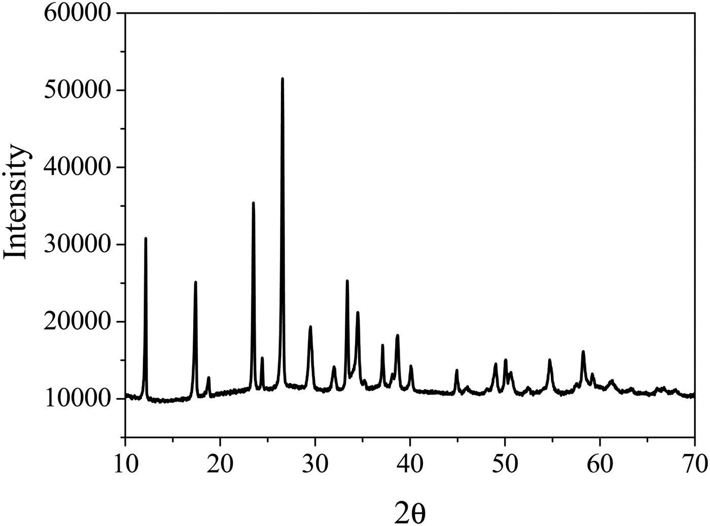
The X-ray powder diffraction pattern for trimetallic compound is shown in Figure 1. X-ray diffraction (XRD) data indicate that the sample is highly crystalline and isostructural with the compound (NH4)HNi2(OH)2(MoO4)2, which is trigonal with hexagonal unit-cell parameters (a = 6.0174 Å and c = 21.8812 Å) as previously reported by Levin et al. (Reference Levin, Soled and Ying1996).
XRD powder diffraction pattern of (NH4)Ni2.4Co0.6O(OH)(MoO4)2•1.5H2O.

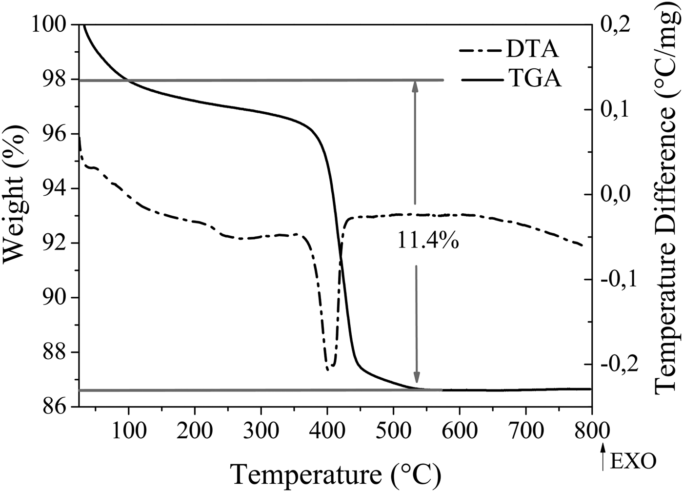
Material composition was determined by the chemical analysis and TGA weight loss measurement. From the AA and TGA experimental results shown in Table I and Figure 2, respectively, the chemical formula for the solid was proposed. As shown in Table I, the experimental composition is agreed with the theoretical composition calculated from the chemical formula.
TGA and DTA measurements of (NH4)Ni2.4Co0.6O(OH)(MoO4)2•1.5H2O.

Elemental compositions for (NH4)Ni2.4Co0.6O(OH)(MoO4)2•1.5H2O.

One endothermic event was observed by TGA and DTA plots (Figure 2), which registered a weight loss of 11.4% between 100 and 400 °C for the layered material. These events correspond to water desorption and ammonium decomposition from the solid; this behavior is explained in the following chemical reaction:
 $$\eqalign{&\lpar {\rm NH}_ 4 \rpar {\rm Ni}_{2.4} {\rm Co}_{0.6} {\rm O}\lpar {\rm OH} \rpar \lpar {\rm MoO}_4 \rpar_ 2 .1.5{\rm H}_2 {\rm O } \mathop{-\!\!\!\!-\!\!\!-\!\!\!\!\longrightarrow}\limits^{400^{\circ}{\rm C}} \cr & \quad 1.4{\rm NiMoO}_4 + 0.6{\rm CoMoO}_4 +{\rm NiO} + 1.0{\rm NH}_3 \uparrow + 2.5{\rm H}_ 2 {\rm O} \uparrow.}$$
The metallic-oxide phases that appear in the right-hand side of the above reaction were identified from the X-ray powder diffraction patterns of solid calcinated at 400 °C. The material treated thermally to 400 °C, showed peaks characteristic of the β −phase described by Mazzocchia et al. (Reference Mazzocchia, Kaddouri, Anouchinsky, Sautel and Thomas1993) and was found in the Powder Diffraction File (PDF-4) database (ICDD, Reference Kabekkodu2009) with the number 00-021-0868.
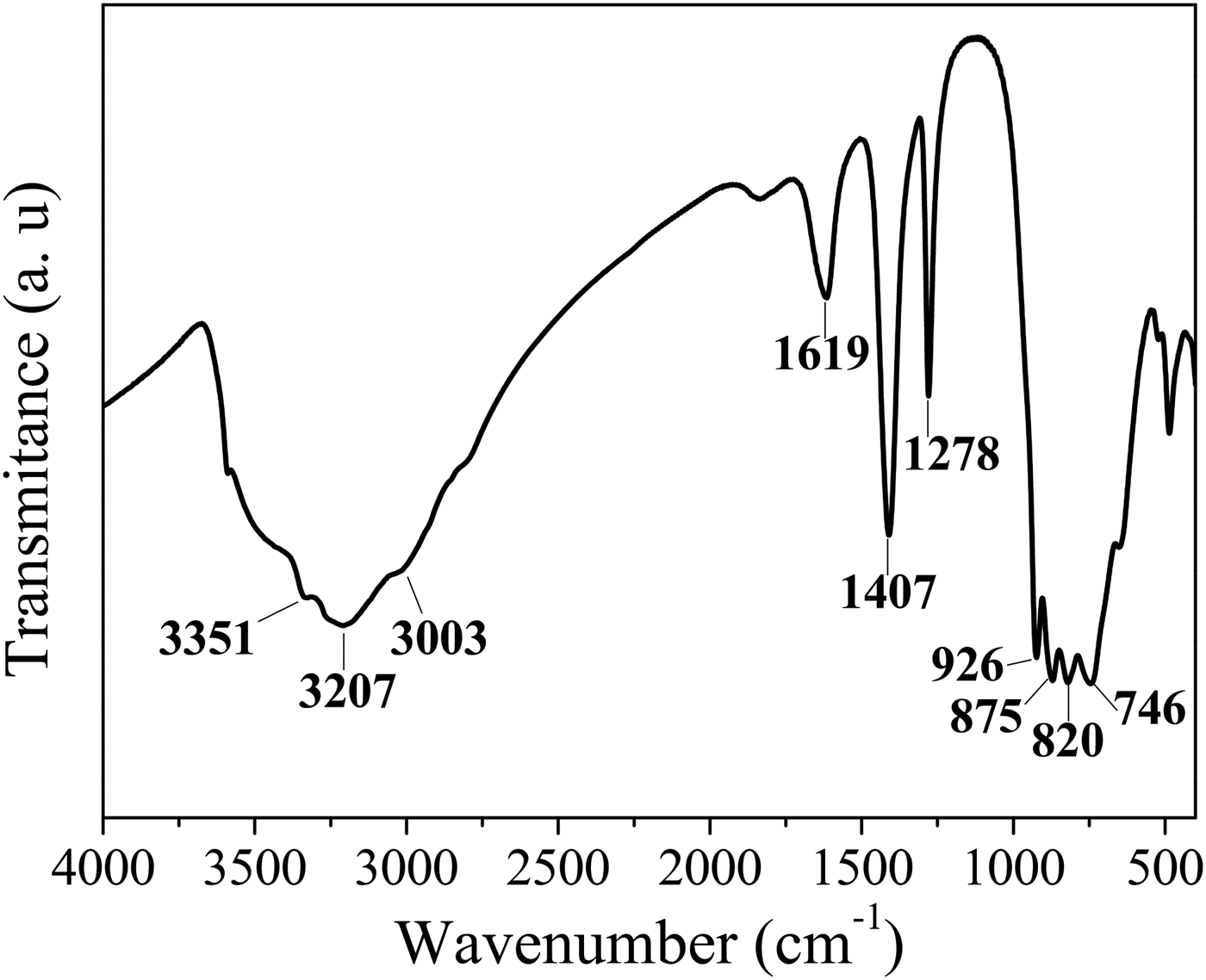
The FTIR spectrum presented in Figure 3, confirmed the presence of ammonium ions in the structure by ν 3 (N–H) asymmetric stretching in the region 3300–3050 cm−1 and the characteristic absorption to 1407 cm−1 by bending ν 4 (H–N–H). The absorption bands attributed to the molybdate ion were observed at 926 cm−1 corresponding to the symmetric stretching vibration mode, ν 1 (O–Mo–O), and 875, 820, and 746 cm−1 for asymmetric stretching, ν 3 (Mo–O) (Nyquist and Kagel, Reference Nyquist and Kagel1971; Levin et al., Reference Levin, Soled and Ying1996; Palacio et al., Reference Palacio, Echavarría, Hoyos and Saldarriaga2005). According to these results, the molybdate ion showed a tetrahedral environmental. Signals attributed to the presence of water were also observed at 1619 and 3351 cm−1 associated with H–O–H bending and O–H stretching, respectively (Nyquist and Kagel, Reference Nyquist and Kagel1971; Palacio et al., Reference Palacio, Echavarría, Hoyos and Saldarriaga2005).
FTIR spectrum of (NH4)Ni2.4Co0.6O(OH)(MoO4)2•1.5H2O.

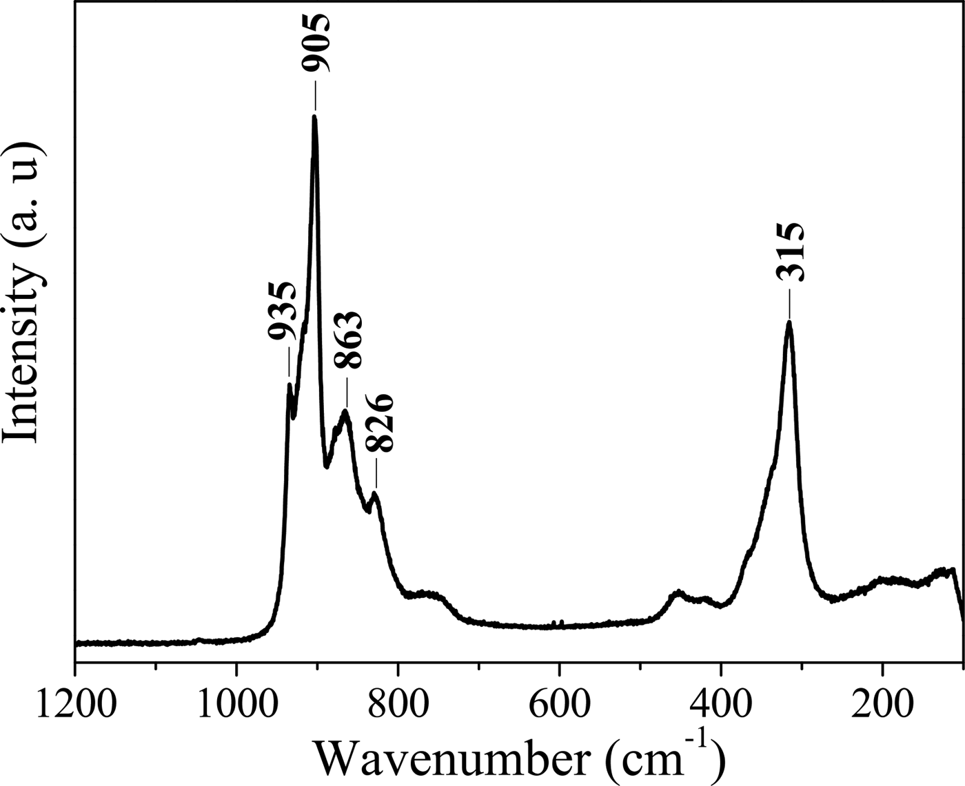
Studies carried out by several authors (Saleem and Aruldhas Reference Saleem and Aruldhas1983; Levin et al., Reference Levin, Soled and Ying1996) confirmed by Raman spectroscopy that normal modes of the molybdate ion are expected to occur in the regions 750–975 and 250–450 cm−1 corresponding to the symmetric stretching and bending, respectively. This affirmation was confirmed in our spectrum (Figure 4) that showed two strong signals at 905 and 315 cm−1, characteristic of the tetrahedral coordination of the molybdate ion, therefore confirming the results obtained in FTIR.
Raman spectrum of (NH4)Ni2.4Co0.6O(OH)(MoO4)2•1.5H2O.

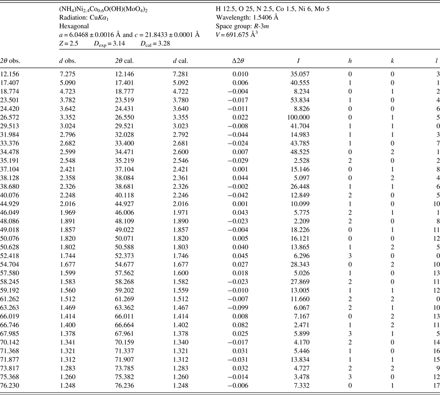
Crystallographic and XRD data of (NH4)Ni2.4Co0.6O(OH)(MoO4)2•1.5H2O are shown in Table II. Systematic absences of reflections limit the crystal lattices of the compound to hexagonal with space group R-3m. Values of unit-cell parameters were determined to be a = 6.0468 ± 0.0016 Å and c = 21.8433 ± 0.0001 Å.
X-ray powder diffraction data for (NH4)Ni2.4Co0.6O(OH)(MoO4)2•1.5H2O.

ACKNOWLEDGEMENTS
We acknowledge financial support by the Universidad de Antioquia and 2013–2014 Sustainability Program; to Gabriel Alonso Núñez (PhD) and Eloísa Aparicio (M.Sc.), Centro de Nanociencias y Nanotecnología, Universidad Nacional Autónoma de México for allowing us to collect powder diffraction data; to Dr. Arnaldo Faro, Universidade Federal de Río de Janeiro, Río de Janeiro, Brazil, for allowing me to collect FTIR and Laser Raman data. S. Amaya acknowledges COLCIENCIAS for her doctoral fellowship.