I. INTRODUCTION
Recently, special nanomaterials as viable alternative to chemical sensors and biosensors have attracted extensive attention due to their associated properties with some molecular recognition. Reference Baptista, Belhout, Giordanib and Quinn1 Numerous methods have been developed to detect chemical and biological species by these nanomaterials, such as electrochemistry, fluorescence, Raman spectrum, and many others. Reference Zhang, Zhou, Liu, Liu, Men, Cai, Duan and Li2–Reference Wu, Mitra, Yan and Zhou7 However, most of these technologies require utilization of sophisticated equipment and professional operators. The ideal sensing technology would realize simple, inexpensive, and quickly detecting. Hydrogel photonic crystals (HPCs) as visualized detecting technologies were popular and attractive because they could detect target analytes by naked eyes. Reference Li, Zhao, Long, Gao, Yang, Tung and Song8–Reference Cai, Natasha, Zhang and Asher13 HPCs utilize a periodic array of colloidal particles embedded in a smart responsive hydrogel matrix which contains chemical or biological recognition in response to external stimuli. Reference Cai, Natasha, Zhang and Asher13 The highly ordered periodic array with a certain lattice-spacing embedded in hydrogel can diffract light in the visible region. The hydrogel matrix in HPCs could be functionalized and the corresponding target analytes with different concentrations could make the functionalized hydrogel swelling or shrinkage in some extent. Hence the lattice of the colloidal crystal embedded in such hydrogel would be increased or decreased, further leading diffraction wave length to red-shift or blue-shift. The shift of diffraction wave length can be read out by naked eyes according to the change of diffraction light color. Reference Zhang, Wang, Luo, Tikhonov, Kornienko and Asher14
More researchers have been devoted to fabricating various HPCs for analytes such as carbonate ions, Reference Li, Zhao, Long, Gao, Yang, Tung and Song8 humidity, Reference Tian, Wang, Zheng, Song, Jiang and Zhu9 temperature, Reference Kang, Moon, Lee, Park, Jang, Yang and Yang10 organic solvent, Reference Wang, Cao, Feng, Yin and Gao11 pH value, Reference Tian, Ma, Cui, Wang, Song and Jiang12 metal ions, Reference Zhang, Wang, Luo, Tikhonov, Kornienko and Asher14,Reference Goponenko and Asher15 glucose, Reference Zhang, Losego and Braun16–Reference Alexeev, Das, Finegold and Asher22 candida albicans, Reference Cai, Kwak, Punihaole, Hong, Velankar, Liu and Asher23 3-pyridinecarboxamide, Reference Yuan, Li, Liu, Gao, Pan and Liu24 blood cells, Reference Zhang, Cai, Shang, Wang, Cheng, Rong, Gu and Zhao25 kinase, Reference MacConaghy, Geary, Kaar and Stoykovich26 light and magnetism. Reference Wang, Hu, Deng, Liang, Li, Liu and Zhu27 In the past years, three-dimensional (3D) and two-dimensional (2D) array have been extensively studied to fabricate HPCs by embedding them into the functional hydrogel films. Reference Sharma, Jana, Kesavamoorthy, Shi, Virji, Finegold and Asher28–Reference Xue, Meng, Wang, Wang, Xue and Xu30 3D array embedded in the hydrogel was mainly obtained by two methods. In the first method, 3D HPCs were fabricated by the electrostatic self-assembly of microspheres in solutions containing polymerizable monomers without easily ionized functional groups, then the monomers were photopolymerized around the 3D hexagonal nonclose packed (hncp) array. Reference Moshe, Alexeev and Asher29,Reference Xue, Meng, Wang, Wang, Xue and Xu30 In this route, the hydrogel precursors were required exclusion of ions to avoid destroying the periodic arrangement of microspheres in array, so the hydrogel film need to be further functionalized to introduce the desired functional groups. Reference Zhang, Wang, Luo, Tikhonov, Kornienko and Asher14 In the second one, 3D HPCs was prepared by casting aqueous solutions of polymerizable monomers into the 3D array prepared in advance, then the monomers were polymerized around the hexagonal close-packed (hcp) 3D array. Reference Wang, Cao, Feng, Yin and Gao11 However, the ordered alignment of 3D array is easily broken in the injecting process to a certain degree. Additionally, the self-assembly of 3D array in this methods is generally time-consuming. Reference Xue, Meng, Wang, Wang, Xue and Xu30 These facts seriously restrict the practical applications of the 3D array/hydrogel composites as sensors.
Most recently, 2D HPCs have been developed owing to rapid reaction process to external stimuli and quick preparation process of the 2D array. Reference Xue, Meng, Wang, Wang, Xue and Xu30–Reference Men, Zhang, Hang, Liu, Li, Cai, Xiong and Li34 To prepare the 2D HPCs, the 2D array was first prepared and then the polymerization solution was layered on the 2D array to form 2D HPCs after polymerization. These 2D HPCs have some advantages compared to 3D ones. For instance, functional monomers could be directly cast onto 2D array to be further polymerized into hydrogel without destruction of 2D periodic structure. Reference Zhang, Chao, Liu and Asher35 Additionally, 2D HPCs allow fast detection to target analytes as sensors because of rapid diffusion of detecting molecules. Previous work mainly focused on applying 2D polystyrene (PS) colloidal crystal to prepare 2D HPCs sensors. Reference Zhang, Wang, Luo, Tikhonov, Kornienko and Asher14,Reference Xue, Meng, Wang, Wang, Xue and Xu30–Reference Men, Zhang, Hang, Liu, Li, Cai, Xiong and Li34 However, the obtained signals (the diffraction light) of such 2D HPCs sensors were very weak because of relatively low scattering cross section of PS itself. To increase diffraction signals, a high reflective mirror (Au, Ag, or Al mirror) was generally used to reflect the forward diffracted light for the diffraction spectra measurement and the color identification. Reference Zhang, Wang, Luo, Tikhonov, Kornienko and Asher14,Reference Xue, Meng, Wang, Wang, Xue and Xu30–Reference Men, Zhang, Hang, Liu, Li, Cai, Xiong and Li34
In the work, we develop a strategy to prepare a free-standing 2D PS colloidal crystal with Au nanoshells/hydrogel composite film (CAuHCF). The 2D PS colloidal crystal was first fabricated by self-assembly and then it was coated with an Au layer on the surface of PS microspheres to form 2D PS colloidal crystal with Au nanoshells. Then it was incorporated into stimulus-responsive hydrogel. Such CAuHCF showed more brilliant diffraction color and much stronger diffraction intensity compared to 2D PS colloidal crystal/hydrogel composite film without Au nanoshells to external stimuli, allowing it to be easily monitored visually. Specifically, the strong diffraction signal of the composite film could be easily detected when it was placed on a transparent substrate (i.e., quartz) without needing a high reflective mirror. The strong diffraction signal drives from the large scattering cross section of Au layer coated on PS microspheres in the 2D colloidal crystal. Here, a CAuHCF sensor response to pH was formed by taking polyacrylic acid (PAA) as hydrogel film through the strategy presented in this paper. The CAuHCF sensor showing strong diffraction intensity and its preparation process is quite simple. In addition, various functional hydrogel films could be prepared into novel optical sensors to detect different analysts by this facile synthesizing route in this paper.
II. MATERIALS AND EXPERIMENTS
A. Materials
Monodispersed PS microspheres with a diameter of 350 nm in aqueous suspension (2.5 wt%) used were purchased from Alfa Aesar Corporation (Shanghai, China). Acrylic acid (AAc), sodium hydroxide, dimethyl sulfoxide (DMSO), acetic acid, phosphoric acid and boric acid were obtained from Sinopharm Chemical Reagent Co, Ltd (Shanghai, China). 2-hydroxy-1-[4-(2-hydroxyethoxy)-phenyl]-2-methyl-1-propanone (Irgacure 2959) was obtained from Tianjin Heowns Biochemical Technology Co, Ltd (Tianjing, China). N-methylenebisacrylamide (BAAm) as crosslinking agent was obtained from Aladdin Industrial Corporation (Shanghai, China). All the reagents were directly used without further purification. Water was used Milli-Q water (18.2 MΩ·cm) obtained from a Millipore ultrafilter system.
B. Fabrication of 2D PS monolayer colloidal crystal
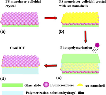
The 2D PS monolayer colloidal crystal with uniform morphology on a large scale was prepared by using an air–water interfacial self-assembly method as previously reported work. Reference Li, Koshizaki and Cai36–Reference Cong, Yu, Tang, Li and Liu41 In brief, a certain amount of PS microspheres in aqueous alcohol was dropped onto a water surface on a cleaned glass slide. The PS monolayer colloidal crystal was obtained by self-assembly at air–water interface with the evaporation of solvent, as shown in Fig. 1(a).
The synthetic procedure of the preparation of CAuHCF: (a) 2D PS monolayer colloidal crystal on a glass slide was fabricated by an air/water interfacial self-assembly method; (b) An Au layer was sputtered on the surface of PS microspheres to form the 2D PS colloidal crystal with Au nanoshells; (c) Polymerization solution was cast into the 2D PS colloidal crystal with Au nanoshells and then the solution was photopolymerized into hydrogel film; (d) The CAuHCF was obtained by peeling it off from the substrates and washed by a lot of water.

C. Fabrication of 2D PS colloidal crystal with Au nanoshells
The 2D PS colloidal crystal with Au nanoshells was prepared by puttering a layer Au on the top of the PS microspheres. The deposition process was carried out for 8 min at a current of 20 mA to the certain thickness by an ion-beam coater in a vacuum of 1 × 10⁻¹ mbar. Through this process, ordered 2D colloidal array with hemispherical Au nanoshells was fabricated, as shown in Fig. 1(b).
D. Preparation of CAuHCF
In a typical experiment, Irgacure 2959 (0.33 g) was dissolved in 1 mL DMSO. Then, 500 µL Irgacure 2959 solution were added into a 10 mL mixture solution containing 50 vol% H2O, 50 vol% AAc, and 0.08 g BAAm followed by vigorously shaking. After that, the aforementioned polymerization solution was carefully deposited on the 2D PS colloidal crystal with Au nanoshells to form a solution film on it. Then a coverslip was gingerly covered onto the polymerization solution surface, followed by photopolymerized with UV light from a UV Lamp (16 W, 365 nm) for 30 min, as shown in Fig. 1(c). The resulting free standing CAuHCF was washed with large amounts of pure water in Fig. 1(d).
E. Characterization
The morphology of as-prepared samples were characterized by a field-emission scanning electron microscope (FE-SEM; Sirion200, FEI Company, Hillsboro, Oregon) after depositing a thin layer of Au on them for good electric conductivity. Energy disperse spectrum (EDS) of Au shell was examined on Oxford INCA analysis system. UV–vis extinction spectra were measured on Cary 500 (Varian Inc., Palo Alto, California). The diffraction spectra of the 2D PS microsphere array with Au nanoshells/hydrogel composite film were recorded at a fixed angle by utilizing an Idea Optics PG2000-Pro-EX Spectrometer, R1-A-UV Support and Halogen & Deuterium Light Source. The wave length of the light source is 215–2500 nm. The diffraction wave length from 2D colloidal crystal would red-shift with the increase of angle of the incident light and would blue-shift with the increase of angle of the incident light. This may make the diffraction wave length beyond the range of visible light. To obtain the diffraction peak in the visible region, we adjusted the angle between the light source and the 2D array/hydrogel composite films normal at 32°. Such 2D array/hydrogel composite films were incubated in B-R buffer solution overnight before diffraction measurements. The photograph of the composite films were taken using a digital camera (Sony, Cyber-shot DSC-N2) at an angle close to 32° between observation direction and the normal of 2D array hydrogel film. The 2D array/hydrogel composite films were placed on a quartz slide during the process of measuring diffraction spectra and taking photographs.
III. RESULTS AND DISCUSSION
A. CAuHCF
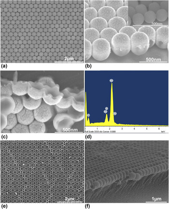
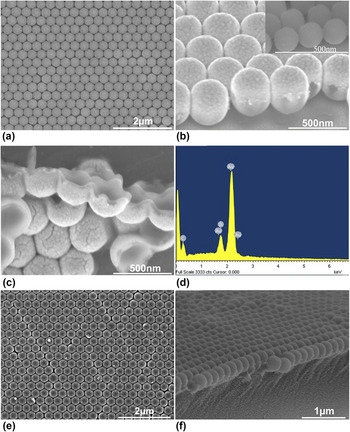
Figure 2(a) illustrates a SEM image of as-synthesized uniform a 2D PS colloidal crystal with Au nanoshells on a glass slide, indicating high ordering. There are obvious Au nanoshells on the PS microsphere in Fig. 2(b). The PS microspheres were dissolved in dichloromethane and the nanoshells could be obviously observed in Fig. 2(c). The thickness of the Au nanoshell is approximately 50 nm. EDS analysis for such shells confirms that the shells consist of Au, as shown in Fig. 2(d) (the Si element comes from silicon substrate and C element comes from residue of PS microspheres). SEM image of the air-dried CAuHCF presents that the integrality of the 2D PS colloidal crystal with Au nanoshells was well maintained during the polymerization and transferring process, as could be found in Fig. 2(e). Figure 2(f) reflects the cross-sectional image of as-synthesized CAuHCF, demonstrating that the 2D PS colloidal crystal with Au nanoshells was monolayerly embedded onto the surface of the hydrogel film.
(a) Top view of SEM image of the 2D PS colloidal crystal with Au nanoshells; (b) Cross-sectional view of SEM images of (a), (Inset: SEM image of PS colloidal crystal); (c) SEM image of Au nanoshells after dissolving PS microspheres with dichloromethane; (d) EDS analysis of Au nanoshells; (e) Top view of SEM image of the CAuHCF; (f) Cross-sectional view of (e).
B. Optical diffraction intensity
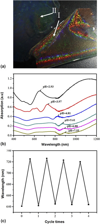
The CAuHCF shows very brilliant diffraction color, but the diffraction color of the 2D PS colloidal crystal/hydrogel composite film is not very obvious in Fig. 3(a). The bright color of the CAuHCF makes it easy to monitor the 2D colloidal crystal diffraction by naked eyes or instrument. Extinction spectra of as-prepared CAuHCF at different pH values are showed in Fig. 3(b). Each curve of the extinction spectra displays one obvious peak, which originates from Bragg diffraction of the ordered periodic structure of the 2D PS colloidal crystal with Au nanoshells. Reference Gu, Horie, Kubo, Yamada, Fujishima and Sato42 The localized surface plasmon resonance (LSPR) of Au nanoshells was too weak to be observed. The diffraction peak was gradually red-shifted from 646 to 891 nm with pH value increasing from 2.93 to 7.55. The redshift is attributed to increase microsphere interspaces due to the swelling of hydrogel caused by ionization of carboxyl groups, which actuates an osmotic pressure inside the gel, resulting in swelling of hydrogel. Reference Zhang, Wang, Luo, Tikhonov, Kornienko and Asher14,Reference Lee and Asher43 The increasing interspaces of 2D PS microsphere colloidal crystal indicates that the interspace among PS microspheres coated Au shells can be increased because of the swelling of hydrogel film, although the Au hemi-nanoshells on adjacent PS spheres are slightly interconnected after a certain amount of deposition of Au. We examined the reversibility of the CAuHCF by immersing it in the buffer solution of pH 2.93, followed by immersing it in the buffer solution of pH 3.97. The whole process can be accomplished in only a few minutes. Figure 3(c) shows the results of four cycles of our sensor based gold nanoshell arrays for its response to pH from 2.93 to 3.97. Little variation in response occurs, indicates good reproducibility.
(a) Photograph of CAuHCF (I) and 2D PS colloidal crystal/hydrogel composite film (II) in water at the same time; (b) Extinction spectra of CAuHCF at different pH: 2.93, 3.97, 4.81, 5.61, 6.88, and 7.55; (c) Reversibility of CAuHCF at pH 2.93 and 3.97.

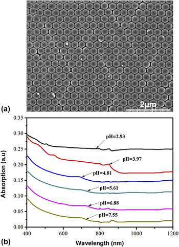
2D PS colloidal crystal/hydrogel composite film as a contrast was prepared, as shown in Supplement Material in Fig. S1. The SEM image of 2D PS colloidal crystal/hydrogel composite film is shown in Fig. 4(a). It shows that the monolayer colloidal crystal is on surface of a hydrogel film and all spaces between neighboring microspheres are full of hydrogel polymer. Their diffraction spectra at different pH value could not be detected by an ordinary UV–vis absorption spectra spectrometer, as demonstrated in Fig. 4(b), which indicates that the diffraction signals may be too weak to be detected.
SEM image (a) and extinction spectra at different pH value (b) of the 2D PS colloidal crystal/hydrogel composite film.

We measured the diffraction spectra of the CAuHCF and the 2D PS colloidal crystal/hydrogel composite film at different pH value by a diffraction spectra spectrometer and a R1-A-UV Support. The support has two arms: one excites white light (Halogen & Deuterium Light Source and the wave length of the light source is 215–2500 nm) and could rotate 180°; one collects the diffraction light by a semi-transparent mirror and could rotate 360°. Reference Men, Zhang, Hang, Liu, Li, Cai, Xiong and Li34 The two arms were at the same angle and measurement angle of the probe to the normal direction of the 2D colloidal crystal hydrogel film sensor was about 32° in measuring process, as shown in Fig. S2. Here, the sensitivity and corresponding diffraction intensity of the composite film in response to pH value was investigated.
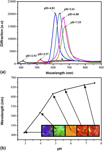
Figure 5(a) displays the diffraction spectra of the CAuHCF at pH value changed from 2.93 to 7.55. Although the sensor based on 2D Au nanoshells array was placed on quartz slide during measuring process, there still remained strong diffraction peak. At pH 2.93, a diffraction peak with a maximum of 408 nm was observed, indicating that it diffracted blue light. The diffraction peak was gradually red-shifted to 674 nm with the pH increasing to 7.55. The PS microsphere interspacings increase due to the swelling of the hydrogel, leading the diffraction peak to red-shift. Figure 5(b) shows the dependence of their diffraction peak position on pH value, indicating the position of diffraction peak increased significantly as the pH value increase. Furthermore, the inset in Fig. 5(b) clearly displays their visual color change on quartz slide with the changing pH value. The color changed from blue to green, to reddish orange, to red and finally to dark red as pH change. Thus, pH can be roughly determined by observing the changes of diffraction color.
(a) Diffraction spectra of the CAuHCF at different value of pH value: 2.93, 3.97, 4.81, 5.61, 6.88, and 7.55; (b) Dependence of the diffraction wave length maximum of the CAuHCF on pH value. The inset in (b) displays corresponding photographs of the CAuHCF placed on quartz slide at different pH value.

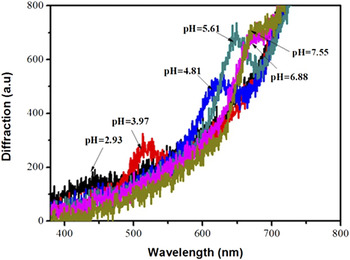
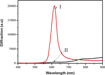
We also probed diffraction spectra of the 2D PS colloidal crystal/hydrogel composite film on quartz substrates, as displayed in Fig. 6. The diffraction wave length also red-shifted with the increase in pH value, but diffraction signals of such 2D PS colloidal crystal/hydrogel composite film on quartz slide were very weak. To improve the diffraction intensity, researchers used high reflective mirror such as Al, Au, and Ag mirror to improve diffraction intensity. Reference Zhang, Wang, Luo, Tikhonov, Kornienko and Asher14,Reference Xue, Meng, Wang, Wang, Xue and Xu30–Reference Men, Zhang, Hang, Liu, Li, Cai, Xiong and Li34 Fig. 7 gives a comparison of the diffraction intensity of CAuHCF and 2D PS colloidal crystal/hydrogel composite film on the quartz slide at pH value of 4.81. The diffraction intensity of CAuHCF was about 30-fold larger than that from the 2D PS colloidal crystal/hydrogel composite film on the transparent substrate, as shown in Fig. 7.
Diffraction spectra of the 2D PS colloidal crystal/hydrogel composite film on quartz substrates at different pH value: 2.93, 3.97, 4.81, 5.61, 6.88, and 7.55.

Comparison of diffraction intensity on quartz slide at pH value of 4.81: CAuHCF (I) and 2D PS colloidal crystal/hydrogel composite film (II).

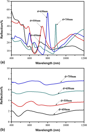
To verify our experimental results, we calculated the reflection spectra of CAuHCF and 2D PS colloidal crystal/hydrogel composite film by finite-difference time domain (FDTD) method, as shown in Fig. 8(a). The diameter of the PS microsphere diameter is 350 nm and the thickness of Au nanoshell is 50 nm. The interspaces are set to 450, 550, 650, and 750 nm, respectively. Periodic boundary conditions are adopted along x and y axes, while perfect matched layer (PML) boundary conditions are adopted along z axis. The incident light is along the z axis with electric field along the x axis. The mesh grid is 2 nm, and the dielectric function of Au is from the Palik’s data. For all of the four cases of CAuHCF, there are strong reflection peaks originated from Bragg diffraction of the ordered periodic structures. We can find that the reflection peak red-shifts with increasing the periodicity of the array. The reflection intensity is about 20–60% in the Vis-NIR wave length range. In this work, their intense reflection signals come from the Au shells with large scattering cross section due to the surface plasmon resonance. Reference Men, Zhou, Hang, Li, Duan, Cai and Li44,Reference Men, Liu and Li45 Large scattering cross-section leads to intense diffraction signal. Reference Weissman, Sunkara, Tse and Asher46 For comparison, we also calculated the reflection spectra of the 2D PS colloidal crystal/hydrogel composite film with the same PS microsphere size like CAuHCF, and the results are shown in Fig. 8(b). There are no obvious reflection peaks because of the low scattering cross section of PS microsphere than Au nanoshells. The calculated results correspond well to our experimental results that corresponding higher diffraction intensity of 2D PS microsphere array with Au nanoshells than that of 2D PS microsphere array. The optical diffraction intensity of CAuHCF sensor was not stronger than that of Au nanosphere array/hydrogel composite, but such sensors based Au nanoshells array with the simple preparation process could be directly used without needing a high reflective mirror. Reference Men, Zhou, Hang, Li, Duan, Cai and Li44
The calculated reflection spectra of CAuHCF (a) and 2D PS colloidal crystal/hydrogel composite film (b) by FDTD. The interspaces are 450, 550, 650, and 750 nm, respectively (the diameter of PS microsphere is 350 nm, the thickness of Au nanoshell is 50 nm).

IV. CONCLUSION
In summary, we develop a simple but effective method to fabricate an interesting, free-standing CAuHCF by photopolymerizing a hydrogel film on the top of the 2D PS colloidal crystal with Au nanoshells. The CAuHCF would swell and the 2D array particle spacing would increase to result in visually evident red shift of the diffraction light and a change of diffraction color with pH increase. Therefore, pH can be determined by observing the changes of diffraction light and diffraction color. Additionally, such CAuHCF displayed strong diffraction signal on ordinary quartz substrate and its diffraction intensity was about 30-fold larger than that from the 2D PS colloidal crystal/hydrogel composite film on the transparent substrate due to large scattering cross section caused by the surface plasmon resonance. The intense optical diffraction intensity could make the detection more easily by monitoring the change of diffraction wave length and color identification. More importantly, it is expected that other sensors based functional hydrogel and 2D PS colloidal crystal with Au nanoshells will be fabricated by the presented strategy in this work.
Supplementary Material
To view supplementary material for this article, please visit https://doi.org/10.1557/jmr.2017.34.
ACKNOWLEDGMENTS
The authors acknowledge the financial support from National Natural Science Foundation of China (Grant Nos. 51371165, 51571189), the State Key Program of National Natural Science Foundation of China (Grant No. 51531006), Anhui Provincial Natural Science Foundation (Grant No. 1508085JGD07), Cross-disciplinary Collaborative Teams Program in CAS, the CAS/SAFEA International Partnership Program for Creative Research Teams.