Introduction
Cystic fibrosis (CF) is an autosomal recessive disease, with an incidence of 1 in 2400 live births in the UK. It is a generalised exocrinopathy that causes electrolyte transport disturbances, resulting in thick mucus formation.Reference Gysin, Alothman and Papsin1 Sinonasal mucous gland function and mucociliary clearance are affected, which can lead to infective sinusitis. Many authors of computed tomography (CT) studies on the CF population have demonstrated the almost universal involvement of the paranasal sinuses.Reference Gysin, Alothman and Papsin1, Reference Eggesbø, Søvik, Dølvik, Eiklid and Kolmannskog2 The underdevelopment of sinus cells, particularly the frontal cells, can predispose the affected individual to sinus disease. The majority of CF patients present with chronic rhinosinusitis, nasal polyposis or both.Reference Mainz and Koitschev3
Pulmonary disease is still the primary cause of premature death in most CF patients.4 The upper airway is a potential site for colonisation, and the paranasal sinuses may become reservoirs for opportunistic bacteria.Reference Mainz and Koitschev3 The ‘single airways’ concept has been reinforced by studies which show that improvements in sinus disease can result in improved lower airway function in asthma.Reference Loebinger, Bilton and Wilson5 Investigations of the effect of sinus disease on CF are lacking, and there is little evidence regarding the surgical management of CF-related chronic rhinosinusitis.Reference Mainz and Koitschev3 Around 10–20 per cent of CF patients will eventually require sinus surgery.Reference Schulte and Kasperbauer6 However, these patients generally develop rapid recurrences of sinus symptoms; the initial results of primary surgery are discouraging. Rowe-Jones and Mackay demonstrated that up to 50 per cent of CF patients experienced recurrence of symptoms within 2 years, which required a second procedure.Reference Rowe-Jones and Mackay7
In 1914, Lothrop reported a combined external and transnasal approach to the frontal sinus.Reference Lothrop8 The endoscopic modified Lothrop procedure described by MayReference May9 and Gross et al. Reference Gross, Zachmann, Becjer, Vickery, Moore and Lindsey10 involves removal of the frontal sinus floor bilaterally, with frontal septectomy and resection of the nasal septum to create a wide median frontal sinus drainage pathway. Cystic fibrosis patients do not have normal mucociliary function and, regardless of the size of the opening, normal mucociliary clearance will not be achieved. The optimal management of chronic rhinosinusitis in this setting has not been firmly established. However, we have performed the modified Lothrop technique in patients with CF at the Royal Brompton Hospital, UK, and this has resulted in overall improvement in patients' quality of life and excellent functional results during follow up of up to five years. The principle of this procedure is to achieve adequate and permanent aeration to the frontal sinus regardless of its anatomical condition.
Materials and methods
Case one
A 38-year-old female with CF, who had previously undergone a lung transplant in 1998, presented with a long-standing history of highly symptomatic, chronic frontal sinusitis and nasal polyposis. This occurred in spite of multiple surgical procedures (10 functional endoscopic sinus surgery (FESS) procedures) performed for frontal sinus clearance. Serial CT scans (Figure 1) demonstrated opacification of the frontal sinuses, particularly at the level of the frontoethmoidal recesses, despite appropriate medical treatment. The frontoethmoidal recesses were subsequently cleared. However, symptoms reappeared the following year, which led to recurrent chronic chest infections.
Coronal computed tomography section demonstrating opacification of the frontal sinuses in case one.

In 2007, the patient underwent an endoscopic modified Lothrop procedure using an image guidance system (BrainLab, Heimstetten, Germany). The frontoethmoidal recess was identified and a superior septal window was removed. This was followed by the drilling of the frontal beak using a 45 degree angle endoscope (Karl Storz Endoscopy, Tuttlingen, Germany) with a Rad® 55 curved bur, high-speed, 3.6 mm drill (Xomed-Medtronics, Jacksonville, Florida, USA). The intersinus septum was identified and drilled in its extension, resulting in patent frontal cavities, with no intra-operative complications. No other surgery was performed on the sinuses. The patient went home the following day with a 10-day course of azithromycin and nasal douches.
Case two
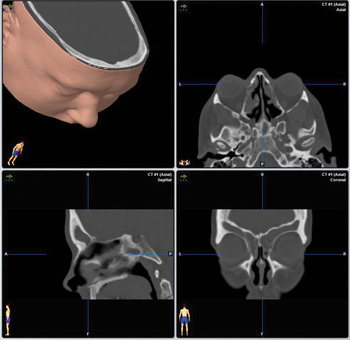
A 44-year-old female with CF presented with a long-standing history of recurrent chest infections, frontal pain and post-nasal drip. Medical treatment, including multiple (six) FESS procedures, had proven ineffective. On examination, polypoid thickening was found in the frontoethmoidal recess with mucopus. A CT scan of the sinuses revealed opacification of the frontal sinuses bilaterally, with marked mucosal congestion and bilateral blockage of the frontoethmoidal recesses.
Due to the persistence of the clinical symptoms, in 2005 the patient underwent endoscopic modified Lothrop surgery under image guidance (BrainLab) for treatment of frontal sinus disease (Figure 2). Drilling of the frontal beak was performed under endoscopic vision (using a 45 degree angle Karl Storz endoscope (Karl Storz Endoscopy)) with a Rad® 55 curved bur, high-speed, 3.6 mm drill (Xomed-Medtronics). A septal window was created, and the intersinus septum and floor of the frontal sinuses were removed bilaterally, achieving large cavities to ensure patency of the frontal sinus. No other sinus surgery was performed. The procedure was completed without any complications. The patient was discharged the following day with a 10-day course of azithromycin and nasal douches.
Tri-planar images as viewed on an image guidance system, demonstrating bilateral opacification of the frontal sinuses in case two.

Results
At follow-up appointments, the first patient reported complete resolution of both upper and lower respiratory tract symptoms, and described how the procedure had significantly affected her quality of life. The patient felt that the impact of the procedure on her quality of life was ‘as good as the lung transplant’.
The second patient was reviewed at follow-up appointments and described a complete absence of frontal symptoms with occasional crusting, and remarkable improvement in terms of chest infection recurrence.
The patients continued using the nasal douches and attended regular follow-up appointments for five years. There was approximately a 20 per cent stenosis of the newly formed frontal sinus ostium over time (in both patients). Neither patient had any significant sinus symptoms or needed further surgery.
Discussion
Cystic fibrosis was, until a few decades ago, a commonly fatal condition for children affected. By 1987, life expectancy had risen to 23 years, and by 2006 the mean survival had further increased to 37 years.Reference Rickert, Banuchi, Germana and April11 This has created a new population of CF patients with chronic rhinosinusitis and severe medical problems, for whom quality of life needs to be addressed.
At least half the patients with CF fulfil the diagnostic criteria for chronic rhinosinusitis, and a further third of patients report intermittent rhinosinusitis symptoms.Reference Mainz and Koitschev3
The single airways concept has been well described in relation to sinus disease and asthma.Reference Bresciani, Paradis, Des Roches, Vernhet, Vachier and Godard12, Reference Ragab, Scadding, Lund and Saleh13 It may also be a useful model to reflect the relationship between chronic rhinosinusitis and chronic infective respiratory conditions such as CF. Chronic pulmonary infection with pseudomonas is central to the progression of CF lung disease.Reference Bresciani, Paradis, Des Roches, Vernhet, Vachier and Godard12, Reference Taylor, Morgan, Nicholson, Mackay, Hodson and Pitt14, Reference Davidson, Murphy, Mitchell, Smith and Light15 Pseudomonas infections are serious complications for post-transplant CF patients; they are thought to originate from the nasal sinuses.Reference Walter, Gudowius, Bosshammer, Römling, Weissbrodt and Schürmann16, Reference Lewiston, King, Umetsu, Starnes, Marshall and Kramer17
In the CF population, sinonasal disease can lead to significant morbidity, increased risk of pneumonia, acute exacerbations and frequent hospitalisations.Reference Kingdom, Lee, Fitzsimmons and Cropp18 Control of upper respiratory tract infections is an important factor in preventing recurrence of lower respiratory tract infections, which is particularly important for CF patients undergoing heart and lung transplantation.
Over the years, different types of sinus surgery have been proposed for CF patients with chronic rhinosinusitis and/or polyps. In 1987, Cepero et al. reported a recurrence rate of 61 per cent following polypectomy alone, but this reduced to 13 per cent when polypectomy was combined with ethmoidectomy.Reference Cepero, Smith, Catlin, Bressler, Furuta and Shandera19 However, not all surgeons were convinced. Marks et al. performed fairly extensive surgery in primary and revision FESS-treated patients.Reference Marks and Kissner20 Procedures included endoscopic sphenoethmoidectomy, the Caldwell–Luc technique and frontal sinus obliteration. Their results indicated that endoscopic surgery failed to control frontal and maxillary sinus disease. It was therefore recommended that surgery only be performed when ‘absolutely necessary’. In 1996, Rowe-Jones and Mackay reported a study conducted at our institution in which total ethmoidectomy and large middle meatal antrostomy were carried out.Reference Rowe-Jones and Mackay7 Six of the 46 patients underwent clearance of the frontoethmoidal recess and sinus ostium for frontal sinus suppuration. All patients gained some initial improvement, with a 50 per cent chance of return to pre-operative severity or the requirement of a further operation within 24 months.
• Paranasal sinus disease affects patients with cystic fibrosis (CF)
• The ‘single airways’ concept may be useful to reflect the relationship between chronic sinus disease and CF
• Successful paranasal sinus management reduces the incidence of tracheobronchitis and pneumonia in these patients
• Surgical management of frontal sinus disease in this population is challenging; many procedures have been described
• Drilling-out surgical techniques have not been reported for management of CF frontal sinus disease
• In select cases, the modified Lothrop procedure can result in lasting symptomatic relief, of both the upper and lower respiratory tract
There are no reports of drilling-out surgical techniques used for the management of CF frontal sinus disease. Some argue that CF patients do not have normal mucociliary function, suggesting that normal mucociliary clearance cannot be achieved, regardless of the size of the surgical opening. However, Wormald's research group has published evidence to indicate that the modified Lothrop procedure has no adverse effects on mucociliary clearance of the frontal sinus in animal models.Reference Rajapaksa, Ananda, Cain, Oates and Wormald21 Irrigation of the frontal sinus in the immediate post-operative period showed a trend towards improved post-operative mucociliary function at three months. We feel that the large drainage pathway created in the modified Lothrop procedure greatly facilitates douching of the frontal sinus and allows more free drainage, thereby preventing the stasis of secretions. Our reports of two patients revealed lasting improvements in symptoms, of both the upper and lower respiratory tract.
Conclusion
Paranasal sinus surgery is the second most frequently performed surgery in CF patients after laparotomy for meconium ileus.Reference Ramsey and Richardson22 Many patients benefit from minimally invasive paranasal sinus surgery, which can result in improvements in nasal symptoms, subsequent weight gain and well-being. Successful sinus management also reduces the incidence of tracheobronchitis and pneumonia. This study shows that the modified Lothrop procedure may improve quality of life for these patients. This procedure has a role in the integrated approach for frontal sinus disease in CF patients, provided the surgical expertise is available and careful patient selection is carried out.