Introduction
In vitro fertilization (IVF) and embryo culture have become widely used technology in bovine research. According to the International Embryo Transfer Society (Viana, Reference Viana2019) most bovine embryos in the world (68.7%) are produced by IVF, but fresh transferred IVF embryos are predominant (overall 73.2%).
It has been suggested that IVF embryos are less resistant to traditional cryopreservation methods (slow freezing) compared with embryos produced in vivo due to the higher amount of lipids (Abe et al., Reference Abe, Yamashita, Satoh and Hoshi2002). After being absorbed by somatic cells, fatty acids are esterified in triacylglycerol (TAG) and cholesterol esters and stored as neutral lipids in droplets (M. Carro et al., Reference Carro, Buschiazzo, Ríos, Oresti and Alberio2013). Neutral lipids serve as major intracellular energy storage molecules (Bertevello et al., Reference Bertevello, Teixeira-Gomes, Seyer, Vitorino Carvalho, Labas, Blache, Banliat, Cordeiro, Duranthon, Papillier, Maillard, Elis and Uzbekova2018). Moreover, beta-oxidation appears to be important for oocyte nuclear maturation (Paczkowski et al., Reference Paczkowski, Schoolcraft and Krisher2014; Valsangkar and Downs, Reference Valsangkar and Downs2013; Ya and Downs, Reference Ya and Downs2013) and oocyte competence acquisition (Dunning et al., Reference Dunning, Anastasi, Zhang, Russell and Robker2014). However, following oxidative stress, lipid peroxidation is increased and oxygen reactive species are released, which is accentuated by cryopreservation (Seidel, Reference Seidel2006).
Abnormal lipid accumulation in bovine first occurs during in vitro maturation (IVM) (Del Collado et al., Reference Del Collado, Saraiva, Lopes, Gaspar, Padilha, Costa, Rossi, Vantini and Garcia2015) and continues during embryo culture (reviewed by Seidel, Reference Seidel2006). The use of metabolic regulators can be used to improve embryo quality. Forskolin enhances cellular lipolytic activity (Paschoal et al., Reference Paschoal, Sudano, Guastali, Dias Maziero, Crocomo, Oña Magalhães, Da Silva Rascado, Martins and Da Cruz Landim-Alvarenga2014). l-Carnitine mediates beta-oxidation, regulating ATP production from lipids, and is a potent antioxidant (Zolini et al., Reference Zolini, Carrascal-Triana, Ruiz de King, Hansen, Alves Torres and Block2019). l-Carnitine increases lipid metabolism and improve rates of development in preimplantation mouse and bovine embryos (Sutton-McDowall et al., Reference Sutton-McDowall, Feil, Robker, Thompson and Dunning2012). Conjugated linoleic acid (CLA) t10,c12 induces LA incorporation and triacylglycerols (TAG) and phospholipids production, improving cytoplasmic and cellular membranes lipid profile (Tominaga et al., Reference Tominaga, Shimizu, Ooyama and Izaike2000). LA has antioxidant properties and can reduce cytoplasmic lipids and improve cryosurvival in cattle (Karaşahin, Reference Karaşahin2019; Pereira et al., Reference Pereira, Baptista, Vasques, Horta, Portugal, Bessa, Silva, Pereira and Marques2007).
Therefore, culture medium supplementation with metabolic regulators can improve embryo quality and cryosurvival, but most studies are focused on one lipid accumulation pathway. As metabolism is an interconnected network, we combined three lipid modulators with distinct targets into a cocktail that modulates lipid metabolism in multiple pathways, to obtain additional gain in embryo cryotolerance. We investigated lipid modulation during oocyte maturation and embryo development, hypothesizing that the supplementation of regulators at IVM, mid (D4, 96 hpi) or late (D6, 144 hpi) embryo culture would have positive impacts on embryo development and cryotolerance.
Materials and methods
All media components were purchased from Sigma-Aldrich (Merck KGaA, Darmstadt, Germany) unless otherwise stated.
Experimental design
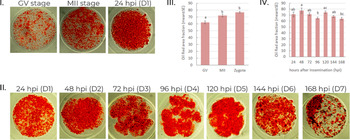
In experiment 1, oocytes before and after IVM and zygotes were stained to investigate if lipid accumulation was relevant during IVM or IVF in our system. Then, embryos from D1 (24 hpi) to D6 (144 hpi) before cavitation were collected and stained to investigate lipid accumulation during embryo culture.
Treatment with lipid modulators cocktail (forskolin, l-carnitine, and linoleic acid) was carried out during IVM in Experiment 2. Comparison between Control and treated groups for lipid content (matured oocytes and D7 blastocysts) and developmental rates (cleavage and blastocyst formation) was performed. Vitrified-warmed D7 blastocysts were cultured and re-expansion and hatching rates were recorded after 4, 24 and 48 h. After 48 h culture, blastocysts were fixed and stained to estimate embryo quality (blastocyst cell numbers and apoptosis rate).
In experiment 3, treatment with lipid modulators cocktail was carried out during embryo culture. Comparison among Control and groups treated at D4 (supplemented at D4–D7) or D6 (supplemented at D6–D7 at double levels) was performed for lipid content (in D7 blastocysts) and developmental rates (blastocyst formation).
In experiments 2 and 3, vitrified-warmed D7 blastocysts were cultured and re-expansion and hatching rates were recorded after 4, 24 and 48 h. After 48 h culture, blastocysts were fixed and stained to estimate embryo quality (blastocyst cell numbers and apoptosis).
Cumulus–oocyte complexes (COC) recovery and IVM
COCs were obtained from slaughterhouse ovaries. Ovary transport was performed in thermic bottle at 37°C and COCs were recovered by follicular aspiration with needle and syringe. Follicular fluid was examined under a stereoscope and COCs displaying compact nonatretic cumulus with more than two layers of cells and homogeneous cytoplasm were selected for the experiment and divided between groups for IVM. IVM was performed in 4-well plates in groups of 80–100 COCs and 500 μl medium per well. TCM199 was supplemented with 10% FCS, 1 μg/ml follicle stimulating hormone (Folltropin™, Bioniche Animal Health, Belleville, Canada), 50 μg/ml hCG (Chorulon™, MSD Saude Animal, São Paulo, Brazil), 1 μg/ml 17β estradiol, 16 μg/ml sodium pyruvate, 10,000 units penicillin and 10 mg streptomycin/ml. Lipid modulators cocktail composed of 2.5 mM l-carnitine, 150 µM CLA and 15 µM forskolin was added to the Lip. Mod. IVM group. Plates were incubated for 24 h at 38.5°C, 5% CO2 in atmospheric air and high humidity.
Embryo production
Following IVM, IVF was performed using frozen straws of bovine semen thawed at 36.5°C for 30 s. Semen was transferred to a 45/90 Percoll gradient and centrifuged at 3600 g for 7 min. After supernatant removal, the pellet was resuspended with 700 μl HEPES-buffered TCM199 medium and centrifuged at 520 g for 5 min. The pellet was collected (50 μl) and transferred to Tyrode’s albumin lactate pyruvate (TALP) medium supplemented with 0.6% bovine serum albumin (BSA), 10 μg/ml heparin, 18 μM penicillamine, 10 μM hypotaurine and 1.8 μM epinephrine. The spermatozoa suspension was adjusted to the proportion of 500 cells per oocyte. Co-culture of oocytes with spermatozoa was conducted in 70-μl drops of TALP medium covered with mineral oil for 18 h (38.5°C, 5% CO2 in atmospheric air and high humidity). After IVF, presumptive zygotes were submitted to vigorous pipetting for cumulus cells removal and transferred in groups of 15 structures to 100-μl drops of synthetic oviductal fluid with amino acids (SOF-AA) medium supplemented with 1.5% FBS and 6 mg/ml BSA medium under mineral oil. Embryos were kept for 7 days (168 h) at 38.5°C, 5% CO2 in atmospheric air and maximum humidity. Feeding was carried out at D4 and D6. Supplementation of lipid modulators cocktail (2.5 mM l-carnitine, 150 µM CLA and 15 µM forskolin) was performed in the D4 group from D4 to D7, and at double amounts (5 mM l-carnitine, 300 µM CLA and 30 µM forskolin) in the D6 group from D6 to D7.
Vitrification and warming
D7 grade 1 blastocysts were selected and transferred to HEPES-buffered SOF-AA. Embryos were vitrified in groups of 3–5 in two steps according to the Vajta et al. (Reference Vajta, Holm, Kuwayama, Booth, Jacobsen, Greve and Callesen1998) protocol with minor modifications. First, embryos were transferred to HEPES-buffered SOF-AA supplemented with 7.5% ethylene glycol and 7.5% DMSO for 3 min. Then they were transferred to HEPES-buffered SOF-AA supplemented with 15% ethylene glycol and 15% DMSO and a minimum drop (0.1–0.5 μl) containing embryos was placed in a vitrification device (WTA, Cravinhos, Brazil) and directly immersed in liquid nitrogen within 30 s. Embryos were kept in liquid nitrogen for at least 1 week. For warming, a vitrification device tip was rapidly placed in 0.3 M sucrose HEPES-buffered SOF-AA for 3 min and then transferred to 0.15 M sucrose HEPES-buffered SOF-AA for additional 3 min. Embryos were washed twice in HEPES-buffered SOF-AA for 5 min and moved in groups of 7–10 to 50 μl SOF-AA drops. Embryos were cultured for 48 h at 38.5°C, 5% CO2 in atmospheric air and maximum humidity. Evaluation of re-expansion and hatching was performed 4, 24 and 48 h after warming.
Oil Red O staining
Enzymatically (TrypLE Express, ThermoFisher) denuded oocytes and blastocysts were fixed in 4% paraformaldehyde for 30 min at 37°C and stored at 4°C in phosphate-buffered saline (PBS). Oocytes and embryos were moved to 50% ethanol in distilled water for 2 min and transferred to 2.45 mg/ml Oil Red O solution in 70% isopropanol 30% distilled water for 30 min, and after they were rapidly washed on 50% ethanol in distilled water and transferred to PBS. Oocytes and embryos were mounted on slides and images were captured under an inverted microscope. Analysis was performed using ImageJ software (NIH, USA) for the percentage of stained area from total cytoplasm on a two-dimensional photograph (lipid area fraction).
Caspase 3 and Hoechst staining
Vitrified-warmed blastocysts were fixed in 4% paraformaldehyde for 30 min at 37°C and stored at 4°C in PBS. Immunofluorescence for caspase 3 and nuclei staining with Hoechst stain was performed as previously described (Oliveira et al., Reference Oliveira, Saraiva, de Lima, Oliveira, Serapião, Garcia, Borges and Camargo2016). Briefly, embryos were permeabilized in 0.5% Triton X-100 solution for 30 min and washed three times in PBS supplemented with 0.2% Tween-20 (PBS-T) for 10 min. Blocking was performed for 30–60 min in 3% BSA at room temperature. Incubation with primary antibody (anti-caspase 3, rabbit, 1:750) was done at 4°C for 12 h. The embryos were then washed three times in PBS-T and incubated with secondary antibody (donkey anti-rabbit IgG conjugated to Alexa Fluor 555, 1:400) for 30–120 min at room temperature. Nuclei were stained with Hoechst 33342 stain for 20 min and embryos were examined under a fluorescence microscope (Alexa Fluor 555: wavelengths Ex 556–Em 573 nm; Hoechst 33342: wavelengths Ex 352–Em 461 nm). Images were taken for each embryo and cells were counted using ImageJ software (NIH, USA) for total cell number and apoptosis assessment.
Statistical analysis
Lipid area fraction comparison was performed using Kruskal–Wallis and Dunn tests for time course analysis, T-test for experiment 1 and analysis of variance (ANOVA) and Tukey for experiment 2. Cleavage, blastocyst, re-expansion, and hatching rates were compared using Fisher’s exact test. Apoptosis rate was compared using chi-squared test with Yates correction. All analysis were performed at 5% level of significance in Instat GraphPad software.
Results
Lipid accumulation during maturation, fertilization and embryo culture
In experiment 1, we wished to check whether the accumulation of lipids occurred stably during culture or if there was a specific time point in early embryo development when lipid accumulation was triggered. First, we evaluated IVM and IVF (n = 112 oocytes/zygotes, 32–43 per time point obtained in three replicates) (Figure 1.I and 1.III). Oil Red O staining (Figure 1.I) revealed that the cytoplasmic lipid fraction was increased (P < 0.05) after IVM [matured oocytes vs germinal vesicle (GV) oocytes] and no difference was found comparing matured oocytes and zygotes (Figure 1.III). For lipid accumulation during embryo culture (n = 133 embryos, 12–28 per time point obtained in four replicates) (Figure 1.II and 1.IV), we detected an increase in lipid content from 96 hpi (D4) to 120 hpi (D5) (Figure 1.IV). Therefore, lipid accumulation occurred during IVM and embryo culture (D4–D5) under our conditions.
Lipid accumulation during IVM and embryo culture. I. Images of oocytes (before and after IVM) and zygotes stained with Oil Red are shown. II. Images of IVF embryos (D1–D6) stained with Oil Red are shown. Structures were collected at each time point as follows: −24 hpi: GV oocyte; 0 hpi: metaphase II (MII) oocyte; 24 hpi: zygote and 2-cell; 48 hpi: 2-, 4- and 8 cell; 72 hpi: 4-, 8- and 16-cell; 96 and 120 hpi: 8- and 16-cell; 144 hpi: 16-cell and morula. III, IV. Graphs show cytoplasmic area fraction (%) of oocytes and zygotes (III) and embryos (IV). Different letters show statistically significant differences.

Supplementation of lipid modulators cocktail during IVM reduces lipid content in oocytes but not in blastocysts
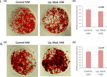
Then we wanted to determine if supplementation of lipid modulators could reduce lipid accumulation during IVM and if this would reflect in D7 blastocysts lipid content. The use of lipid modulators during IVM reduced (P < 0.05) the cytoplasmic lipid fraction compared with oocytes matured in the Control medium (n = 88–104 matured oocytes per group obtained in four replicates) (Figure 2.I). However, comparison between blastocysts developed after fertilization of treated oocytes showed no difference in lipid content (n = 15–25 blastocysts obtained in three replicates) (Figure 2.II). Our results demonstrated that lipid content drop in oocytes is not maintained after IVF and embryo culture.
Lipid modulators supplementation during IVM: effects in lipid levels of matured oocytes and blastocysts. I. (a) Matured oocytes stained with Oil red O from Control and Lip. Mod. IVM groups. (b) Graph shows cytoplasmic area fraction (%). II. (a) Blastocysts stained with Oil red O from Control and Lip. Mod. IVM groups. (b) Graph shows cytoplasmic area fraction (%).

Supplementation of lipid modulators cocktail during IVM decreased embryo development and cryosurvival
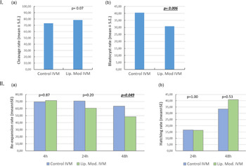
In Figure 3.I, we show the development of fertilized oocytes matured in Control or Lip. Mod. medium (n = 451–514 oocytes per group, obtained in six replicates). Cleavage rate was similar (P = 0.07) between Lip. Mod. IVM or Control IVM (Figure 3.Ia), and a tendency for higher rates was observed for Lip. Mod. IVM, suggesting a positive effect on oocyte quality. However, blastocyst rate was lower (P = 0.006) in the Lip. Mod. IVM group (Figure 3.Ib).
Lipid modulators supplementation during IVM: embryo development and cryosurvival. I. Cleavage rate (a) and blastocyst rate (b) are shown. II. Survival of vitrified-warmed blastocysts produced with oocytes matured in Control or Lip. Mod. media. Re-expansion rates (a) and hatching rate (b) after warming are shown. Comparison between groups was performed at each time point and P-values are shown.

D7 blastocysts produced from oocytes matured in Lip. Mod. or Control IVM medium were vitrified and warmed for cryosurvival analysis (n = 86–91 per group, obtained in six replicates). No difference (P > 0.05) was detected between groups for blastocyst re-expansion rate 4 or 24 h after warming, but a decreased re-expansion rate was found 48 h after warming (Figure 3.IIa). Hatching rates remained similar (P > 0.05) between groups at 24 and 48 h (Figure 3.IIb). After the 48 h culture, vitrified-warmed embryos were fixed and stained for nuclei (Hoechst 33342 stain) and immunostained for active caspase 3 enzyme (n = 28–18 per group, obtained in two replicates). No difference was detected (P > 0.05) in mean cell number (Figure 4a) or apoptosis rate (Figure 4b) between Control and Lip. Mod. IVM blastocysts.
Lipid modulators supplementation during IVM: quality of vitrified-warmed blastocysts. Graphs show total cell number (a) and apoptosis rate (b).

Therefore, lipid content drop in matured oocytes did not reflect the improvements in embryo production rates, cryosurvival or embryo quality. In contrast, lower blastocyst rates and decreased re-expansion rates were detected for the treated group.
Supplementation of lipid modulators cocktail during embryo culture increases lipid content in blastocysts
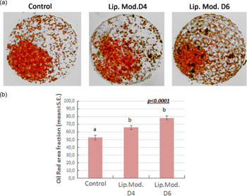
Next, we wanted to determine if supplementation of lipid modulators during embryo culture could reduce lipid accumulation and affect lipid content in D7 blastocysts. The use of lipid modulators during embryo culture increased (P < 0.05) the cytoplasmic lipid fraction compared with the Control, for both D4 and D6 groups (Figure 5, n = 17–24 per group obtained in two replicates).
Lipid modulators supplementation during embryo culture: lipid levels. (a) Images of Oil red O stain of Control, Lip. Mod. D4 and D6 groups. (b) Graph shows stained area fraction of blastocysts. Different letters show statistically significant differences.

Our results showed a clear effect of lipid modulator supplementation during embryo culture, in the opposite direction that was expected.
Supplementation of lipid modulators cocktail during embryo culture does not alter embryo development or cryosurvival
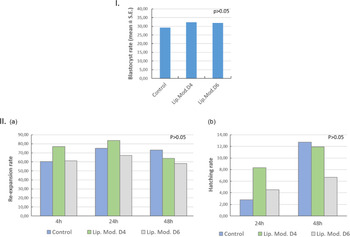
In Figure 6.I, we show the development of embryos cultured in Control or Lip. Mod. medium (n = 380–393 oocytes per group, obtained in four replicates). Cleavage rate was assessed before treatment, as a quality control of the experiment, and was 77.99%. Blastocyst rate was similar (P > 0.05) among groups (Figure 6.Ib).
Lipid modulators supplementation during embryo culture: embryo development and cryosurvival. I. Blastocyst rates after lipid modulators supplementation. II. Re-expansion rates (a) and hatching rate (b) after warming are shown. Comparison among groups was performed at each time point and P-values higher than 0.05 were found in all analyses.

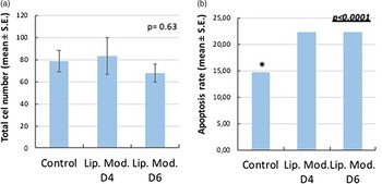
D7 blastocysts produced from Lip. Mod. D4, Lip. Mod. D6 and Control groups were vitrified and warmed for cryosurvival analysis (n = 40–52 per group, obtained in three replicates). No difference (P > 0.05) was detected among groups in terms of blastocyst re-expansion rates 4, 24 or 48 h after warming (Figure 6.IIa). Hatching rates also remained similar (P > 0.05) 24 and 48 h among groups (Figure 6.IIb). After 48 h of culture, vitrified-warmed embryos were fixed and stained for nuclei (Hoechst 33342 stain) and immunostained for active caspase 3 enzyme (n = 28–12 per group, obtained in two replicates). No difference was detected (P > 0.05) in mean cell numbers (Figure 7a), but apoptosis rate was increased in Lip. Mod. D4 and D6 groups compared with the Control (Figure 7b).
Lipid modulators supplementation during embryo culture: quality of vitrified-warmed blastocysts. Graphs show total cell number (a) and apoptosis rate (b). Asterisks indicate statistical difference of marked group.

Therefore, increased lipid content detected after lipid modulators supplementation did not reflect improvements in embryo production rates or cryosurvival, but negatively affected embryo quality.
Discussion
In this report, we combined for the first time three well known lipid modulators for intensive effects on oocyte and embryo lipid profiles and investigated their role in early embryo development and cryopreservation. Three routes of lipid modulation were affected: enzymatic hydrolysis (forskolin); metabolism increase (l-carnitine); and lipid profile improvement (CLA). Concentrations of the modulators used in the present study were based on previous standardization experiments from our group (de Lima et al., Reference de Lima, Saraiva, Oliveira and Garcia2017; Garcia et al., Reference Garcia, de Lima, Saraiva and Oliveira2017). Concentrations added to the embryo culture that led to the best results for cryosurvival after vitrification were selected for this experiment. All supplements were already well characterized alone for their positive effects on early embryo development and lipid modulation.
Forskolin treatment in oocytes reduced oocyte lipid content (Fu et al., Reference Fu, Wu, Li, Hou, Zhou, Lun-Suo, Wang and Zhu2011) and increased unsaturated lipids in developed blastocysts (Razza et al., Reference Razza, Sudano, Fontes, Franchi, Belaz, Santos, Castilho, Rocha, Eberlin, Machado and Nogueira2018). Forskolin supplementation during the final stages of early embryo development led to improved cryotolerance (Paschoal et al., Reference Paschoal, Sudano, Guastali, Dias Maziero, Crocomo, Oña Magalhães, Da Silva Rascado, Martins and Da Cruz Landim-Alvarenga2014) and reduced lipid content in blastocysts (Paschoal et al., Reference Paschoal, Sudano, Schwarz, Maziero, Guastali, Crocomo, Magalhães, Martins, Leal and Landim-Alvarenga2017). Reduced lipid content and improved cryosurvival was also reported in porcine embryos (Men et al., Reference Men, Agca, Riley and Critser2006).
l-Carnitine supplementation during IVM reduced oxidative stress, improved embryo quality (Sovernigo et al., Reference Sovernigo, Adona, Monzani, Guemra, Barros, Lopes and Leal2017), and affected lipid droplets localization in the cytoplasm (Chankitisakul et al., Reference Chankitisakul, Somfai, Inaba, Techakumphu and Nagai2013). During embryo culture, l-carnitine reduced lipid content and improved survival rates after slow freezing (Takahashi et al., Reference Takahashi, Inaba, Somfai, Kaneda, Geshi, Nagai and Manabe2013). In previous studies from our group, supplementation of l-carnitine at D4 increased blastocyst production (62.0 vs 53.6 Control), and improved expansion (60.7 vs 19.6 Control) and hatching rates (41.4% vs 14.3 Control) of vitrified-warmed embryos (Garcia et al., Reference Garcia, de Lima, Saraiva and Oliveira2017).
The t10,c12 CLA isomer increased neutral lipids storage when added to IVM medium (M. Carro et al., Reference Carro, Buschiazzo, Ríos, Oresti and Alberio2013), affecting oocyte lipid profile and improving the morphology of embryos developed after fertilization of treated oocytes (Phongnimitr et al., Reference Phongnimitr, Liang, Srirattana, Panyawai, Sripunya, Treetampinich and Parnpai2013). Supplementation of t10,c12 CLA during embryo culture has had controversial effects. CLA supplementation during embryo culture reduced cytoplasmic lipid deposition and improved cryosurvival (Pereira et al., Reference Pereira, Baptista, Vasques, Horta, Portugal, Bessa, Silva, Pereira and Marques2007), but lower embryo survival was also reported using the same concentration (Al Darwich et al., Reference Al Darwich, Perreau, Petit, Papillier, Dupont, Guillaume, Mermillod and Guignot2010). In another study, lipid content was increased in embryos supplemented with CLA, and no positive effects were reported for vitrification (Absalón-Medina et al., Reference Absalón-Medina, Bedford-Guaus, Gilbert, Siqueira, Esposito, Schneider, Cheong and Butler2014). Under our conditions, supplementation of t10,c12 CLA at D4 and D6 of embryo culture increased the expansion rate (70.4 vs 34.7 Control) and hatching rate (50.0% vs 16% Control) following vitrification and warming, with best results when supplementation was performed at D4 (de Lima et al., Reference de Lima, Saraiva, Oliveira and Garcia2017).
Our first finding was that lipid accumulation occurred during IVM under our conditions. Although in some studies lipid content was found to decrease during IVM (Ferguson and Leese, Reference Ferguson and Leese1999; Kim et al., Reference Kim, Kinoshita, Ohnishi and Fukui2001), other studies corroborated our findings and demonstrated lipid accumulation at this stage (Aardema et al., Reference Aardema, Vos, Lolicato, Roelen, Knijn, Vaandrager, Helms and Gadella2011; Del Collado et al., Reference Del Collado, Saraiva, Lopes, Gaspar, Padilha, Costa, Rossi, Vantini and Garcia2015). IVM also affected lipid patterns in oocytes, altering diacylglycerols (DGs) and triacylglycerols (TGs) patterns compared with in vivo matured oocytes (del Collado et al., Reference del Collado, Da Silveira, Oliveira, Alves, Simas, Godoy, Coelho, Marques, Carriero, Nogueira, Eberlin, Silva, Meirelles and Perecin2017). As neutral lipids are important intracellular energy storage molecules and are also involved in membrane biosynthesis during early embryonic development (Hizaki et al., Reference Hizaki, Segi, Sugimoto, Hirose, Saji, Ushikubi, Matsuoka, Noda, Tanaka, Yoshida, Narumiya and Ichikawa1999; Hochi et al., Reference Hochi, Kimura and Hanada1999), and oocyte quality is associated with a superior lipid profile (Hochi et al., Reference Hochi, Kimura and Hanada1999), lipid modulation at this stage could have positive effects.
The combined use of forskolin, l-carnitine and CLA in our study decreased lipid accumulation in oocytes during IVM. However, this effect was transient as it was not detected in blastocysts. In addition, no improvement on embryo production and cryosurvival was detected. In addition to that, blastocyst rate and re-expansion rate was decreased in treated groups. Therefore, the oocyte lipid profile seems to be less relevant than embryo lipid profile for bovine blastocysts, and the expected benefits of lipid modulation during IVM were not detected at blastocyst stage.
Lipid accumulation also occurred during embryo culture, between 96 hpi (D4) and 120 hpi (D5). Corroborating our results, Sudano and colleagues (Sudano et al., Reference Sudano, Rascado, Tata, Belaz, Santos, Valente, Mesquita, Ferreira, Araújo, Eberlin and Landim-Alvarenga2016) found that ACSL3 transcripts (activates long-chain fatty acids for both synthesis of cellular lipids) and cytoplasmic lipid droplets increased at the morula stage, followed by a decrease at the blastocyst stage. For this reason, supplementation with lipid modulators was carried out from D4 onwards. The addition of a higher concentration of lipid modulators from D6 to D7 aimed to promote intense modulation only at the final stage of early embryo development. This is usually the stage when blastocyst number is estimated, which represents a strategic stage for the treatment of embryos that would be vitrified.
Under our conditions, supplementation of embryo culture medium increased lipid content both in D4 (1.25 times the control) and D6 (1.5 times the control). Despite the enhanced lipid content, lipid modulators had no detrimental effects on blastocyst rates or cryosurvival. Considering the metabolism of CLA, lipid accumulation based on TAG and phospholipids possibly occurred (T. Carro et al., Reference Carro, Buschiazzo, Ríos, Oresti and Alberio2013). As discussed above, Absalón-Medina reported increases in lipid content following CLA supplementation during embryo culture (Absalón-Medina et al., Reference Absalón-Medina, Bedford-Guaus, Gilbert, Siqueira, Esposito, Schneider, Cheong and Butler2014) which was not the case for forskolin and l-carnitine. However, this accumulation could have positive effects for embryo quality as it suggests the incorporation of neutral lipids and establishment of a superior lipid profile, which has an important association with cryotolerance (Sudano et al., Reference Sudano, Santos, Tata, Ferreira, Paschoal, Machado, Buratini, Eberlin and Landim-Alvarenga2012).
In our study, no positive effect of lipid modulators was seen on embryo development or cryosurvival. Additional studies to determine intracellular and membrane lipid composition, and post-transfer embryo development, would be of great interest and could reveal the benefits of intense lipid modulation. This is because, although lipid reduction observed after forskolin supplementation has not improved early embryo development, pregnancy rate increases of approximately 30% were reported after the culture for 48 h of embryos in forskolin-supplemented medium (Sanches et al., Reference Sanches, Marinho, Filho, Pontes, Basso, Meirinhos, Silva-Santos, Ferreira and Seneda2013). Therefore, it is possible that positive effects of lipid modulators cocktail are detectable only after embryo transfer.
We reported an effect on embryo quality, considering that vitrified-warmed blastocysts from treated groups presented increased apoptosis. It is possible that the metabolism of high lipid content embryos produced an excess of oxygen reactive species after vitrification-warming, when lipid modulator supplement was absent, as l-carnitine attenuates the oxidative stress (Sovernigo et al., Reference Sovernigo, Adona, Monzani, Guemra, Barros, Lopes and Leal2017). In this regard, no changes in cell numbers were seen and, as apoptosis is a physiological event and can represent a survival mechanism under stressful conditions (Paula-Lopes et al., Reference Paula-Lopes, Lima, Satrapa and Barros2013), its effects on further development remain to be elucidated.
We conclude that lipid modulators cocktail supplementation during IVM and embryo culture affected lipid accumulation in oocytes and embryos. However, no positive effects on early embryo development or cryosurvival could be detected. Further studies are needed to define the effects of this supplement on lipid profile and post-transfer embryo development.
Funding/Acknowledgements
This work was supported by Embrapa (grant 12.13.06.020.00.00).
Ethics
All procedures followed ethical guidelines for experimentation.
Conflict of interest
The authors declare that this scientific work was conducted following the ethical guidelines of research and there is no conflict of interest.