Research within the domain of developmental psychopathology has increasingly evaluated immediate and longer term consequences of chronic stress exposure among children. Moreover, a prominent theme of recent research on children's stress response is that stress impacts both biological and psychological functioning throughout development. The hypothalamic–pituitary–adrenal (HPA) axis is the primary neuroendocrine stress response system in mammals (McEwen & Stellar, Reference McEwen and Stellar1993; Sapolsky, Reference Sapolsky1994). As such, the HPA axis has received increased attention as research on biological aspects of children's stress response evolves.
To date, children's HPA axis functioning has most often been assessed in the context of an isolated traumatic event (Goenjian et al., Reference Goenjian, Yehuda, Pynoos, Steinberg, Tashjian and Yang1996), repetitive trauma (Cicchetti & Rogosch, Reference Cicchetti and Rogosch2001; DeBellis, Reference DeBellis2001), or chronic stressful life circumstances (Ashman, Dawson, Panagiotides, Yamada, & Wilkinson, Reference Ashman, Dawson, Panagiotides, Yamada and Wilkinson2002; Lupien, King, Meaney, & McEwen, Reference Lupien, King, Meaney and McEwen2000). Few studies have examined the cumulative effects of trauma and life stress on HPA axis functioning. The present study evaluates the independent and combined effects of recent trauma and life stress as well as previous trauma exposure on basal morning and afternoon cortisol levels among school-aged children.
HPA Axis Stress Response
The HPA axis is a complex system in which components work together under both nonstressful and stressful conditions (Lupien & McEwen, Reference Lupien and McEwen1997; Sapolsky, Krey, & McEwen, Reference Sapolsky, Krey and McEwen1984). Various inputs converge upon neurons of the hypothalamic paraventricular nucleus and activate increased production of corticotropin-releasing hormone (CRH). In combination with other hormones such as vasopressin, CRH stimulates the production of adrenocorticotropin hormone (ACTH) in the anterior pituitary. ACTH secreted into circulation stimulates cells of the adrenal cortex to produce and release cortisol. HPA axis hormones regulate their own release through a complex negative feedback system (Sapolsky et al., Reference Sapolsky, Krey and McEwen1984). Under nonstressful conditions, the HPA axis acts with a normal circadian rhythm, reaching daily morning peaks and early evening nadirs in humans. Basal levels of cortisol are necessary for normal brain growth and support metabolic activity necessary to sustain general functioning.
Under stressful conditions, perceptions of physical or psychological stress activate the HPA axis and resultant elevations in cortisol levels maximize an organism's ability to manage stressful experiences competently, both physiologically and behaviorally (Sapolsky, Reference Sapolsky1994). Although short-term elevations in cortisol levels can be critical for survival in stressful circumstances, prolonged exposure to elevated cortisol levels can present serious health risks including muscle atrophy, decreased sensitivity to insulin, hypertension, arterial disease, impaired growth and tissue repair, as well as neurological damage (Arnaldi, Mancini, Polenta, & Boscaro, Reference Arnaldi, Mancini, Polenta and Boscaro2004; Brotman et al., Reference Brotman, Girod, Garcia, Patel, Gupta and Posch2005; Sapolsky, Reference Sapolsky2004b). In addition, hyperactivity of the HPA axis is one of the best replicated findings in the neurobiology of depression (Bremner & Vermetten, Reference Bremner and Vermetten2001; Guechot et al., Reference Guechot, Lepine, Cohen, Fiet, Lemperiere and Dreaux1987).
Like high levels, chronic exposure to low levels of cortisol is associated with health risks including asthma, allergies, disorders related to hyperactivity of the immune system, and neuronal damage (Heim, Ehlert, & Hellhammer, Reference Heim, Ehlert and Hellhammer2000; Kruger & Spiecker, Reference Kruger and Spiecker1994; McEwen, Reference McEwen1998). Lack of cortisol may increase vulnerability to both bodily stress-related disorders such chronic pain syndromes (Crofford, Pillemer, & Kalogeras, Reference Crofford, Pillemer and Kalogeras1994; Griep, Boersma, deKloet, Reference Griep, Boersma and deKloet1993; Heim, Ehlert, Hanker, & Hellhammer, Reference Heim, Ehlert, Hanker and Hellhammer1998) and posttraumatic stress disorder (PTSD) in adults and adolescents (Goenjian et al., Reference Goenjian, Yehuda, Pynoos, Steinberg, Tashjian and Yang1996; Mason, Giller, Kosten, Ostroff, & Podd, Reference Mason, Giller, Kosten, Ostroff and Podd1986; Yehuda, Boisoneau, Mason, & Giller, Reference Yehuda, Boisoneau, Mason and Giller1993; Yehuda, Boisoneau, Lowy, & Giller, Reference Yehuda, Boisoneau, Lowy and Giller1995; Yehuda et al., Reference Yehuda, Southwick, Nussbaum, Wahby, Giller and Mason1990). Because chronic exposure to both high and low cortisol concentrations are associated with a host of physical and psychological symptoms, it is important to understand conditions under which these alterations of basal HPA axis functioning are most likely to occur.
Stress and Basal HPA Axis Functioning
Beginning with seminal studies conducted by Selye (Reference Selye1936), stress has been associated with activation of the HPA axis culminating in elevated cortisol levels. In early research, stress and increased cortisol secretion became synonymous such that the presence of hypercortisolism, the condition in which cortisol levels remain high over the day and into evening hours was used to define states of stress (Heim et al., Reference Heim, Ehlert and Hellhammer2000). Many studies demonstrate positive associations between stress exposure and both concurrent cortisol levels and secretion of cortisol in response to subsequent stressors (Cicchetti & Rogosch, Reference Cicchetti and Rogosch2001; Gunnar, Morison, Chisholm, & Schuder, Reference Gunnar, Morison, Chisholm and Schuder2001).
Thus, exposure to chronic stress, which includes both earlier and current stressful events, may result in heightened activation of the HPA axis, culminating in elevated basal cortisol levels (Cicchetti & Rogosch, Reference Cicchetti and Rogosch2001; Gunnar et al., Reference Gunnar, Morison, Chisholm and Schuder2001). Indeed, it has recently been suggested that the duration of stress exposure may be an important factor in the association between stress and hypercortisolism (DeBellis et al., Reference DeBellis, Baum, Birmaher, Keshavan, Eccard and Boring1999; Essex, Klein, Cho, & Kalin, Reference Essex, Klein, Cho and Kalin2002; Gunnar & Vazquez, Reference Gunnar and Vazquez2001). Basal cortisol hypersecretion has been observed among children from low socioeconomic status (SES) families compared to those from medium and high SES families (Lupien et al., Reference Lupien, King, Meaney and McEwen2000; Lupien, King, Meaney, & McEwen, Reference Lupien, King, Meaney and McEwen2001), infants and children whose mothers are severely depressed (Ashman et al., Reference Ashman, Dawson, Panagiotides, Yamada and Wilkinson2002; Field et al., Reference Field, Healy, Goldstein, Perry, Bendall and Schanberg1988; Hessl et al., Reference Hessl, Dawson, Frey, Panagiotides, Self, Yamada, Hann, Huffman, Lederhendler and Minecae1998), child victims of sexual and physical abuse (Cicchetti & Rogosch, Reference Cicchetti and Rogosch2001; Hart, Gunnar, & Cicchetti, Reference Hart, Gunnar and Cicchetti1996; Kaufman, Reference Kaufman1991), and children reared in Romanian orphanages that pose multiple risks to healthy development (Carlson & Earls, Reference Carlson and Earls1997; Gunnar et al., Reference Gunnar, Morison, Chisholm and Schuder2001).
Although many theorists identify the duration of stress exposure as a potentially important contributor to its impact on HPA axis functioning, only one study has systematically evaluated the combined effects of early and concurrent stress on children's basal cortisol levels. Essex et al. (Reference Essex, Klein, Cho and Kalin2002) examined the relation between high levels of concurrent maternal stress (e.g., maternal depression, role overload, financial stress) and maternal stress in infancy and afternoon/evening salivary cortisol levels among 282 children who were 4.5 years old. Children exposed to only high levels of concurrent or early stress had cortisol levels that did not significantly differ from those never exposed to stress. However, children exposed to high levels of stress at both time points had significantly higher cortisol levels than children never exposed to maternal stress. Thus, repetitive exposure to maternal stress was associated with elevated afternoon/evening cortisol concentrations, which suggests that stress exposure in infancy may sensitize children to the deleterious effects of later stress exposures.
Despite the long-time emphasis on hypercortisolism, neurobiological studies are increasingly supporting a different and more complex pattern of HPA axis functioning in which individuals exposed to a wide array of stressors exhibit basal hypocortisolism, exhibited by lower than expected cortisol concentrations particularly near the height of circadian rhythm (for review, see Gunnar & Vazquez, Reference Gunnar and Vazquez2001; Heim et al., Reference Heim, Ehlert and Hellhammer2000). Insecure attachment and less responsive parental care during infancy are associated with lower morning cortisol levels (Spangler & Grossman, Reference Spangler and Grossman1993) and blunted cortisol response to stressors in early childhood (Marsland et al., Reference Marsland, Epel, Brown, Anderson, Kogen and Little2000). Similarly, a flattening of the expected daily rhythm resulting from relatively low early morning basal cortisol has been observed among infants with colic (Finsterward, Selig, Schieche, Wurmser, & Papousek, Reference Finsterward, Selig, Schieche, Wurmser and Papousek2000) and 2-year-old children living under neglectful conditions in Romanian orphanages (Carlson et al., Reference Carlson, Dragomir, Earls, Farrell, Macovei and Nystrom1995a, Reference Carlson, Dragomir, Earls, Farrell, Macovei and Nystrom1995b; Carlson & Earls, Reference Carlson and Earls1997).
Based on experimental data and theoretical considerations, several authors have proposed theories to explain the development of hypocortisolism and the physiological mechanisms behind the phenomenon (DeBellis et al., Reference DeBellis, Baum, Birmaher, Keshavan, Eccard and Boring1999; Dienster, Reference Dienstbier1989; Hellhammer & Wade, Reference Hellhammer and Wade1993; McEwen, Reference McEwen1998; Yehuda, Resnick, Kahana, & Giller, Reference Yehuda, Resick, Kahana and Giller1993). According to several of these theories, initial exposure to significant stressors result in hypersecretion of basal HPA axis hormones and hyperactivity of HPA axis responses to subsequent stressors. However, a longer term and possibly developmental consequence of exposure to significant stress involves hyposecretion of HPA axis hormones, presumably because frequent elevations in cortisol result in the downregulation of HPA axis components and ultimately, decreased adrenocortical secretion (Gunnar & Vazquez, Reference Gunnar and Vazquez2001; Yehuda, Giller, Southwick, Lowy, & Mason, Reference Yehuda, Giller, Southwick, Lowy and Mason1991). Mechanisms potentially associated with hypocortisolism include reduced biosynthesis at various levels of the axis, downregulation of pituitary CRH receptors secondary to chronic elevations or hypersecretion of CRH, downregulation of ACTH receptors in response to elevated ACTH secretion, and increased feedback regulation of cortisol, perhaps through increased cortisol receptors (Heim et al., Reference Heim, Ehlert and Hellhammer2000; Yehuda et al., Reference Yehuda, Levengood, Schmeidler, Wilson, Guo and Berber1996).
Because it is presumed that hypocortisolism is a long-term consequence of prolonged HPA axis hyperactivity, it may be most likely to occur under two conditions. First, exposure to an initial stressor must be associated with significant and prolonged HPA axis hyperactivity, which may be contingent on the perceived severity of the stressor and/or its duration. This criterion demands that the stressor is perceived as significant enough by the organism to facilitate a prolonged physiological response. Second, there is some indication that hypocortisolism is most likely to be evidenced if basal cortisol levels are measured when a significant period of time has passed since the initiation of stress exposure. Indeed, research conducted with rodents, nonhuman primates, and humans demonstrate that stress exposure may affect basal hypocortisolism in a time-dependent manner such that stress reactivity involves initial increases, but subsequent decreases in basal glucocorticoid levels (DeBellis et al., Reference DeBellis, Baum, Birmaher, Keshavan, Eccard and Boring1999; Goenjian et al., Reference Goenjian, Yehuda, Pynoos, Steinberg, Tashjian and Yang1996; Houchyar, Cooper, & Woods; Reference Houchyar, Cooper and Woods2001; Mason, Brady, & Tolliver, Reference Mason, Brady and Tolliver1968; Natelson et al., Reference Natelson, Ottenweller, Cook, Pitnam, McCarthy and Tapp1988; Rivier & Vale, Reference Rivier and Vale1987; Sanchez et al., Reference Sanchez, Noble, Lyon, Plotsky, Davis and Nemeroff2005).
Sanchez et al. (Reference Sanchez, Noble, Lyon, Plotsky, Davis and Nemeroff2005) investigated the effects of maternal separation on rhesus monkeys' cortisol secretion at the time of stress exposure, as well as 12 and 22 months later. Maternal separation occurred repetitively when the monkeys were between 3 and 6 months of age. Elevations in basal cortisol levels were evidenced immediately after maternal separation. However, analysis of diurnial plasma cortisol concentrations measured at 12 months of age revealed that monkeys who underwent maternal separation between 3 and 6 months of age had lower a.m. cortisol levels than those not exposed to maternal separation. Hypocortisolism detected at 12 months was still evident at 22 months, although suppression of basal levels was smaller, suggesting that the flattened cortisol rhythm of macaques previously exposed to repetitive maternal separation may be transient and a normal pattern may reemerge over time (Sanchez et al., Reference Sanchez, Noble, Lyon, Plotsky, Davis and Nemeroff2005).
Similarly, the length of time since active stress exposure may have influenced HPA axis functioning among sexually abused girls recruited within 6 months of abuse disclosure compared with nonabused sociodemographically matched girls (Putnam, Trickett, Helmers, Dorn, & Everett, Reference Putnam, Trickett, Helmers, Dorn and Everett1991). Augmented plasma cortisol concentrations among sexually abused girls relative to controls soon after abuse disclosure suggest that CRH hypersecretion may have occurred among abused girls during the emotional and physical stress associated with the traumatic assaults. Prolonged CRH hypersecretion may have led to an adaptive downregulation of CRH receptors in the anterior pituitary of the sexually abused girls, resulting in blunted basal cortisol secretion or return to normal cortisol levels in the long term (DeBellis et al., Reference DeBellis, Baum, Birmaher, Keshavan, Eccard and Boring1999). Indeed, several years after abuse disclosure, a subsample of these same sexually abused girls exhibited blunted ACTH secretion in response to exogenous CRH challenge. Further, sexually abused girls no longer exhibited elevated basal cortisol levels as they did not differ from nonabused controls on 24-hr urinary-free cortisol (UFC) secretion and cortisol responses to CRH stimulation (DeBellis et al., Reference DeBellis, Baum, Birmaher, Keshavan, Eccard and Boring1999).
Thus, the timing of stress exposure may be an important contributor to basal HPA axis activity. Hypocortisolism is more likely evidenced when a significant period of time has passed since the occurrence of a significant or prolonged stressor. Because hyportisolism may be a longer term consequence of trauma exposure that occurs after prolonged hyperactivity of the HPA axis, young children may not evidence blunted HPA axis activity, even if they are exposed to severe and repetitive stressors early in life. In fact, DeBellis et al. (Reference DeBellis, Baum, Birmaher, Keshavan, Eccard and Boring1999) found that prepubescent children with PTSD (mean age = 10.4 years) and a history of chronic and overwhelming adversity throughout their development, excreted significantly greater concentrations of 24-hr UFC than children matched for age, gender, sexual maturation, height, and weight who had neither PTSD nor a history of trauma or maltreatment. Further, duration of maltreatment experiences was significantly positively correlated with UFC concentrations. Despite chronic maltreatment and a significant period of time since the last maltreatment incident, history of trauma was associated with HPA axis hyperactivity. Thus, a blunted pattern of HPA axis activity may be more frequently evidenced among adolescents compared to children.
To summarize, patterns of basal cortisol secretion under conditions of repetitive stress exposure are poorly defined. Currently, there is evidence to support both hyper- and hyposecretion of basal cortisol in circumstances of repetitive stress exposure. Manifestation of specific patterns of HPA axis functioning in stressful circumstances may be contingent on factors that have remained unmeasured in many previous evaluations including duration and severity of stress exposure and the amount of time since active exposure to stressors. In the present study, the independent and cumulative effects of recent life stress, previous trauma, and recent trauma exposure on salivary cortisol levels were examined.
Method
Participants
Sixty-eight elementary and middle school students (mean age = 10.7 years, range = 7.6–13.8 years) from three schools located in a large southern city were recruited to represent diverse racial and economic groups. Schools included a public elementary school, a parochial elementary school, and a parochial school serving children in kindergarten through eighth grade (K–8). Forty-four percent of participants were male. Parents whose children attended the public school reported significantly lower annual family incomes than parents of children who attended either parochial school, χ2 (14, 54) = 28.92, p < .05. Parents of public school children were also, on average, less likely to have completed high school than parents of children who attended parochial schools, χ2 (8, 51) = 19.40, p < .05. Both elementary schools were racially homogeneous, with the vast majority of students being African American. In contrast, the K–8 school was more racially diverse, χ2 (12, 67) = 60.44, p < .001, and was composed of 47.1% African American, 35.3% multiracial, 11.8% Hispanic, and 5.9% White students. Children who attended the parochial elementary school were significantly younger than students from the other settings, F (3, 69) = 7.63, p < .001. Due to demographic differences across school contexts, regression analyses were conducted after controlling for school.
Procedure
Parents were informed about the study through letters sent home with their children at the school. These letters explained confidentiality, informed parents of limits to confidentiality, and indicated that results would be used for research purposes only. Parents provided written consent prior to their child's participation in the study. Children were offered a small reward (e.g., pencils, stickers) for returning consent forms, regardless of whether or not parents consented to study participation. Parents were contacted by phone to schedule an appointment for data collection. In circumstances when parents could not be contacted by telephone, letters were sent home with the child to schedule an appointment.
Doctoral students interviewed each child separately. Interviews lasted approximately 2 hr. Preceding the administration of questionnaires, child assent was obtained and children were informed about confidentiality and its limits. Each questionnaire was read aloud to accommodate a broad range of reading levels. Participants received a small treat (e.g., school supplies) for their participation in the study. Upon completion of data collection, parent names were entered into a drawing for $75.00 at each data collection site.
Morning and afternoon saliva samples were collected on a different day than interviews. Morning samples were collected from participants at their schools between 7:45 and 8:10 a.m. (collection time: M = 8:00 a.m., SD = 2.81 min). Afternoon samples were collected at school between 2:15 and 3:00 p.m. (collection time: M = 2:48 p.m., SD = 21.95 min). There were no differences in collection times by school. Fifteen minutes prior to sampling, children rinsed their mouths out with water. Saliva was collected using the Salivette devise, which consisted of a cotton roll and a test tube, manufactured by Sarstedt, Newton, NC. Children were asked to chew gently on the cotton for approximately 60 s to stimulate saliva production. They then inserted the cotton role into the Salivette tube, which was stored in a cooler and transported to the university laboratory where they were stored in a freezer at –70ºC until transported for assay.
Measures
Life stressors
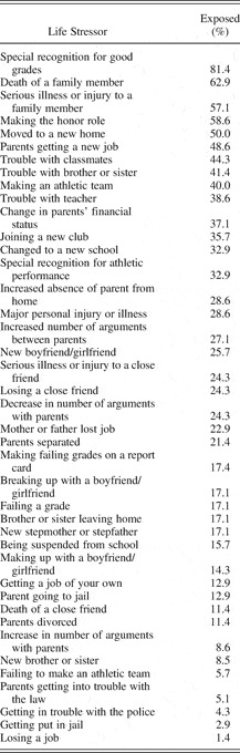
The Life Events Checklist (LEC; Johnson & McCutcheon, Reference Johnson, McCutcheon, Sarason and Spielberger1980) contains 46 life stressors including events that are typically perceived as both “positive” (e.g., parents getting a new job, making an athletic team) and “negative” (e.g., death of a family member, trouble with teacher). Children indicated whether events occurred in the past year. A sum of life events represents frequency of exposure to life stressors. The LEC has been used with youth of varying socioeconomic and ethnic backgrounds (DuBois, Felner, Brand, Adan, & Evans, Reference DuBois, Felner, Brand, Adan and Evans1992). It shows reasonable test–retest reliability over 2 weeks and correlates with a variety of relevant measures (Brand & Johnson, Reference Brand and Johnson1982; Johnson & McCutcheon, Reference Johnson, McCutcheon, Sarason and Spielberger1980).
Trauma exposure
Trauma exposure was assessed using the UCLA PTSD Index for DSM-IV Child- and Parent-Report Versions (Pynoos, Rodriguez, Steinberg, Stuber, & Frederick, Reference Pynoos, Rodriguez, Steinberg, Stuber and Frederick1998). These measures have three components. On the first component, children or caregivers were asked to indicate whether or not the child had ever experienced a specific traumatic event. One item, “Having your private sexual body parts touched by someone older or being forced to have sex,” was removed from the original scale because of its sensitive nature. However, victimization through sexual abuse may have been captured within the item, “Having any kind of very stressful thing happen, where you were really scared, or where you thought that your or someone else would be hurt badly or killed.” Children and caregivers were asked to indicate how old the child was when the traumatic event occurred. Two scores were derived from each version of this measure: child and parent reports of frequency of previous trauma exposure (number of traumatic events that occurred prior to 12 months before the assessment) and frequency of recent trauma exposure (number of traumatic events that occurred within past 12 months). Children are their caregivers demonstrated moderately high levels of agreement in their reports of previous (r = .26, p < .05) and total (r = .37, p < .05) traumas. As a result, child reports of traumatic events were used in the present study.
Salivary cortisol
Saliva samples were assayed for salivary cortisol in duplicate using a highly sensitive enzyme immunoassay (Salimetrics, PA). This technique has a range of sensitivity from 0.007 to 1.8 µg/dl. The average intra- and interassay coefficients of variation are 4.13 and 8.89%, respectively. The test uses 25 µl of saliva (for singlet determinations). The a.m. and p.m. cortisol levels were determined in micrograms per deciliter by calculating the mean of duplicate assay results.
Results
Descriptive information and relations among study variables
Exposure to life stressors
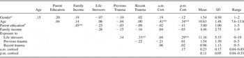
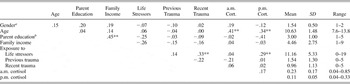
Within 12 months prior to measurement, children in the present sample experienced an average of 11.1 life stressors. Table 1 presents percentages of children who experienced each life stressor. Children who attended the public school experienced a higher frequency of life stressors (M = 12.4, SD = 5.7) than those from both parochial schools (M = 9.5, SD = 4.2), F (2, 65) = 7.13 p < .01. Frequency of exposure to life stressors was positively related to frequency of exposure to recent traumatic events (r = .33, p < .01). Table 2 presents descriptive information on the study variables including mean, standard deviation, range of values, and zero-order correlations among variables.
Frequency of exposure to life stressors and perceived impact

Descriptive information and correlation among study variables
a1 = male, 2 = female.
bHighest level of education attained by parent: 1 = some high school, 2 = graduated high school/GED, 3 = some college or vocational school, 4 = college degree, 5 = professional degree.
** p < .01.
Exposure to trauma
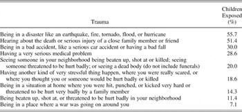
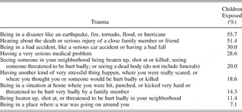
Children reported experiencing an average of 2.4 traumatic events during their lifetimes. On average, they experienced one traumatic event within 12 months of measurement (recent trauma) and 1.5 traumatic events prior to 12 months before measurement (previous trauma). Table 3 presents percentages of children who experienced traumatic events during their lifetimes.
Frequency of exposure to traumatic events
The a.m. and p.m. cortisol concentration
Mean cortisol values for a.m. and p.m. measurement were 0.23 and 0.11 µg/dl, respectively. Children's age was significantly positively correlated with both a.m. (r = .41, p < .01) and p.m. cortisol levels (r = .34, p < .01). Frequency of exposure to life stressors was significantly positively correlated with p.m. cortisol level (r = .29, p < .05), but not with a.m. cortisol level (r = .04, p = .77).
Stress, trauma, and cortisol levels
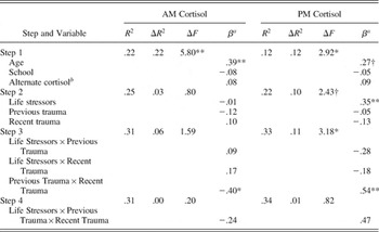
Regression analyses were conducted to evaluate independent and cumulative effects of stress and trauma exposure on basal cortisol levels. Independent regression analyses were conducted for the criteria a.m. and p.m. cortisol levels. For both regression equations, control variables were entered in the first step: age, school, and alternate cortisol level (a.m. cortisol in the equation with p.m. cortisol criterion and p.m. cortisol in the equation with a.m. cortisol criterion).
Main effects of life stress, previous trauma, and recent trauma were entered in the second step of regression equations. Two-way interaction terms were entered in the third step and a three-way interaction term was entered in the fourth step. Variables were centered around their means to facilitate interpretation of the interaction term and to control for multicollinearity (Aiken & West, Reference Aiken and West1991). Results of multiple regression analyses are presented in Table 4.
Regression analyses testing the moderating effects of life stressors, previous trauma, and recent trauma on cortisol concentrations

Note: For entire model, a.m. cortisol, F (10, 65) = 2.48*; p.m. cortisol, F (10, 65) = 2.88**.
aStandardized regression weights for the step in which main effect or interaction was entered.
bAlternate cortisol is the p.m. cortisol level in the model predicting a.m. cortisol and the a.m. cortisol level in the model predicting p.m. cortisol.
† p < .10.
* p < .05.
** p < .01.
Consistent with zero-order correlations, age significantly predicted a.m. cortisol levels (β = .39, p < .01) and tended to predict p.m. cortisol levels (β = 27, p < .10). Frequency of exposure to life stressors significantly predicted p.m. cortisol level (β = .35, p < .01). Neither the Life Stressors × Previous Trauma nor Life Stressors × Recent Trauma interaction terms significantly predicted a.m. cortisol levels. However, the Previous Trauma × Recent Trauma interaction significantly predicted a.m. cortisol (β = −.40, p < .05) and p.m. cortisol levels (β = .54, p < .01).
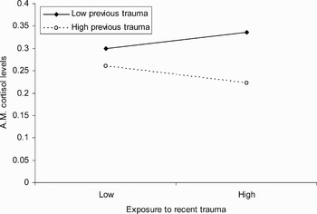
The relation between recent trauma exposure and a.m. cortisol level differed among children with lower and higher levels of exposure to previous trauma. As exposure to recent trauma increased, a.m. cortisol levels decreased for those with higher previous trauma exposure, but remained the same for those with lower previous trauma exposure. In circumstances of high recent trauma exposure, children with higher previous trauma exposure exhibited lower a.m. cortisol levels than children with lower previous trauma exposure (see Figure 1).
Regression lines for the regression of exposure to recent trauma on a.m. cortisol level (µg/dl) by previous trauma exposure.

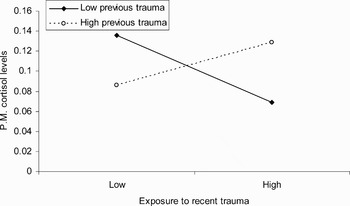
The relation between exposure to recent trauma and p.m. cortisol also differed among children with lower and higher levels of exposure to previous trauma. As exposure to recent trauma increased, p.m. cortisol levels decreased among children who reported lower previous trauma exposure, but increased among those with higher previous trauma exposure. In circumstances of low recent trauma exposure, children with higher previous trauma exposure exhibited lower p.m. cortisol levels. However, in conditions of high recent trauma exposure, children with higher previous trauma exposure exhibited higher p.m. cortisol levels (see Figure 2).
Regression lines for the regression of exposure to recent trauma on p.m. cortisol level (µg/dl) by previous trauma exposure.

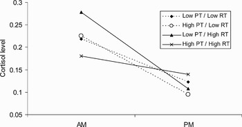
To facilitate interpretation of basal cortisol secretion patterns in the context of previous and recent trauma exposure, a.m. and p.m. cortisol levels were analyzed using repeated-measures multiple regression. Repeated-measures multiple regression begins by partitioning the total variance into within- and between-groups components (Cohen & Cohen, Reference Cohen and Cohen1983; Gully, Reference Gully1994; Hollenbeck et al., Reference Hollenbeck, Ilgen and Sego1994). This procedure thus provides the power advantages of repeated-measures analysis of variance (ANOVA) while maintaining regression's advantages of allowing continuous predictor variables (Rusher & Kaplan, Reference Ruscher and Kaplan2002). The regression proceeds in hierarchical steps, regressing the dependent variables (cortisol values) on all within terms (time of day of cortisol samples), all between terms (previous and recent trauma exposure), and finally all between–within interactions (of particular interest here, Time of Day × Previous Trauma × Recent Trauma interaction). Age served as a covariate in the analysis. Results revealed that the three-way interaction between time of day, previous trauma, and recent trauma was significant, F (1, 63) = 4.0, p < .05, indicating that the pattern of cortisol across the day varied depending upon the degree of previous and recent traumatic experiences (see Figure 3).
The a.m. and p.m. cortisol levels (µg/dl) by frequency of exposure to previous and recent trauma.

Follow-up repeated-measures multiple regression analyses were conducted to determine more specifically which patterns of cortisol differed from each other. Cutpoints were set for the continuous trauma exposure variables and four separate follow-up analyses were conducted, each one holding one of the trauma exposure variables constant (e.g., low previous exposure) while examining the interaction between time of day and the alternate exposure variable (e.g., recent exposure). Low previous exposure was defined as one or fewer traumatic exposures, high previous trauma as two or more exposures, low recent trauma was defined as zero exposures in the past year, and high recent trauma as one or more traumatic exposures in the past year.
For children exposed to high frequencies of previous trauma, there was a significant Time of Day × Recent Trauma Exposure interaction, F (1, 30) = 4.3, p < .05. In addition, for children exposed to high frequencies of recent trauma, there was a significant Time of Day × Previous Exposure interaction, F (1, 33) = 4.1, p = .05. Thus, as illustrated in Figure 3, children exposed to higher levels of both previous and recent trauma exhibited a flatter profile of cortisol secretion than children exposed to only high frequencies of previous trauma or high frequencies of recent trauma. No other significant differences in cortisol patterns were observed.
Discussion
The present study evaluated associations among exposure to life stress, previous trauma, recent trauma, and cortisol levels among school-aged children from a community sample. Results revealed that repetitive trauma exposure (higher levels of both previous and recent trauma) is related to blunted a.m. cortisol levels and elevated p.m. cortisol levels. For both a.m. and p.m. cortisol, the effects of previous trauma exposure on cortisol levels were most apparent as exposure to recent trauma increased.
In addition, life stress within the past 12 months was positively associated with p.m. cortisol levels. This finding is consistent with prior research documenting positive associations between exposure to nontraumatic early adversity and afternoon cortisol levels. Elevations in afternoon cortisol levels have been observed among children exposed to high levels of maternal stress (Essex et al., Reference Essex, Klein, Cho and Kalin2002) and maternal depression (Ashman et al., Reference Ashman, Dawson, Panagiotides, Yamada and Wilkinson2002).
Despite the long-time association between stress and hypercortisolism, the present study supports accumulating evidence for a different pattern of HPA axis functioning in contexts of stress. Hypocortisolism, defined as lower than expected cortisol levels at the height of the cortisol circadian rhythm, has received increasing attention, and may be a fairly common pattern associated with adverse early life conditions and chronic stress exposure (Gunnar & Vazquez, Reference Gunnar and Vazquez2001; Heim et al., Reference Heim, Ehlert and Hellhammer2000).
Blunted a.m. cortisol levels contribute to flattening or loss of expected daily cortisol rhythm, which has been recognized as a significant pathology in HPA axis functioning (Gunnar & Vazquez, Reference Gunnar and Vazquez2001; Heim et al., Reference Heim, Ehlert and Hellhammer2000).Although flattened cortisol patterns are more commonly thought to occur from “lower values near the early morning peak of the circadian cortisol cycle than from higher or elevated values near the through of the cycle” (Gunnar & Vazquez, Reference Gunnar and Vazquez2001, p. 516), elevations in p.m. cortisol levels also contribute to a loss of daily cortisol rhythm. The present study suggests that exposure to previous and recent trauma is associated with a flattening of cortisol rhythm, due to both relatively suppressed a.m. cortisol levels and elevated p.m. cortisol levels. Further, exposure to life stress contributed to flattened daily patterns due to its positive association with p.m. cortisol levels.
Currently, there is debate over the influence of stressor characteristics including severity, duration, and timing on basal HPA axis activity as well as HPA axis reactivity to subsequent stress exposure. Some studies suggest that duration of stress exposure is an important contributor to HPA axis response. For example, basal corticosterone in rats decreased gradually over 5 weeks of repeated stress exposure (Natelson et al., Reference Natelson, Ottenweller, Cook, Pitnam, McCarthy and Tapp1988). Similarly, monkeys exposed to multiple stress experiments over several years exhibited the lowest levels of 17-hydroxycorticosteroid (a cortisol metabolite) over time when exposed to repeated shock avoidance sessions (Mason et al., Reference Mason, Brady and Tolliver1968). In humans, basal hypocortisolism has been evidenced among children exposed to some chronic stressors including insecure attachment (Spangler & Grossman, Reference Spangler and Grossman1993), prolonged colic (Finsterward et al., Reference Finsterward, Selig, Schieche, Wurmser and Papousek2000), and exposure to neglectful rearing conditions (Carlson et al., Reference Carlson, Dragomir, Earls, Farrell, Macovei and Nystrom1995a, Reference Carlson, Dragomir, Earls, Farrell, Macovei and Nystrom1995b; Carlson & Earls, Reference Carlson and Earls1997). Essex et al. (Reference Essex, Klein, Cho and Kalin2002) found that exposure to repetitive maternal stress was associated with elevated afternoon cortisol levels among preschool children, a pattern that contributes to the flattening of daily cortisol diurnal rhythm. In contrast, hypercortisolism has been observed in the context of other chronic stressors such as low SES (Lupien et al., Reference Lupien, King, Meaney and McEwen2000, Reference Lupien, King, Meaney and McEwen2001) and exposure to maternal depression (Ashman et al., Reference Ashman, Dawson, Panagiotides, Yamada and Wilkinson2002; Field et al., Reference Field, Healy, Goldstein, Perry, Bendall and Schanberg1988; Hessl et al., Reference Hessl, Dawson, Frey, Panagiotides, Self, Yamada, Hann, Huffman, Lederhendler and Minecae1998).
The present study supports the relation between chronic stress exposure and hypocortisolism, and indicates that the duration of stress exposure is an important contributor to its relations with basal HPA axis functioning. Thus, exposure to chronic or repetitive stressors may result in different and perhaps more profound alterations in basal HPA axis activity than exposure to isolated stressful events.
The present findings support current theories of hypocortisolism that stipulate that exposure to significant stressors may initially result in elevated basal cortisol levels. However, frequent elevations in basal cortisol levels may result in downregulation of HPA axis components or enhanced negative feedback inhibition of the pituitary for cortisol, thereby resulting in lower basal cortisol secretion or reduced cortisol secretion in response to subsequent stressors (DeBellis et al., Reference DeBellis2001; Hellhammer & Wade, Reference Hellhammer and Wade1993; Heim et al., Reference Heim, Ehlert and Hellhammer2000; Yehuda, Boisoneau, et al., Reference Yehuda, Boisoneau, Mason and Giller1993; Yehuda, Resick, et al., Reference Yehuda, Levengood, Schmeidler, Wilson, Guo and Berber1993). Children in the present sample exposed to repetitive trauma (both previous and recent) may have exhibited elevated basal cortisol levels around the time they experienced an initial traumatic events. However, at the time of measurement, they may have been exhibiting a longer term consequence of these initially heightened basal cortisol levels, basal cortisol hyposecretion, in response to recent trauma.
Indeed, prior research demonstrates that a history of trauma exposure may be associated with attenuation of acute HPA axis response to a subsequent trauma (Meaney, Aitken, Sharma, & Sarrieau, Reference Meaney, Aitken, Sharma and Sarrieau1989), especially when prior stressors are similar to the current stressor (Yehuda et al., Reference Yehuda, Giller, Southwick, Lowy and Mason1991). For example, acute cortisol levels were assessed among 37 adult female rape victims within 51 hr of the sexual assault. Women with a history of previous assault had lower mean cortisol levels than women without a prior history of assault. Further, only women with no history of sexual assault showed the expected association between greater severity of rape and higher cortisol levels after the rape (Resnick, Yehuda, Pitman, & Foy, Reference Resnick, Yehuda, Pitman and Foy1995).
From the present analyses, it is unclear if the development of hypocortisolism was preceded by periods of frequent or chronic activation of the HPA axis. However, it is notable that inspection of mean a.m. and p.m. cortisol concentrations among children exposed to varying frequencies of trauma reveals that children exposed to low levels of previous trauma and higher levels of recent trauma tended to exhibit the highest a.m. cortisol level. Although merely a trend, this finding lends support to theories that describe elevations in basal cortisol levels that are evident in close temporal proximity to initial stress exposure.
It is notable that there are some reports of relations between exposure to isolated traumatic events and hypcortisolism “long after trauma exposure” (DeBellis, Reference DeBellis2001, p. 548). Some theorists argue that a substantial amount of time after stress exposure is necessary for physiological events related to hypocortisolism to take place (DeBellis et al., Reference DeBellis, Baum, Birmaher, Keshavan, Eccard and Boring1999). However, many studies that evaluate the longer term correlates of isolated traumatic events involve high intensity stressors such as exposure to combat-related atrocities (Yehuda et al., Reference Yehuda, Giller, Southwick, Lowy and Mason1991) or catastrophic natural disasters (Goenjian et al., Reference Goenjian, Yehuda, Pynoos, Steinberg, Tashjian and Yang1996). Significant traumatic events are often associated with psychological symptoms including recurrent memories of the event or other concomitant stressors so that responses to these “isolated” events may actually represent responses to chronic stress exposure.
The present study indicates that hypocortisolism and the resultant flattening of the daily cortisol rhythm is associated with repetitive stress exposure; however, the meaning of hypocortisolism as a phenomenon is an issue of debate (Heim et al., Reference Heim, Ehlert and Hellhammer2000). Some theorists consider hypocortisolism a specific correlate of psychiatric disorder (Yehuda, Resick, et al., Reference Yehuda, Levengood, Schmeidler, Wilson, Guo and Berber1993) or an indicator of the abnormal and maladaptive response of a generally hypoactive system (McEwen, Reference McEwen1998).
Other theorists conceptualize hypocortisolism as an adaptive strategy that allows for increased stress tolerance, better performance, and the maintenance of physical health under stressful conditions (DeBellis et al., Reference DeBellis, Baum, Birmaher, Keshavan, Eccard and Boring1999; Deinstbier, Reference Dienstbier1989). Given the deleterious effects of chronically elevated or blunted cortisol levels, organisms may strive to inhibit basal hyper- or hypocortisolism in distressing situations by “resetting” a.m. and p.m. basal cortisol to moderate levels. Thus, a flattened profile of cortisol secretion may represent compensatory adaptation to recurrent stress exposure or a physiological attempt to buffer the individual from dysregulation of HPA axis functioning in circumstances of repetitive stress exposure (DeBellis et al., Reference DeBellis, Baum, Birmaher, Keshavan, Eccard and Boring1999).
Although flattened basal cortisol levels may reflect ongoing, adaptive attempts to cope with chronic stress (Cicchetti & Rogosch, Reference Cicchetti and Rogosch2001), the long-term physiological consequences of loss of daily rhythm are unknown. To date, there is no evidence to suggest that loss of daily rhythm is associated with resilient outcomes (Gunnar & Vazquez, Reference Gunnar and Vazquez2001). Further research is needed to delineate both consequences of and physiological mechanisms associated with flattening of circadian rhythm in contexts of recurrent stress and trauma exposure. This research should focus on HPA axis functioning at and above the level of cortisol secretion. Conducted longitudinally, such research may contribute to identifying critical periods, longer term physiological and mental health correlates, and contributors to alterations in basal HPA axis functioning (i.e., frequency of trauma exposure, time since trauma exposure, and physiological development).
Study Limitations and Future Directions
Limitations of the current study illustrate areas for future growth and reflect some of the general weaknesses in research on HPA axis functioning. First, each participant provided only one a.m. and one p.m. saliva sample. Reliability of these samples would have been improved by repeated collection over multiple days. Second, to facilitate interpretation of cortisol data, daily patterns of cortisol release are inferred from a.m. and p.m. values. However, alternative daily patterns of cortisol release may not have been detected with only two daily measurements. Third, an exclusive focus on cortisol levels that reflect a downstream effect of HPA axis functioning suggests that more complicated effects involving higher levels of neuroendocrine function may have gone unmeasured.
Fourth, a weakness of the current study is our unawareness of children's wake-up time. A high level of variability in children's time of awakening could contribute to differences in cortisol levels at the time of measurement. However, all children in the present sample attended neighborhood schools where no before-school care was offered. Similarity in wake-up time was inferred because all schools began the day at approximately the same time and there were no differences between children attending the different schools in the times of cortisol collection.
Fifth, children the present sample (ages 7.6–13.8 years) are presumed to be heterogeneous with regard to physical/sexual maturation, and a positive correlation between age and cortisol levels has been robustly documented in this and prior research (Kiess et al., Reference Kiess, Meidert, Dressendorfer, Scheiver, Kessler and Koing1995; Lupien et al., Reference Lupien, King, Meaney and McEwen2001; Walker, Walder, & Reynolds, Reference Walker, Walder and Reynolds2001). Despite this association, significant limitations in normative data on cortisol levels among children of varying developmental levels render it difficult to know what constitutes normal or abnormal patterns of cortisol release among youth (Cicchetti & Rogosch, Reference Cicchetti and Rogosch2001; Gunnar & Vazquez, Reference Gunnar and Vazquez2001). More specifically, some theorists suggest that compensatory strategies, which may include hypocortisolism in the context of chronic stress, may be “more likely after maturation” (DeBellis, Reference DeBellis2001, p. 548). However, other studies illustrate hypocortisolism in the context of chronic stress among infants and toddlers (Carlson & Earls, Reference Carlson and Earls1997; Finsterward et al., Reference Finsterward, Selig, Schieche, Wurmser and Papousek2000). Developmental level should be further evaluated as researchers continue to identify moderating factors in the relations between stress exposure and basal HPA axis functioning.
Results of the present study indicate that organisms may utilize protective compensatory adaptation strategies in high-risk contexts. To further explore whether flattening of daily cortisol patterns represent a physiological attempt to prepare an organism for chronic stress exposure, cortisol response to acute stressors should be examined. Among children with a history of chronic stress exposure, if cortisol levels fail to rise or rise only slightly in response to an acute stressor, this blunted response may reflect chronic adaptation to stress exposure (Gunnar & Vazquez, Reference Gunnar and Vazquez2001).
As research on child development moves toward evaluating complex interactions between biology and experience, the physiological correlates of stress exposure will be defined further. The present study suggests that although exposure to recurrent stress and trauma may be associated with changes in physiological functioning, not all children exposed to stressful experiences respond in the same fashion (Cicchetti & Walker, Reference Cicchetti and Walker2001; Sapolsky, Reference Sapolsky1994). The effects of significant stress exposure on basal HPA axis activity among children are influenced by the frequency, severity, and duration of stress exposure, time since stress exposure, and developmental level. Future research should strive to further disentangle these complex interactional processes.