Introduction
Inferior turbinate hypertrophy is a common cause of chronic nasal obstruction. It is most frequently caused by vasomotor dysfunction, allergic rhinitis, chronic sinusitis or a deviated septum.Reference Back, Hytonen, Malmberg and Ylikoski1 Many patients seek surgical intervention for this condition. The optimal surgical approach requires successful volumetric reduction of tissue while avoiding damage to the mucosal surface.
During surgical treatment of patients with inferior nasal concha hypertrophy, protection of the ciliated conchal epithelium is very important. Therefore, in this study we aimed to examine the histological appearance of the ciliated conchal epithelium and underlying tissues in patients with nasal obstruction due to hypertrophy of the inferior turbinate who had undergone routine clinical radiofrequency thermal ablation.
The benefits of radiofrequency thermal ablation for conchal reduction in suitable patients have been demonstrated.Reference Back, Hytonen, Malmberg and Ylikoski1, Reference Smith, Correa, Kuo and Reinisch2 The radiofrequency energy is directed beneath the mucosa, in order to create local damage to the inferior concha subepithelium without harming the conchal bone or mucosal surface. In the area surrounding the resultant necrosis, the new tissue formed during wound healing is replaced by fibroblasts, and the consequent wound contraction reduces the volume of the concha.Reference Back, Hytonen, Malmberg and Ylikoski1–Reference Lee, Hwang and Kingdom3 In addition, the fibrosis and decreased tissue volume cause adhesions between the mucosa and the periosteum of the concha, decreasing blood flow and minimising oedema.Reference Lin, Lin, Su and Chang4
Materials and methods
Patients were given verbal and written information on the study, and written, informed consent was obtained. The study was reviewed by the local ethics committee.
Between November 2002 and January 2008, radiofrequency thermal ablation and tissue biopsy were performed in our hospital in six patients with chronic nasal obstruction caused by inferior concha enlargement, who had not responded to regular medical treatment applied for at least one month. Two of the patients were female and the remaining four male. The patients' average age was 29 years.
The six patients shared the following characteristics: enlarged inferior concha; response to local decongestants; no systemic disease; and no previous conchal surgery.
All patients were prepared for surgery in the supine position. Topical anaesthesia was administered, followed by injection of Jetocaine® (Lidokain HCl 20mg/ml – Adeka Drug Company, Turkey) without adrenaline into each inferior concha (to a total of 3 ml administered to the anterior edge, middle and posterior edge of the concha). Radiofrequency energy was delivered by a Reflex 45 probe mounted on an ENTec Coblator® device (Coblator Plasyma Surgery System, ArthroCare, USA). The probe was inserted longitudinally into the anterior surface, middle surface and posterior surface of the inferior concha, under the mucosa, and radiofrequency energy was applied for a period of about 20 seconds, in four to six steps; approximately 450–480 J was administered. As the probe was withdrawn, radiofrequency energy was applied at a level to induce coagulation, in order to reduce bleeding. Nasal packing was not applied.
Two hours after the procedure, patients were discharged with a prescription for antibiotics, decongestants, analgesics, nasal irrigation and intranasal steroids.
In each patient, punch biopsy tissue samples approximately 1 mm3 in volume were taken from the anterior part of the inferior concha, just before radiofrequency treatment and four, 30, 48 and 60 months after treatment. Tissue sections were prepared by haematoxylin and eosin staining The histological effects of radiofrequency thermal ablation on the conchal epithelium and subepithelium were examined, and pre- and post-treatment appearances compared.
Since the current study focussed on histological changes relating to routine doses of radiofrequency thermal ablation applied to treat inferior turbinate hypertrophy, we did not assess patient symptoms.
Results and analysis
In the pre-radiofrequency treatment period, all patients had used intranasal steroids, antihistamines and decongestants. Following radiofrequency treatment, patients used intranasal steroids for a period of one month.
All patients tolerated radiofrequency treatment well. Bleeding requiring nasal packing was not encountered in any patient. Repeated radiofrequency treatment was applied when necessary. No mucosal damage, infection or adhesion was seen.
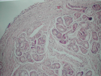
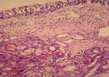
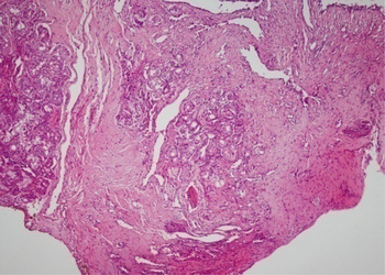
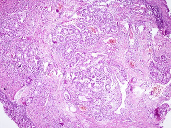
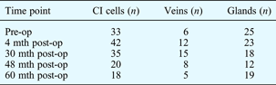
Histological samples prepared four months after radiofrequency treatment showed a proliferation of blood vessels, an increased number of inflammatory cells and a slightly decreased number of glands. Thirty months after treatment, the number of inflammatory cells and glands had decreased but there were signs of increased vascular proliferation, fibrosis and granulation (compared with normal mucosa). At 48 and 60 months post-treatment, the number of inflammatory cells and blood vessels had decreased significantly, the number of glands had increased, and lobulation was observed (Table I; Figures 1–5).
Photomicrograph showing pre-treatment inferior concha histology (H&E; ×200).

Photomicrograph showing inferior concha histology at four months post-treatment (H&E; ×200).

Photomicrograph showing inferior concha histology at 30 months post-treatment (H&E; ×200).

Photomicrograph showing inferior concha histology at 48 months post-treatment (H&E; ×200).

Photomicrograph showing inferior concha histology at 60 months post-treatment (H&E; ×200).

Results of histopathological examination
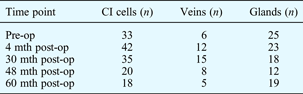
Data represent: Mean number of items per medium-power field; CI = chronic inflammatory; pre-op = before radiofrequency thermal ablation; post-op = after radiofrequency thermal ablation; mth = months
Discussion
The nasal conchae are covered with pseudo-stratified, cylindrical, ciliary epithelium containing goblet cells. However, the anterior end of the inferior concha is covered with non-keratinised squamous epithelium, as in the nasal vestibule. The mucus covering the mucosa is secreted by the goblet cells (found in the lamina propria) and the submucosal glands, and moved to the nasopharynx by ciliary action.Reference Onerci and Ünal5
An additional, important characteristic of conchal mucosa is the existence of many thin-walled venous sinuses surrounded by smooth muscle. These venous sinuses make the conchal mucosa much thicker than other nasal mucosa.Reference Onerci and Ünal5 In the inferior concha, venous sinuses predominate; in the middle concha, submucosal glands predominate. Following parasympathetic innervation and exposure to neuropeptides, the venous sinuses fill with blood and the mucosa becomes much thicker. Consequently, the conchal size increases, resulting in varying degrees of nasal obstruction.Reference Onerci and Ünal5, Reference Sapcı, Sahin, Karavus and Akbulut6
Following radiofrequency thermal ablation treatment, conchal mucosa shows vein proliferation, granulation tissue development, an increase in the number of chronic inflammatory cells and a slight decrease in the number of glands, compared with normal mucosa. Tissue biopsy two years post-treatment shows a decrease in the number of inflammatory cells but also an increase in vascular proliferation and a decrease in the number of glands, indicating a fibrotic process with increased granulation.Reference Sargon, Çelik, Uslu, Yücel, Denk and Ceylan7 Scanning electron microscopy of Mucosa surface and subeptelium has shown no difference in cilia number or morphology, comparing appearances before and after radiofrequency treatment. Furthermore, no ultrastructural pathology has been detected.Reference Sargon, Çelik, Uslu, Yücel, Denk and Ceylan7
In the current study, the final biopsy (60 months after radiofrequency treatment to the anterior part of the inferior concha) clearly showed a decreased number of inflammatory cells, decreased vascularisation, decreased number of glands and increased amount of fibrous tissue, suggesting minimal impairment of normal function. In our small group of patients, tissue biopsies indicated that there was no damage to the conchal mucosa or submucosal glands.
• Turbinate reduction by radiofrequency thermal ablation has become a popular treatment for inferior nasal concha hypertrophy
• This study examined inferior nasal concha epithelium and subepithelium before and after such radiofrequency treatment, in six patients with inferior nasal concha hypertrophy
• Initial changes included proliferation of blood vessels, an increased number of inflammatory cells and a slightly decreased number of submucosal glands
• Later changes included a decreased number of inflammatory cells and blood vessels, and an increased number of glands
We recommend that radiofrequency thermal ablation should be directed towards the anterior part of the inferior concha, as this is the region harbouring most of the erectile tissue which causes increased nasal resistance and hence airway obstruction.Reference Li, Powel, Riley, Troell and Guilleminault8, Reference Elwany, Gaimaee and Fattah9
Conclusion
Radiofrequency thermal ablation is a reliable surgical method capable of reducing the volume of the inferior concha without harming the nasal mucosa, and with only minimum discomfort. The procedure is fast, simple and relatively painless, and may be used without post-operative nasal packing, thus improving patient comfort. The procedure does not cause carbonisation or osteitis of the concha. Our patients' post-treatment tissue biopsies showed that treatment-induced fibrosis resulted in contraction of the concha, with only minor tissue destruction (as indicated by the existence of secretion glands), compared with pre-treatment appearances.
It is still too early to assess the long-term consequences of radiofrequency thermal ablation used to treat inferior conchal nasal hypertrophy. If the procedure is reliable in the long term, it may offer an alternative to other techniques used for this purpose. This would be beneficial, especially considering the procedure's potential for use in the out-patient setting, its minimal associated patient discomfort, and its facility for re-application as needed.