INTRODUCTION
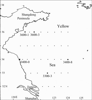
In July 2008, undisturbed sediments were obtained from a grid of 33 sampling stations between 32°N and 38°50′N 120°20′E and 124°10′E during the Open Research Cruise by RV ‘KE XUE SAN HAO’ in the Yellow Sea, China (Figure 1). Many species of free-living marine nematodes were determined and some new species were discovered. The present paper describes two new species from this region which belong to the genus Paramarylynnia Huang & Zhang, Reference Huang and Zhang2007.
Map of sampling stations in the Yellow Sea.

Paramarylynnia was established in the family Cyatholaimidae by the authors in 2007. The type species of the genus is Paramarylynnia ventralseta Huang & Zhang. Since then Paramarylynnia ventralseta is the only valid species of the genus and no other new member has been added. Paramarylynnia is characterized by a cuticle with transverse rows of even dots; lateral differentiation absent; gubernaculum large and dilated at the distal end, without distal dentate; tail conico-cylindrical.
MATERIALS AND METHODS
Undisturbed sediment samples were taken using a 0.1m2 improved Gray–O'Hara box corer in July 2008, and meiofauna samples were obtained using a syringe (2.6 cm diameter). Samples were fixed with 5% formalin in filtered seawater. In the laboratory, sorting and slide mounting were carried out as detailed by Huang & Zhang (Reference Huang and Zhang2010) and Huang & Wu (Reference Huang and Wu2010). Type specimens have been deposited in the type collections of the museum of the Institute of Oceanology, Chinese Academy of Sciences.
Abbreviations are as follows: a, body length/ maximum body diameter; a.b.d., anal body diameter; b, body length/pharynx length; c, body length/tail length; c.b.d., corresponding body diameter; Spic, spicule length along arc; V, distance of vulva from the anterior end of body; V%, V/body length × 100%.
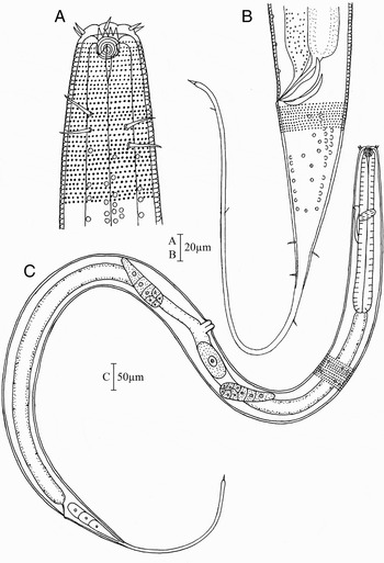
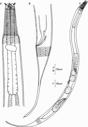
Paramarylynnia filicaudata sp. nov. (A) Lateral view of male anterior end; (B) lateral view of male posterior end; (C) lateral view of female body.

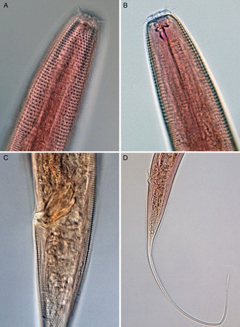
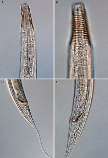
Paramarylynnia filicaudata sp. nov. (A) Lateral surface view of male anterior end, showing cephalic setae, amphidial fovea and cuticle punctation; (B) lateral optical section of female head end, showing buccal cavity with dorsal tooth and ventrosublateral teeth; (C) lateral optical section of male posterior body part, showing copulatory apparatus; (D) lateral view of female posterior end, showing filiform tail end.

TYPE MATERIAL
Four males and three females collected and studied.
Holotype: ♂1 on slide 126194-401; paratype: ♀1 on slide 126194-221; paratypes: three males and two females on slides 126194-401 and 126194-221 respectively.
TYPE LOCALITY AND HABITAT
Subtidal muddy sediment in the Yellow Sea. Station 3400-8: 34°N, 124°E, water depth 80 m.
ETYMOLOGY
The new species name refers to the filiform tail shape.
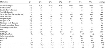
MEASUREMENTS (SEE TABLE 1)
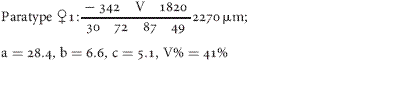
Individual measurements (and average) of Paramarylynnia filicaudata sp. nov. (in µm).

DESCRIPTION
Males: body long, cylindrical, maximum diameter 51–59 µm. Head diameter 25–29 µm, about 53% of diameter at the base of pharynx. Cuticle transversally annulated and punctuated (Figures 2A & 3A). The dots about the same size and closely arranged in transversal rows, beginning from level of cephalic setae to tail end. At the pharyngeal region, the dots are more conspicuous than that on the rest of the body. Six longitudinal rows of simple rounded cuticle pores, but fewer in cervical part. No lateral differentiation, but numerous lateral modified punctuations (LMPs) present in cluster in pharyngeal and cloacal region. Four longitudinal files of two to three sub-lateral cervical setae.
Amphidial fovea round, multispiral with 5 turns, 13–17 µm (about 0.4 c.b.d.) wide, situated at a distance near the apex. Mouth opening surrounded by six conical lips with hardly visible inner labial sensilla. Outer labial and cephalic sensilla short, arranged in one circle of ten setae. Four cephalic setae more or less equal to six outer labial setae, about 10 µm long.
Buccal cavity with a distinct large dorsal tooth and two small ventrosublateral teeth (Figures 2A & 3B). Pharynx cylindrical, slightly broadened at base. Nerve ring at 43% of pharyngeal length from anterior end. Ventral gland not clearly defined. Excretory pore situated anterior to the nerve ring. No cardia.
Tail elongated, 420–460 µm (8.4–9.0 a.b.d.), with a long filiform part constituting about two thirds of total tail length, and with a delicate terminal spinneret (Figure 3D). No subventral setae on the conical part. Three caudal gland cells situated within the conical part.
Spicules 42 µm measured along arc (mean value), boat-shaped, swollen in the middle and gradually tapering towards both ends (Figure 2B). Gubernaculum curved, 30 µm as arc, without apophysis. Precloacal supplements not clear. Testes are indistinct.
Females: similar to males in most respects, but the tail relatively longer than that of males. Didelphic, ovaries antidromously reflexed; anterior ovary situated subventrally to the right of the intestine, posterior one subventrally to the left of the intestine. Vulva at 40% of body length from anterior end. Eggs oval.
DIFFERENTIAL DIAGNOSIS AND DISCUSSION
Paramarylynnia filicaudata sp. nov. are characterized by long filiform tail and absence of subventral setae on conical part of tail; spicules boat-shaped, swollen in the middle and gradually tapering towards both ends. This new species is closely related to P. subventrosetata Huang & Zhang, Reference Huang and Zhang2007, but differs from it by the following characters: longer filiform tail, without subventral setae on the conical part of the tail, and spicules taper proximally compared to two rows of subventral setae present on the conical tail part and spicules cephalated in P. subventrosetata.
Paramarylynnia stenocervica sp. nov. (A) Lateral view of male anterior end; (B) lateral view of male posterior end; (C) lateral view of female body.

Paramarylynnia stenocervica sp. nov. (A) Lateral optical section of male head end, showing cephalic setae, buccal cavity, dorsal teeth and pharynx; (B) lateral surface view of female head end, showing cephalic setae, amphidial fovea and cuticle punctation; (C) lateral view of male posterior end, showing spicules, gubernaculum and tail; (D) lateral view of male posterior body part, showing spicules, gubernaculum and precloacal supplements.

TYPE MATERIAL
Five males and five females collected and studied.
Holotype: ♂1 on slide 3600-101; paratype: ♀1 on slide 3400-002; paratypes: four males and four females on slides 3300-302 and 3600-302, 303 respectively.
TYPE LOCALITY AND HABITAT
Subtidal muddy sediment in the Yellow Sea. Station 3600-1: 36°N 120°50′E, water depth 30 m; 3600-3: 36°N 121°30′E, water depth 30 m and station 3300-3: 33°N 122°30′E, water depth 20 m.
ETYMOLOGY
The new species name refers to the strongly narrower anterior end of the body.
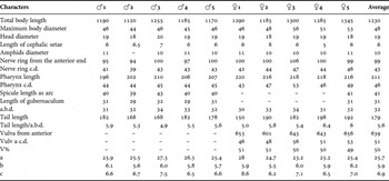
MEASUREMENTS (SEE TABLE 2)
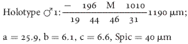
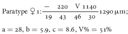
Individual measurements (and average) of Paramarylynnia stenocervica sp. nov. (in µm).

DESCRIPTION
Males: body spindle-shaped, sharply tapering towards the anterior end from the anterior third of pharynx onwards (Figure 4C). Cuticle heterogeneous with transverse rows of punctuations, the lateral ones slightly larger. The punctuations of anterior narrower portion larger and more widely spaced than on the rest of the body (Figures 4A & 5B).
Amphidial fovea slightly oval transversally, multispiral with 5 turns, 10–11 µm wide (50% c.b.d.) (Figure 5B). Anterior border of amphideal fovea 9 µm from anterior end. Head diameter 19 µm, 21% of diameter at the base of pharynx. Inner labial papillae not observed, six outer labial setae short, four cephalic setae, 6 µm long.
Buccal cavity with a distinct large dorsal tooth and two small ventrosublateral teeth (Figures 4A & 5A). Pharynx cylindrical, with slightly widened base, no true bulb (Figure 5A). Nerve ring at 46% of pharyngeal length from anterior end. Ventral gland small, located posterior to pharynx. Excretory pore anterior to the nerve ring. No cardia.
Tail conico-cylindrical, 6 a.b.d. long, with a long filiform part, longer than half the total tail length, and with delicate terminal spinnerete (Figure 5C). Caudal glands not clear.
Spicules 41 µm along arc, curved and cephalate proximally. Gubernaculum 31 µm, boat-shaped, swollen in the middle and gradually tapering towards both end. Five small tubular precloacal supplements (Figures 4B & 5D).
Females: similar to males in most respects, but slightly longer in body length. Didelphic, ovaries antidromously reflexed. Vulva at 49% of body length from anterior end. Eggs round.
DIFFERENTIAL DIAGNOSIS AND DISCUSSION
Paramarylynnia stenocervica sp. nov. is characterized by a marked narrower anterior part, cuticle heterogeneous with transverse rows of punctuations, larger and more spaced anteriorly and slight lateral differentiation. Spicules curved and cephalate. Gubernaculum boat-shaped, dilate in the middle and gradually tapering towards both ends. Five small tubular precloacal supplements. It is easily differentiated from other two species of the genus by the marked narrower anterior part of the neck region and heterogeneous cuticle.
ACKNOWLEDGEMENTS
This work was supported by the National Science Foundation of China (No. 30770250), the Knowledge Innovation Program of the Chinese Academy of Sciences (No. KZCX2-YW-417) to K. Xu, and the Open Research Cruise offshore China by RV ‘KE XUE SAN HAO’, Institute of Oceanology, Chinese Academy of Sciences (IOCAS) in July 2008. The authors are very grateful to Mr Renhai Dai and Ms Xiuqin Wu, IOCAS, for their help with sampling and Ms Zhongying Shan for her help with slide making. Two anonymous referees provided valuable comments and revision for the paper.