Isolated ventricular septal defect is the most common CHD.Reference Marelli, Mackie, Ionescu-Ittu, Rahme and Pilote 1 Approximately one-third of symptomatic children with isolated ventricular septal defect and heart failure due to left-to-right shunt undergo surgical closure of the defect.Reference Gersony 2 The risk of pulmonary vascular disease owing to left-to-right shunting is one of the main arguments for early closure of the defect. The prevalence of pulmonary arterial hypertension in adult patients with a closed ventricular septal defect ranges from 3 to 13% in different studies.Reference Duffels, Engelfriet and Berger 3 – Reference Roos-Hesselink, Meijboom and Spitaels 5 We previously demonstrated an abnormal right ventricular systolic pressure response during exercise in 50% of adolescents (age 13–25 years) after surgical closure of an isolated ventricular septal defect.Reference Moller, Brun and Fredriksen 6 A progressive pulmonary vascular disease was suggested. The aim of the present study was to define the prevalence of abnormal right ventricular systolic pressure response in adults operated on for ventricular septal defect early in life when investigated following the same protocol and tests as in our previous study.Reference Moller, Brun and Fredriksen 6 We also investigated whether an abnormal right ventricular systolic pressure response to exercise is associated with impaired cardiac function, pulmonary function, or exercise capacity.
Materials and methods
Patients
We identified consecutive patients aged 30–45 years operated on at Oslo University Hospital (Rikshospitalet) for isolated ventricular septal defect before 5 years of age. Patient data were collected from the hospital’s database of thoracic surgery, DATACOR.Reference Svennevig, Bech, Karlsen, Amlie and Olsen 7 Patients included in the study lived <4 hours travel distance from Oslo University Hospital and were able to perform an exercise test. We excluded patients with other known CHD at the time of the ventricular septal defect closure, and those with current unrelated medical conditions affecting heart or lung function, chromosomal abnormalities, or autoimmune diseases. Attention was paid to exclude patients with right ventricular outflow tract obstruction, as this condition interferes with the assessment of right ventricular function and non-invasive pressure estimation.
The study complies with the Declaration of Helsinki and was approved by the Norwegian Regional Committee for Medical Research Ethics. All participants provided written informed consent. The authors had full access to the data and take responsibility for its integrity. All authors have read and agreed to the manuscript as written.
Measures
The study participants underwent a full resting baseline echocardiographic assessment. Left ventricular systolic function was assessed by fractional shortening in M-mode, and biplane ejection fraction by Simpson’s method from two-dimensional greyscale imaging.Reference Lang, Bierig and Devereux 8 We used pulsed-wave tissue Doppler of the lateral mitral valve annulus to assess the peak systolic velocity and isovolumic acceleration.Reference Roche, Vogel and Pitkanen 9 , Reference Ruan and Nagueh 10 Left ventricular diastolic function was assessed by peak early diastolic velocity and the ratio of early diastolic flow velocity through the mitral valve to the peak tissue velocity of the lateral annulus.Reference De Sutter, De Backer, Van de Veire, Velghe, De Buyzere and Gillebert 11 , Reference Nagueh, Appleton and Gillebert 12 Right ventricular systolic function was measured using M-mode for the tricuspid annular plane systolic excursion,Reference Kaul, Tei, Hopkins and Shah 13 and we measured right ventricular fractional area change from two-dimensional greyscale imaging.Reference Rudski, Lai and Afilalo 14 We assessed the peak tricuspid annular plane systolic motion velocity and isovolumetric acceleration from pulsed-wave tissue Doppler of the tricuspid annulus.Reference Rudski, Lai and Afilalo 14 The combined systolic and diastolic function of either the left or right ventricle was assessed using the myocardial performance index, as measured with pulsed-wave tissue Doppler from the basal segment of the free wall of the respective ventricle.Reference Pellett, Tolar, Merwin and Kerut 15 , Reference Tei, Ling and Hodge 16 The right atrial pressure at rest was estimated by the caval vein inferior index.Reference Kircher, Himelman and Schiller 17 All echocardiographic recordings were obtained with a Vivid E9 scanner (GE Healthcare, Horten, Norway). Echocardiographic studies were saved digitally (still frame and loops) for off-line analysis.
The patients were examined with a cardiopulmonary exercise test on a treadmill ergometer with gas-exchange analysis and electrocardiogram (VMax29; SensorMedics Corp., Yorba Linda, CA, United States of America) following a modified Bruce protocol.Reference Fredriksen, Ingjer, Nystad and Thaulow 18 , Reference Fredriksen, Ingjer and Thaulow 19 Maximum oxygen uptake was expressed in milligrams per kilogram per minute, and the individual results were compared with reference values in healthy adults and expressed as a z score for the age-specific mean.Reference Edvardsen, Scient, Hansen, Holme, Dyrstad and Anderssen 20 All tests were supervised by a cardiologist and performed until exhaustion. Early termination was determined according to common guidelines Reference Paridon, Alpert and Boas 21 based on patient fatigue or the occurrence of predefined adverse events: arrhythmia, ischaemia, chest pain, clinical signs of circulatory compromise, progressive fall in systolic blood pressure >10%, and severe hypertension.
Two hours after treadmill testing, semi-supine cycle ergometry stress echocardiography (Ergoselect 1200 EL; Ergoline GmbH, Bitz, Germany) was performed as described previously.Reference Moller, Peersen, Pettersen, Thaulow, Holmstrom and Fredriksen 22 A stepwise exercise protocol was used with a starting load of 25 W, with a 25 W increase every third minute until 80% of the maximum heart rate from the cardiopulmonary exercise test was reached. Off-line analysis of echocardiographic recordings was performed as described previously.Reference Moller, Peersen, Pettersen, Thaulow, Holmstrom and Fredriksen 22 The maximal velocity of the tricuspid regurgitation jet was measured and averaged from as many image frames as possible during each workload stage. The right ventricular systolic pressure was calculated from each recording using the modified Bernoulli’s equation, adding the right atrial pressure at rest to the calculated pressure gradient between right ventricle and right atrium.Reference Yock and Popp 23 On the basis of previous work by Grunig et alReference Grunig, Weissmann and Ehlken 24 and our own previous studies on the normal right ventricular systolic pressure response to exercise,Reference Moller, Peersen, Pettersen, Thaulow, Holmstrom and Fredriksen 22 we defined a maximal right ventricular systolic pressure ⩾52 mmHg during exercise as abnormal.
Pulmonary function tests included the measurement of static and dynamic lung volumes and gas-diffusing capacity. All tests were performed according to the guidelines of the European Respiratory Society using the Vmax V6200 automated system (SensorMedics Corp.). Total lung capacity, forced vital capacity, forced expiratory volume in one second, and gas-diffusing capacity for carbon monoxide were recorded and reported in absolute values and as percent of predicted. The reference values recommended by the European Respiratory Society were used.Reference Quanjer, Tammeling, Cotes, Pedersen, Peslin and Yernault 25 Whenever airway obstruction was suspected by the supervising pulmonologist, reversibility was tested by inhalation of a β2 receptor agonist.
Data analysis
Parametric data are presented as means with 95% confidence intervals (± 2×standard error of the mean) and as medians with ranges. Unpaired Student’s t-tests were performed to compare measurements under different conditions when the data were normally distributed. Pearson’s correlation coefficient, linear regression, and logistic regression analysis were used to determine relationships between different variables. To determine functional predictors of the pathological pressure response to exercise, we computed four compound variables of cardiac and pulmonary function and performed a logistic regression analysis between all four variables and the presence or absence of a pathological pressure response. Compound variables were computed by averaging the standardised functional variables.
Statistical analysis
p-Values < 0.05 were considered significant. Statistical analysis was performed in Microsoft Excel 2013 (Microsoft Corporation, Redmond, WA, United States of America) and IBM SPSS Statistics version 21 (IBM Corporation, New York, NY, United States of America).
Results
Patients
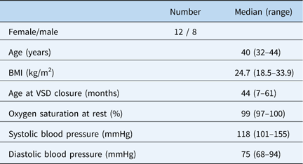
We identified 57 consecutive patients operated on between 1972 and 1984 for isolated ventricular septal defect with a last registered residency within the target area. Of these patients, 13 had died: six deaths (10%) before 2 years of age, four deaths (7%) between 1 and 10 years of age, none between 10 and 20 years of age, three deaths (5%) between 20 and 30 years of age, and none thereafter. Review of the medical charts of the survivors led to the exclusion of nine patients owing to Down syndrome (n=2), other known CHD (n=3), or chronic muscular/skeletal issues precluding exercise testing (n=2). Of the remaining 33 eligible patients, 13 declined to participate or failed to attend their study appointments. Therefore, we could include 20 patients (Table 1), 16 of whom completed all parts of the examinations. Four patients did not perform the cardiopulmonary exercise test owing to reasons unknown at the time of inclusion: technical failure, neurological impairment, recent muscular/skeletal injury, or anxiety. In addition, one patient who did not perform the cardiopulmonary exercise test also did not cooperate sufficiently during the pulmonary function test.
Basic patient characteristics (N=20).

BMI=body mass index; VSD=ventricular septal defect
The median age at surgical closure of the ventricular septal defect was 44 months; a patch had been used in nine patients, direct suture in four patients, and documentation of the surgical technique was not available for the remaining seven patients. Two patients had been operated on for late diagnosed coarctation of the aorta with left thoracotomy several years after ventricular septal defect closure, without signs of recoarctation on follow-up. Both patients were normotensive without medication. One patient was diagnosed during the study visit with a borderline hypoplastic arch, which did not require intervention. One female patient had developed paroxysmal atrial fibrillation and was on permanent treatment with a beta-blocker and flecainide. Two patients had received permanent pacemaker treatment owing to third-degree atrioventricular block many years after the ventricular septal defect closure. Two patients had recently been through a medical investigation for palpitations or chest pain without pathological findings. One patient had been treated for mild bronchial asthma and presented no reversibility on the pulmonary function test.
Findings
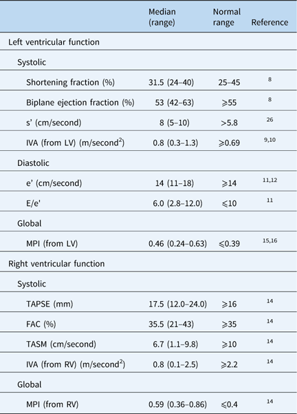
Left ventricular systolic and global function expressed as the median left ventricular ejection fraction and myocardial performance index were slightly abnormal (Table 2). In contrast, the less load-dependent median peak systolic lateral mitral annulus velocity and median left ventricular isovolumetric acceleration were within normal limits. The left ventricular diastolic function expressed as peak early diastolic lateral mitral annulus velocity and the ratio of early diastolic mitral flow velocity and peak early diastolic lateral mitral annulus velocity was normal. Right ventricular systolic function at rest showed divergent results. The median tricuspid annular plane systolic excursion and fractional area change were within normal limits, whereas the median tricuspid annular systolic motion velocity and median right ventricular isovolumetric acceleration were abnormal. The global right ventricular function measured by the median right ventricular myocardial performance index was also abnormal.
Echocardiographic functional data at rest (N=20).

E=early diastolic mitral flow velocity; e'=peak early diastolic lateral mitral annulus velocity; FAC=fractional area change; IVA=isovolumetric acceleration; LV=left ventricle; MPI=myocardial performance index; RV=right ventricle; s'=peak systolic lateral mitral annulus velocity; TAPSE=tricuspid annular plane systolic excursion; TASM=tricuspid annular systolic motion velocity
Red marking of median values out of normal range
Electrocardiograms showed that two patients had a paced rhythm, with one in atrial fibrillation, and the remaining 17 patients were in sinus rhythm. Three patients had a right bundle branch block, but the remaining had no conduction disturbance.
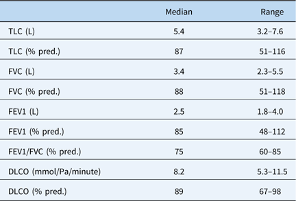
The ratio of forced expiratory volume in one second and forced vital capacity were below the normal ranges (median 75% of predicted) (Table 3). Otherwise, lung volumes, dynamic parameters, and gas-diffusing capacity covered broad ranges but were within normal limits.Reference Quanjer, Tammeling, Cotes, Pedersen, Peslin and Yernault 25 There was no correlation between lung volumes, dynamic airway measurements, or diffusion lung capacity for carbon monoxide and the right ventricular systolic pressure response to exercise.
Pulmonary function at rest (N=19).

DLCO=diffusion lung capacity for carbon monoxide; FEV1=forced expiratory volume in 1 second; FVC=forced vital capacity; TLC=total lung capacity
Red marking of median value below 80% of predicted
Cardiopulmonary exercise tests revealed normal exercise capacity. Compared with age- and gender-specific normal values, the average z score was −0.4 (95% confidence interval −2.8–0.3) for the whole group. The average peak oxygen uptake was 32.1 ml/kg/minute (95% confidence interval 25.7–38.6) for females and 36.8 ml/kg/minute (95% confidence interval 31.2–42.4) for males. Owing to the inclusion of two pacemaker-dependent individuals, we did not analyse the maximal heart rate during exercise.
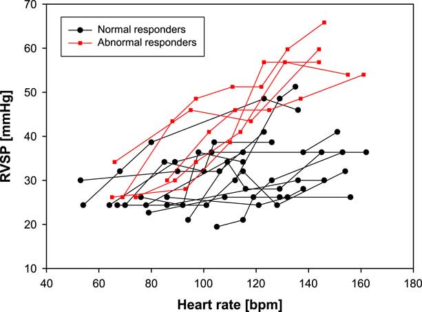
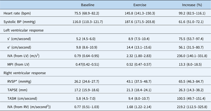
Five of 19 patients (26%) presented with an abnormal right ventricular systolic pressure response to exercise, defined as a maximal right ventricular systolic pressure ⩾ 52 mmHg during semi-supine cycle ergometry stress echocardiography (Table 4, Fig 1). We found no difference in peak oxygen uptake or pulmonary function between patients with or without an abnormal right ventricular systolic pressure increase during exercise.
Scatterplots of serial measurements of right ventricular systolic pressure (RVSP) during exercise. Colour code depending on normal non-responders=black (RVSP <52 mmHg) or abnormal responders = red (RVSP ⩾ 52 mmHg) by means of maximal RVSP during exercise.

Responses to exercise during semi-supine cycle ergometry stress echocardiography (N=19) as mean values with 95% confidence intervals.

BP=blood pressure; e'=peak early diastolic lateral mitral annulus velocity; IVA=isovolumetric acceleration; LV=left ventricle; MPI=myocardial performance index; RV=right ventricle; RVSP=right ventricular systolic pressure; s'=peak systolic lateral mitral annulus velocity; TAPSE=tricuspid annular plane systolic excursion; TASM=tricuspid annular systolic motion velocity
* Upper normal limit of right ventricular systolic pressure during maximal exercise <52 mmHg
Logistic regression
Logistic regression analysis did not identify any predictive value for left ventricular systolic or diastolic function, right ventricular systolic function, or pulmonary function in terms of the odds ratio for an abnormal right ventricular systolic pressure response to exercise. Normal and abnormal responders did not differ in their exercise capacity or in relative changes in left ventricular (peak systolic lateral mitral annulus velocity, peak early diastolic lateral mitral annulus velocity, isovolumetric acceleration, myocardial performance index) or right ventricular (tricuspid annular plane systolic excursion, peak systolic lateral mitral annulus velocity, isovolumetric acceleration) functional parameters during exercise.
Discussion
Abnormal right ventricular pressure response to exercise
Our results demonstrate an abnormal right ventricular systolic pressure response to exercise in 26% of adults with a surgically closed ventricular septal defect, independent of ventricular function, pulmonary function, or exercise capacity. The prevalence of an abnormal right ventricular systolic pressure response to exercise was lower in our group of adult patients in their fourth and fifth decades compared with our previous study in adolescents aged 14–25 years.Reference Moller, Brun and Fredriksen 6 Consequently, our data do not support the concern of a common progressive pulmonary vascular disease after defect closure early in life. Echocardiography did not show any indications of pulmonary arterial hypertension at rest, which should be expected in 4–13% of cases according to the literature.Reference Engelfriet, Duffels and Moller 4 , Reference Roos-Hesselink, Meijboom and Spitaels 5
Our own previous work in adolescents with isolated ventricular septal defects demonstrated normal right ventricular systolic pressure at rest but an abnormal increase in right ventricular systolic pressure during semi-supine cycle ergometry stress echocardiography when compared with a healthy age- and gender-matched control group.Reference Moller, Brun and Fredriksen 6 An abnormal right ventricular systolic pressure response to exercise and, thus, exercise-induced pulmonary hypertension could theoretically represent an early stage of pulmonary vascular disease. The upper normal limits for the right ventricular systolic pressure response to exercise in healthy non-athletic adults measured non-invasively have been published previously.Reference Moller, Peersen, Pettersen, Thaulow, Holmstrom and Fredriksen 22 , Reference Grunig, Weissmann and Ehlken 24 Our data do not indicate a higher prevalence of exercise-induced pulmonary hypertension in an older ventricular septal defect population compared with previous results in adolescents. Therefore, a theoretically progressive character of pulmonary vascular changes late after defect closure is not supported by our data. However, in light of substantial pre-inclusion mortality and the enrolment of only 20 of 33 eligible patients, selection bias towards healthier individuals cannot be excluded.
Ventricular function
As in adolescents, the pressure curves during incremental exercise continued to rise in individuals with an abnormal pressure response, whereas the curves of normal responders reached a plateau (Fig 1). However, the pressure phenomenon obviously does not separate the two groups in terms of ventricular myocardial reserve, and we were not able to demonstrate any relationship between the abnormal right ventricular systolic pressure response to exercise and systolic or diastolic right or left ventricular function at rest or during exercise. Hypothetically, the impaired right ventricular systolic function at rest is due to general defect-specific cardiac injury or surgical trauma rather than being a direct consequence of chronically increased afterload for the right ventricle owing to subclinical pulmonary vascular disease and elevated pulmonary vascular resistance.
Echocardiography at rest confirmed normal or only mildly impaired left ventricular function, both systolic and diastolic, which was also reported by Roos-Hesselink et al in adults late after surgical ventricular septal defect closure.Reference Roos-Hesselink, Meijboom and Spitaels 5 In contrast, right ventricular performance at rest was not consistently subnormal in our study population, at least partly confirming our previous findings of reduced right ventricular systolic function in adolescents after surgical ventricular septal defect closure.Reference Moller, Brun and Fredriksen 6 A recent MRI study by Heiberg et al showed structural and functional changes in the right ventricles of adults operated on for a ventricular septal defect very early in life.Reference Heiberg, Ringgaard, Schmidt, Redington and Hjortdal 27
Pulmonary function
Impaired pulmonary function and its impact on mortality have been demonstrated for heterogenic groups of adults with CHD.Reference Opotowsky 28 – Reference Alonso-Gonzalez, Borgia and Diller 31 In our study group, pulmonary function in terms of dynamic parameters (ratio of forced expiratory volume in one second and forced vital capacity) was in the lower-normal range, whereas lung volumes and gas-diffusing capacity presented a wide range suggestive of a possible impact of repeated thoracotomy and neurological injury. In theory, an abnormal right ventricular systolic pressure response to exercise could represent exercise-induced pulmonary hypertension and early pulmonary vascular disease, which should reduce the gas-diffusing capacity. However, our data do not support this theory. Pulmonary function appears to not be a major cause of impaired exercise capacity in our study group.
Exercise capacity
The aerobic exercise capacity as measured by peak oxygen uptake was normal relative to recent values from a healthy Norwegian population,Reference Edvardsen, Scient, Hansen, Holme, Dyrstad and Anderssen 20 which contradicts previous studies involving patients with isolated ventricular septal defects.Reference Diller, Dimopoulos and Okonko 32 , Reference Dimopoulos, Diller, Piepoli and Gatzoulis 33 Furthermore, our peak oxygen uptake results are better than those reported for ventricular septal defect patients in Kempny et al’s diagnosis-specific reference data for adults with CHD.Reference Kempny, Dimopoulos and Uebing 34 Because previous reports of aerobic capacity in CHD have commonly omitted the age at defect closure, we must suspect that our inclusion criterion of early defect closure before 5 years of age may have diminished the exercise intolerance. Another explanation for the normal peak oxygen uptake in our patients may be selection bias owing to reservations against study participation in patients with exercise intolerance.
Late morbidity and mortality
Our patients exhibited significant late morbidity, and even mortality, which is in line with recent outcome studies.Reference Duffels, Engelfriet and Berger 3 , Reference Engelfriet, Duffels and Moller 4 , Reference Engelfriet, Boersma and Oechslin 35 , Reference Gabriels, De Backer and Pasquet 36 The electrophysiological late morbidity (pacemaker implantation for sinus node disease or high-degree atrioventricular block, atrial fibrillation) is in line with other reports.Reference Roos-Hesselink, Meijboom and Spitaels 5 , Reference Gabriels, De Backer and Pasquet 36 Gabriels et al recently reported late occurrence of left ventricular outflow tract obstruction at the valvar level as a significant late complication,Reference Gabriels, De Backer and Pasquet 36 whereas several patients in our study were diagnosed after defect closure with an obstruction in the aortic arch, with or without the need of intervention.
A substantial number of surgical patients from the data set used for recruitment had died at an early age and after the early post-operative period. Even if mortality analysis was outside the scope of our study, this finding is worrying. Other authors previously confirmed the occurrence of sudden death in adults with a closed ventricular septal defect, representing a large group among patients with CHD.Reference Roos-Hesselink, Meijboom and Spitaels 5 , Reference Meijboom, Szatmari and Utens 37 Thus, the general perception of ventricular septal defects as benign and simple cardiac defects can be questioned and the common practice of terminating clinical follow-up a few years after defect closure may be inadequate.Reference Baumgartner, Bonhoeffer and De Groot 38 However, data allowing risk stratification for cardiac events in these patients do not exist.
Limitations
Our study group was limited in size. However, being able to recruit from a surgical database covering all patients in the south-eastern region of Norway at the time of surgery, we consider it a representative sample without bias towards more severe cases. A majority of our participants were not undergoing congenital cardiac follow-up.
The study did not include a healthy control group. Thus, based on the present data, we are unable to judge age-dependent normality or abnormality of right ventricular pressure response during exercise compared with healthy controls. However, as significant worsening from adolescent age was not found, we do not consider natural pressure increase by ageing as a strong confounder.
In light of the substantial late mortality, there may be bias towards fewer changes in the pulmonary circulation in our group of surviving patients.
In conclusion, our study demonstrated an abnormal right ventricular systolic pressure response to exercise in 26% of adults with surgically closed ventricular septal defects. The prevalence of an abnormal right ventricular systolic pressure response to exercise is comparable to previous data in adolescents with ventricular septal defects. The hypothesis of progressive pulmonary vascular disease after defect closure early in life is not supported by our data. The abnormal right ventricular systolic pressure response to exercise was not associated with impaired ventricular function at rest or during exercise, pulmonary function, or exercise capacity.
Acknowledgements
The authors want to thank Harald Weedon-Fekjær, Oslo Center for Biostatistics and Epidemiology, Research Support Services, Oslo University Hospital, for his assistance with the statistical analysis.
Financial Support
This project was financed by a personal post-doctoral grant from EXTRA funds from the Norwegian Foundation for Health and Rehabilitation (grant no. 2009/2/0340).
Conflicts of Interest
None.
Ethical Standards
The authors assert that all procedures contributing to this work comply with the ethical standards of the relevant national guidelines on human experimentation in Norway and with the Helsinki Declaration of 1975, as revised in 2008, and has been approved by the regional committees for medical and health research, South-Eastern Norway.