INTRODUCTION
Marshallagia marshalli (Orloff, Reference Orloff1933) is an Ostertagiine nematode occurring in the abomasum of sheep, goats and wild ruminants from tropical and subtropical climates, including southern Europe, USA, South America, India and Russia (Taylor et al. Reference Taylor, Coop and Wall2007). Prevalence of this Ostertagiine nematode has been reported ranging from 0·72 to 84·1% in domestic animals from various parts of the world (Anderson, Reference Anderson2000; Tariq et al. Reference Tariq, Chishti, Ahmad and Shawl2008; Khan et al. Reference Khan, Sajid, Khan, Iqbal and Hussain2010; Khalafalla et al. Reference Khalafalla, Elseify and Elbahy2010). Studies in the Middle East such as Saudi Arabia, Syria and Turkey reflect a high prevalence of this nematode (El-Azazy, Reference El-Azazy1995; Nishikawa et al. Reference Nishikawa, Gruner, Giangaspero and Tabbaa1995; Altaş et al. Reference Altaş, Sevgili, Gökçen, Aksin and Bayburs2009). Among different species of Ostertagiine that can infect sheep, M. marshalli is currently the major cause of parasitic gastroenteritis in ruminants in Iran (Eslami et al. Reference Eslami, Meydani, Maleki and Zargarzadeh1979).
In terms of morphological characters, the Ostertagiine subfamily is divided into two groups, those characterized by a bursal formula of 2-2-1 (Teladorsagia) and those in which the lateral rays indicate a 2-1-2 pattern (Marshallagia, Ostertagia), (Lichtenfels et al. Reference Lichtenfels, Pilitt and Lancaster1988).
The life cycle of M. marshalli is direct and similar to Ostertagia spp. except that L2 can hatch from eggs. Following ingestion, larvae burrow into the abomasal mucosa and the young L5 emerge from the nodules around 16 days post-infection (dpi), and egg-laying is apparent soon after (Taylor et al. Reference Taylor, Coop and Wall2007).
Many studies have shown that Ostertagia spp. were largely responsible for the marked alterations in the abomasal physiology of infected sheep (Lawton et al. Reference Lawton, Reynolds, Hodgkinson, Pomroy and Simpson1996). In contrast, there are limited published data about M. marshalli pathogenesis. The available knowledge about M. marshalli in existing research includes a few studies that have been conducted on DNA evidence and morphological characterization based on scanning electron microscopy (Dallas et al. Reference Dallas, Irvine and Halvorsen2001; Borji et al. Reference Borji, Raji and Naghibi2011).
Moreover, reports that suggest a high prevalence of this parasite in livestock in Iran and other countries shows the need for additional research in this field. Therefore, this study was proposed to evaluated pathogenicity of M. marshalli in experimentally infected lambs.
MATERIALS AND METHODS
Preparing infective L3 M. marshalli
Marshallagia marshalli was collected from the abomasum. Identification of the species was confirmed on the basis of light microscopic examination with reference to keys (Yamaguti, Reference Yamaguti1961). Female M. marshalli was fragmentized and eggs incubated at 27 °C for a week, and then larvae were collected and counted. During this period, larvae were fed with Escherichia coli. The infective larvae were taken and transplanted into abomasum of a lamb through a stomach tube. Infective L3 were cultured from feces of donor sheep and stored at 4 °C prior to use.
Animals
Ten male Baluchi sheep were reared indoors in helminth-free conditions from birth. At approximately 6 months of age, all sheep were treated with Albendazol (7·5 mg kg−1 oral dose) and were embedded surgically with abomasal cannula placed at the junction of the fundic and pyloric mucosa. Rectal fecal samples were examined for parasites before the beginning of the experiment. A month after antiparasitic treatment, when no effects of the drug were left, lambs in group A (n = 5) were infected intraruminally with 5000 M. marshalli infective larvae. Group B lambs received no larvae.
Blood and abomasal fluid sample
Jugular blood samples and abomasal fluid were collected from each lamb and this continued for pepsinogen on day 3 before inducing infection, 0 (1 h before inducing infection), 1, 7, 13, 14, 15, 16, 21, 24 and 30. For measuring pH abomasal contents were collected on day 0, 7, 13, 14, 16, 21 and 30. About 20 mL of abomasal fluid was collected through the cannula for measurement of pH. Five millilitres of blood samples was collected in a plain evacuated tube, centrifuged at 800 g for 10 min (Behdad universal centrifuge, Iran), and the serum was stored at −20 °C for later determination of pepsinogen concentrations.
Fecal samples
Fecal samples were collected daily during the post-infection period. Fecal egg count per gram of feces (EPG) was determined using a modified McMaster method (Conceição et al. Reference Conceição, Durão, Costa and da Costa2002; Levecke et al. Reference Levecke, Rinaldi, Charlier, Maurelli, Bosco, Vercuysse and Cringoli2012).
Since M. marshalli is a low egg producer, in order to improve the diagnostic accuracy with this technique, we increased the sample from 10 to 30 g of feces.
Serum pepsinogen
Serum pepsinogen concentrations were measured using a modified method (Berghen et al. Reference Berghen, Dorny and Vercruysse1987). Pepsinogen concentration was determined from the conversion of pepsinogen to pepsin with HCL and assaying the amount of tyrosine released from a protein substrate.
Abomasal pH
About 20 mL of abomasal contents were collected by spontaneous flow from the opened cannula for measurement of pH with a PHM LE438 Standard pH Electrode (Mettler Toledo, China).
Abomasal examination
All 10 sheep were weighed weekly and slaughtered after 30 days. The abomasa were taken to the Parasitology Laboratory and standard parasitological procedures were used to determine the species and estimate the number of parasites in the abomasa (Yamaguti, Reference Yamaguti1961).
Tissue samples were collected immediately from the fundic and pyloric regions for histological examination and were immersed immediately in the 10% neutral buffer formalin as fixative.
Histology
Tissue specimens of fundus and pylorus of each of the 10 sheep were fixed in 10% neutral buffered formalin, processed routinely and embedded in paraffin wax. Sections were cut at 5 μm and stained with haematoxylin and eosin (H&E) and were examined microscopically.
Statistical analysis
For statistical evaluation of the results, SPSS 19 (SPSS for windows Ver. 19) was used. Repeated-measures ANOVA test was used for comparison of pH with pepsinogen values among lambs (Mead and Curnow, Reference Mead and Curnow1983).
RESULTS
Fecal samples
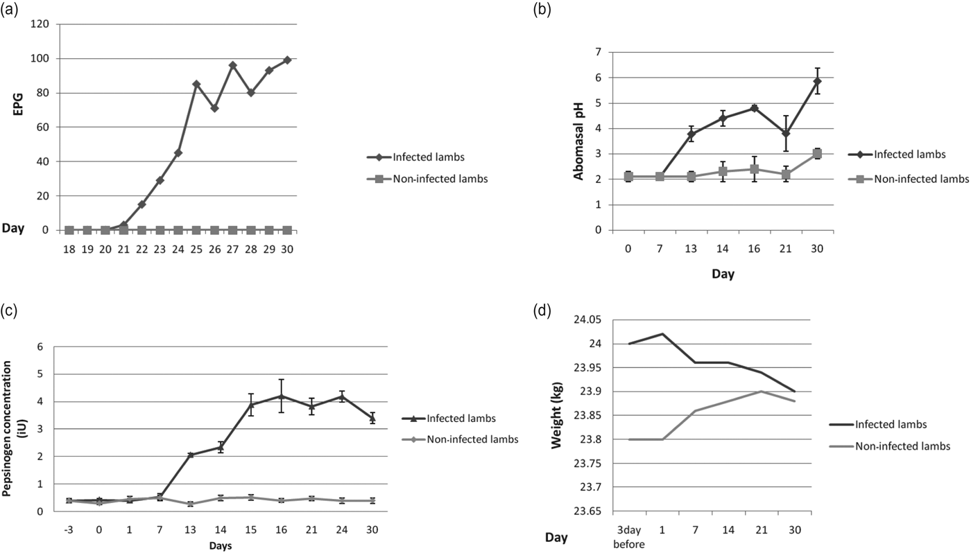
Fecal samples were collected daily post-infection. By 21 dpi patency was observed in all lambs, and the average number of eggs appearing in feces was three. The average number of EPG reached up to 61·6 ± 35·72 per day by 30 dpi. At time of death, the average number of collected M. marshalli worms from abomasa at necropsy was 3450 worms (Fig. 1).
(a) Mean fecal egg counts (EPG) in non-infected lambs and infected lambs which were infected on day 0 with 5000 infective L3 M. marshalli; comparison of abomasal pH (b) and serum pepsinogen (c) concentrations (iU) (mean ± s.d.) in lambs receiving 5000 infective L3 M. marshalli via stomach tube on day 0 with non-infected lambs; (d) Mean body weight (kg) in non-infected lambs and infected lambs which were infected with 5000 infective L3 M. marshalli.

Measurement of pH and pepsinogen concentration
The upper limits of the normal ranges of abomasal pH and the serum pepsinogen concentrations in non-parasitized animals were 2·3 and 0·5 iU respectively. The abomasal pH and serum pepsinogen concentrations of monitored animals were within the normal range during the experiment and did not exceed normal levels.
Dramatic changes in abomasal pH and serum pepsinogen concentrations were seen in larval-infected lambs. The serum pepsinogen concentration and abomasal pH were significantly increased in the infected group and there were noticeable differences among individual animals (P < 0·001). The effects of parasitism on abomasal pH and serum pepsinogen concentration (iU) in infected lambs and in non-infected lambs are shown in Fig. 1. In Fig. 1, comparison of pH and serum concentrations in the two groups is evaluated through 30 days.
Acidity of the abomasal contents and serum pepsinogen concentration varied between 2–2·5 and 0·35–0·5 iU prior to the infection (3 dpi) in infected sheep respectively. In all treated lambs, abomasal pH rose at 7 dpi and remained above normal limits until 16 dpi when values exceeded 4·4. The maximal mean value for the pH was 5·8 at necropsy which seemed to be the result of the stress of transport or attempt to inhibit acid secretion. Plasma pepsinogen elevated on 7 dpi and reached its maximal value at 16 dpi. The changes were due to mean values of about 0·4 iU to over 4·04 iU in each sheep. Abomasal pH and serum pepsinogen began to decrease after 16 dpi though above normal even at time of death. In the case of the monitored sheep, abomasal pH and serum pepsinogen concentration remained constant between 2–3·2 and 0·3–0·5 iU during the entire observation period, respectively.
Body weight
The body weights were very similar between the two groups throughout the infection and there were no significant differences among individual lambs (Fig. 1).
Macroscopic and microscopic lesions
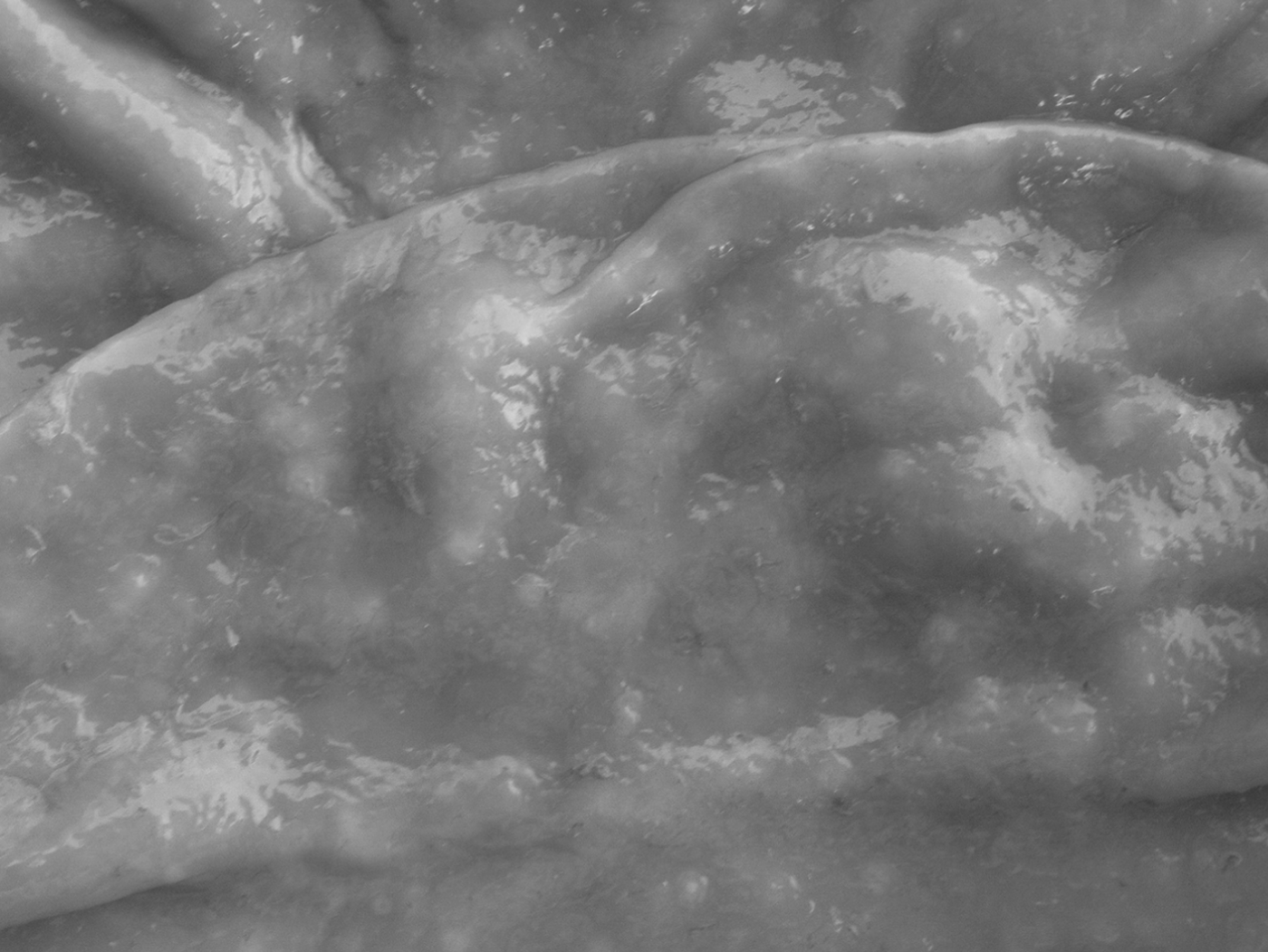
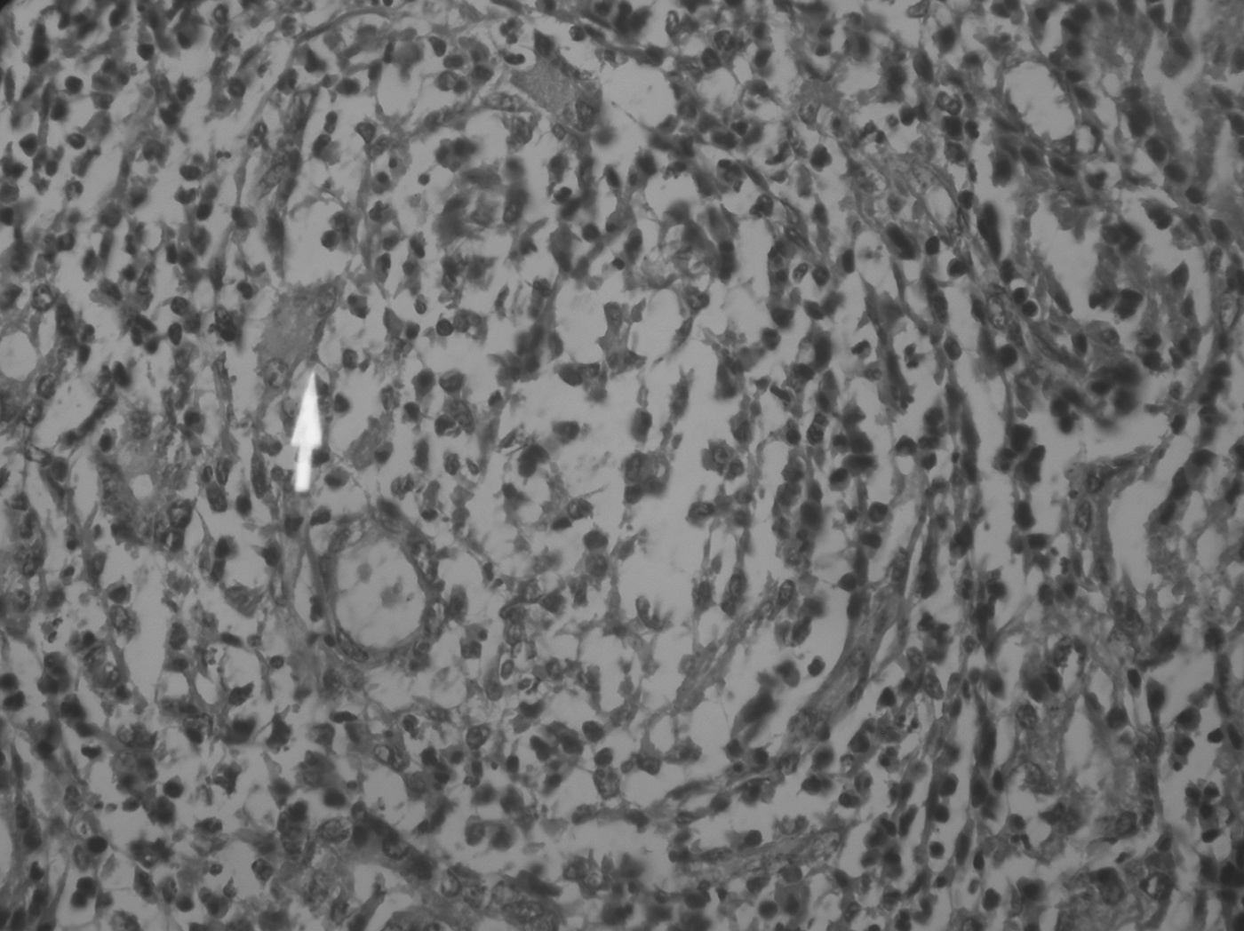
Mucosal lesions were similar in all infected animals, with some variations in their intensity. Macroscopic white nodules were observed in numerous areas of abomasal tissue sections (Fig. 2). The nodules were firm and of a meaty texture. These nodules indicated inflammatory reaction around the parasite aggregations. Microscopic observations consisted of two distinct reactions. The first showed hyperaemia and infiltration of eosinophil and mononuclear cells (Fig. 3) and the latter revealed inflammatory cells containing lymphocytes, macrophages, eosinophils, giant cells as well as fibroblasts, fibrocytes, granulation tissues, and collagen fibres between gastric glands in the infected lambs (Figs 4 and 5). Despite the fact that most of the lesions were observed in mucosal tissues, these abomasal lesions were also seen in the sub-mucosa and muscle areas. Mucosal hyperplasia and cellular necrosis were also seen. In addition atrophic and destroyed glands were replaced with connective tissues.
Thickening, edema and the presence of white nodules in abomasums of M. marshalli-infected lambs.
Histopathology of the M. marshalli-infected abomasums. Interstitial space of mucosa, infiltration of inflammatory cells, some atrophic glands. Arrowhead (cross parasite section). (H&E × 160).
Histopathology of the M. marshalli-infected abomasums. Cross parasite section (arrow head), infiltration of inflammatory cells, widespread atrophic glands, connective tissue formation (H&E × 640).
Histopathology of the M. marshalli-infected abomasums. The presence of large numbers of mononuclear cells, significant number of eosinophil, Giant cells (arrowhead), fibrocytes, fibroblasts, connective tissue and collagen fibres (H&E × 320).
DISCUSSION
Pathophysiological changes resulting from M. marshalli infection caused an increase in abomasal pH and serum pepsinogen concentrations. Reductions in acid secretion are likely to be a direct consequence of the accelerated loss of parietal cells. Increased abomasal pH occurs when parasites emerge from the gastric glands into the abomasal lumen but was not preceded by the increase in serum pepsinogen.
In all infected lambs, serum pepsinogen increased parasitism and there were typical effects on abomasal pH. The presence of fully-grown worms increased serum pepsinogen and abomasal pH while the early infective larval stages had minimal effects until the abrupt rise in all parameters during 14–21 dpi. Abomasal pH and serum pepsinogen concentrations increased at 7 dpi and reached maximal values at 16 dpi. Due to almost simultaneous increases in serum pepsinogen and abomasal pH just at the time of parasite emergence into the lumen along with maximal changes at day 16 dpi, it could be suggested that the emergence of M. marshalii was at 16 dpi.
In all other models of abomasal parasitism, serum pepsinogen and gastrin and abomasal pH increase almost simultaneously at the time of parasite emergence into the lumen (around 18 dpi for Ostertagia ostertagi and Ostertagia leptispicularis and 7–11 dpi for Haemonchus contortus) (Rahman and Collins, Reference Rahman and Collins1990; Taylor et al. Reference Taylor, Coop and Wall2007).
The results of our study are similar to the results in previous experiments with transfer of Teladorsagia circumcincta to uninfected animals resulting in rapid changes in the abomasal function (Lawton et al. Reference Lawton, Reynolds, Hodgkinson, Pomroy and Simpson1996; Simpson et al. Reference Simpson, Lawton, Simcock, Reynolds and Pomroy1997; Scott et al. Reference Scott, Hodgkinson, Khalaf, Reynolds, Pomroy and Simpson1998).
Histopathological examination has demonstrated that the abomasal dysfunction matches the morphological changes within the organ by the accumulation of large numbers of inflammatory cells. In infected lambs, the normal architecture of the gastric mucosa is altered by interstitial inflammation along with mucosa metaplasia and hyperplasia of the epithelium lining glands. The secretory activity and morphology of the stomachs of infected lambs were relatively unchanged until the parasites were expected to emerge from tissues. As a result, it would appear that the major disturbances in abomasal physiology in M. marshalli occur when the parasites are no longer confined to the glands and that larval development and the process of emergence itself may be relatively unimportant. The similarity of serum pepsinogen profiles, pH, and histological changes with ostertagiasis support the suggestion in a previous study (Dunn, Reference Dunn1978) that this nematode is pathogenic. Histological changes were typical of infection with T. circumcincta, particularly loss of parietal cells, the presence of many vacuolated parietal cells, infiltration of inflammatory cells and a thickened mucosa (Armour et al. Reference Armour, Jarrett and Jennings1966; Anderson et al. Reference Anderson, Reynolds and Titchen1988; Fox et al. Reference Fox, Carroll, Hughes, Uche, Jacobs and Vaillant1993; Scott et al. Reference Scott, Khalaf, Simcock, Knight, Reynolds, Pomroy and Simpson2000). Inflammatory reaction around the parasite was found in the interstitial tissue by microscopic observation. Physical stress and ischaemia-induced inflammatory reaction around the parasite in the interstitial tissue should be considered an effective cause in gastrointestinal endocrine dysfunction. Granulation tissue and connective tissue formation surrounding focal inflammatory lesions showed extensive destruction of glandular tissue and replaced the progressive healing process.
In conclusion, M. marshalli produced pathophysiological changes following infections. Parietal cell loss appeared to be by necrosis and was coincident with both changes in abomasal physiology and the rapid accumulation of eosinophils. This suggests that inflammation makes an important contribution to the functional and structural alterations of the abomasum similar to that which occurs in ostertagiasis.
Marshallagia marshalli may act as the major cause of impaired or reduced production of meat, wool and milk even where infection levels are subclinical, but due to the short duration of the study, we found no changes in weight of the two groups, and more research is needed in this area.
ACKNOWLEDGEMENTS
The authors are grateful to Professor H. V. Simpson for providing the pepsinogen measurement method and Dr Azizzadeh for the data analysis. We thank Mr Eshrati and Mr Azari for their technical assistance during data collection and Mr Mohammad Nejad for taking pictures. This work was sponsored by Ferdowsi University of Mashhad.