

INTRODUCTION
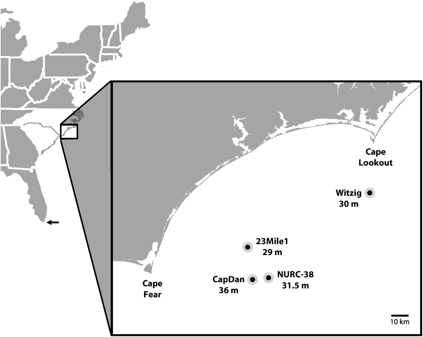
North Carolina waters are biogeographically important as they represent a sharp transition zone between the southern extent of the cool temperate and northern extent of the warm temperate zones along the United States east coast (Searles & Schneider, Reference Searles and Schneider1980). Onslow Bay is the area of continental shelf waters extending from Cape Lookout south along the North Carolina coast to Cape Fear (Figure 1), and it contains the most extensive offshore rock outcrops or ‘hard bottoms' in the mid-Atlantic because there are no erosional river outfalls depositing sediments on the shelf (Riggs et al., Reference Riggs, Snyder, Hine and Mearns1996, Reference Riggs, Ambrose, Cook and Synder1998). These hard bottoms provide the most extensive marine algal habitat between New England and southern Florida, and in combination with the varying temperatures of this transition zone create a large variety of environments suitable to algal growth. This is reflected in the large number of marine algal species reported from North Carolina and especially Onslow Bay (Searles & Schneider, Reference Searles and Schneider1978; Schneider & Searles, Reference Schneider and Searles1991).
Onslow Bay, North Carolina hard bottom ledge sites and depths, where Cottoniella species were collected. The previous northern distribution limit of C. filamentosa along the United States east coast is indicated with an arrow.

The exploration of seaweeds along the mid-Atlantic coast of the United States, and especially North Carolina, has a long history, but investigations of the offshore flora have been sporadic. The first deepwater collections from North Carolina were the result of dredges made by Hoyt in Onslow Bay (Hoyt, Reference Hoyt1920). Later, Williams (Reference Williams1951) published an investigation of the algae of a shallow hard bottom that included the first specimens collected by SCUBA divers. Craig Schneider, Rick Searles and their students made numerous SCUBA and dredge collections in the offshore waters of North Carolina that were published during the 1970s and 1980s (see Schneider & Searles, Reference Schneider and Searles1991 for list of citations). These studies provide a baseline of data for comparison with new explorations of North Carolina's offshore marine algae, a flora that has not been systematically surveyed for over 35 years.
Recent opportunistic marine algal collections from North Carolina's offshore hard bottoms have been made as part of the University of North Carolina Wilmington's Coastal Oceans Research and Monitoring Program (CORMP) and the United States National Oceanographic and Atmospheric Administration's (NOAA) lionfish invasion research programme. These collections have revealed the presence of marine algae unreported in North Carolina waters. This paper reports the collection of two species of Cottoniella Børgesen, a genus previously unknown for the State.
MATERIALS AND METHODS
Specimens of marine algae were collected from various hard bottom locations in Onslow Bay, North Carolina using SCUBA (Figure 1) and either quick dried in silica gel desiccant (Chase & Hills, Reference Chase and Hills1991) or preserved in 5% formalin–seawater solution. Formalin fixed material was prepared as herbarium specimens or stained with aniline blue (Millar & Wynne, Reference Millar and Wynne1992) and mounted on 50% Karo slides. Collections were labelled with a North Carolina marine algal database number (NCweed), and herbarium specimens and slides deposited in the herbarium at the University of North Carolina Wilmington (WNC).
DNA was extracted from a silica gel dried specimen following the protocol of Hughey et al. (Reference Hughey, Silva and Hommersand2001). Methods outlined in Freshwater et al. (Reference Freshwater, Kyhn-Hansen, Sarver and Walsh2000) and Olson et al. (Reference Olson, Melton, Yaudes, Norwood and Freshwater2004), and primers F753 and RrbcSstart of Freshwater & Rueness (Reference Freshwater and Rueness1994) were used to generate sequence data for the large subunit of ribulose-1,5-bisphosphate carboxylase/oxygenase (rbcL). The resulting sequence was used as the query in BLAST searches of the National Center for Biotechnology Information's nucleotide database (www.ncbi.nlm.nih.gov).
RESULTS AND DISCUSSION
SYSTEMATICS
Class FLORIDEOPHYCEAE Cronquist, Reference Cronquist1960
Subclass RHODYMENIOPHYCIDAE Saunders & Hommersand, Reference Saunders and Hommersand2004
Order CERAMIALES Oltmanns, Reference Oltmanns1904
Family SARCOMENIACEAE Womersley, Reference Womersley2003
Genus Cottoniella Børgesen, Reference Børgesen1919
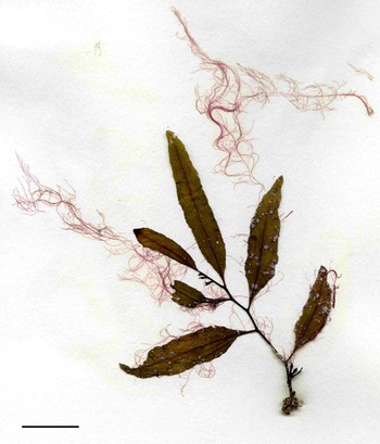
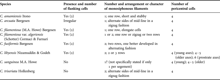
Cottoniella includes seven currently recognized species and one variety (Guiry & Guiry, Reference Guiry and Guiry2013). They are fine filamentous algae in habit and often grow epiphytically on other macroalgae (Figure 2). The genus is identified by the particular construction of main axes and presence of determinate monosiphonous filaments (Womersley, Reference Womersley2003). Cottoniella species have large apical cells that undergo transverse divisions giving rise to short disc-shaped cells (Figure 3A). These disc-shaped cells enlarge and then divide longitudinally to form four pericentral cells (Figure 3A, B). Five pericentral cells have been reported to sometimes occur in older axes of C. sanguinea M.A. Howe and C. libyensis Nizammudin & Godeh, and six are reported in the prostrate axes of the latter species (Howe, Reference Howe1928; Nizamuddin & Godeh, Reference Nizamuddin and Godeh1990). Five of the seven Cottoniella species have flanking cells. The two lateral pericentral cells in these species divide obliquely at their distal ends and then longitudinally at their proximal ends to cut off a pair of flanking cells (Figure 3C, D). Monosiphonous, determinate filaments (one or two, rarely three) occur on the distal portions of erect axes with the number, position and arrangement of these filaments varying among species. The initial monosiphonous filament develops endogenously from the central axial cell distal to the adaxial pericentral cell (Figure 4A, B). When a second monosiphonous filament develops they are often on alternate sides of the mid-line of the axis in successive segments (Figure 4C). Character states for pericentral cells, flanking cells and monosiphonous filaments are used to distinguish species (Table 1). Reproductive structures have only rarely been reported. Hollenberg (Reference Hollenberg1967) describes cystocarps in C. triserata, and tetrasporphytes are described by Cormaci et al. (Reference Cormaci, Furnari and Scammacca1978).
Cottoniella filamentosa growing epiphytically on Sargassum filipendula C. Agardh collected at ‘23 mile reef’ hard bottom site 16 June 2008 (NCweed 1245). Scale bar: 2 cm.

Cottoniella filamentosa vegetative development and structure: (A) adaxial view of apical tip; (B) lateral view of apical tip; (C) adaxial view showing development of flanking cells; (D) adaxial view showing pericentral and flanking cells. BP, abaxial pericentral cell; CA, central axial cell; DP, adaxial pericentral cell; LP, lateral pericentral cell; 1F, distal flanking cell; 2F, proximal flanking cell. Scale bars: A, B, 5 µm; C, 10 µm; D, 20 µm.

Cottoniella filamentosa (A, B) and C. fusiformis (C): (A) lateral view showing development of adaxial pericentral cell and initiation of monosiphonous filaments; (B) lateral view showing development of monosiphonous filaments; (C) adaxial view showing basal cells of secondary monosiphonous filaments on alternate sides of the mid-line of the axis in successive segments. BP, abaxial pericentral cell; CA, central axial cell; DP, adaxial pericentral cell; LP, lateral pericentral cell; MFI, monosiphonous filament initial; 1F, distal flanking cell; 2F, proximal flanking cell; 1M, first monosiphonous filament basal cell; 2M, second monosiphonous filament basal cell. Scale bars: A, 5 µm; B, 10 µm; C, 20 µm.

Character states for flanking cells, monosiphonous filaments and pericentral cells in Cottoniella species.

Four species of Cottoniella (C. arcuata Børgesen, C. filamentosa (M.A. Howe) Børgesen, C. fusiformis Børgesen and C. sanguinea M.A. Howe) have been reported in the tropical and subtropical western Atlantic (Hollenberg, Reference Hollenberg1967; Wynne, Reference Wynne2011), but none have been reported on the United States east coast north of the Florida Keys. Two species of Cottoniella have been collected since July 2007 at four sites in Onslow Bay (Figure 1).
Cottoniella filamentosa (Howe) Børgesen, Reference Børgesen1920
(Figures 2, 3 & 4A, B)
BASIONYM
Sarcomenia filamentosa Howe, Reference Howe1905.
SYNONYM
Cottoniella arcuata var. filamentosa (M.A. Howe) Schotter, Reference Schotter1951.
TYPE LOCALITY
Cape Florida, Key Biscayne, Florida, USA.
REPORTED DISTRIBUTION
Tropical and subtropical waters of the Atlantic, east and west Indian, and south-west Pacific Oceans.
NORTH CAROLINA COLLECTIONS
‘Witzig Rock’ hard bottom, Onslow Bay, NC, (34°16.829′N 076°35.006′W), 30 m depth, NCweed 1246 & WNC2007-s101 through to s109, 1 August 2007, Coll: D.W. Freshwater, Det: D.W. Freshwater and M.H. Hommersand; ‘23 mile reef’, Onslow Bay, NC, (34°00.221′N 077°22.238′W), 29 m depth, NCweed 1245 & WNC2008-s189, 16 June 2008, Coll & Det: D.W. Freshwater; ‘Cap Dan 2’ hard bottom, Onslow Bay, NC, (33°48.202′N 077°18.793′W), 36 m depth, NCweed 261 & WNC2009-s663 through to s665, epiphyte on Botryocladia, 24 June 2009, Coll: D.W. Freshwater & B. Degan, Det: D.W. Freshwater & J.N. Idol; NCweed 926 & WNC2012-s136, growing on hard bottom, Coll: J.N. Idol, D. Wells & M. Dionesotes, 2 May 2012, Det. J.N. Idol; NCweed 1022 & WNC33505 and 33506, growing on hard bottom, Coll: E. Arb, M. Dionesotes & J.N. Idol, 15 August 2012, Det: J.N. Idol; NCweed 1062 & WNC2012-s294 and s295, Coll: J.N. Idol & E. Arb, 20 September 2012, Det: J.N. Idol.
REMARKS
Cottoniella filamentosa is characterized by the consistent development of flanking cells from the lateral pericentral cells of each segment (Figure 3C, D); having only a single monosiphonous filament per segment with these filaments originating distal to the adaxial pericentral cell and arranged in a straight series (Figure 4B), and branch tips that are not strongly curved.
BLAST searches of a 672 base pair segment of the 3′ end of rbcL revealed a closest homology (96%) with Sarcomenia delesserioides Sonder (GenBank Accession No. AF257443, published by Lin et al., Reference Lin, Fredericq and Hommersand2001). The next closest sequences are only 88–90% similar and represent a variety of Ceramiales taxa within the Delesseriaceae and Rhodomelaceae. This result supports the classification of Cottoniella within the Sarcomeniaceae but there is no context within the currently available sequence data to assess potential relationships.
Although common at the ‘Witzig Rock’ collection site, C. filamentosa was not seen during 14 other dives at various Onslow Bay hard bottom sites during a 30 July–9 August 2007 NOAA cruise. The June 2008 ‘23 mile reef’ specimen was the first time this species was seen at that location despite marine algae having been collected there on 12 dives from 2004–2008. Similarly, marine algal collections from the ‘Cap Dan 2’ study site between 2006 and 2009 included no C. filamentosa, but it was consistently found in multiple collections made during 2012.
The previously reported northern distribution limit of C. filamentosa along the United States east coast was the Florida Keys (Dawes & Mathieson, Reference Dawes and Mathieson2008). These reports from Onslow Bay, North Carolina represent a range expansion of 1300 km. Cottoniella filamentosa is also reported from Bermuda (Taylor, Reference Taylor1960; Schneider, Reference Schneider2003), 1000 km east of North Carolina. The Gulf Stream current acts as a barrier to a direct connection between North Carolina and Bermuda, but both sites are sinks for marine organisms distributed from areas further south and there is evidence for biogeographical connectivity between Bermuda and the United States east coast (e.g. Betancur-R et al., Reference Betancour-R, Hines, Acero, Ortí, Wilbur and Freshwater2011).
Cottoniella fusiformis Børgesen, Reference Børgesen1930
(Figures 4C & 5–8)
SYNONYMS
Cottoniella arcuata var. fusiformis (Børgesen) Schotter, Reference Schotter1951; Cottoniella filamentosa var. fusiformis (Børgesen) Cormaci & Furnari, Reference Cormaci and Furnari1987.
TYPE LOCALITY
Las Palmas, Gran Canaria, Canary Islands.
REPORTED DISTRIBUTION
Tropical and subtropical waters of the Atlantic, Mediterranean, South Australia and Pakistan.
NORTH CAROLINA COLLECTIONS
‘NURC-38’ hard bottom, Onslow Bay, NC, (33°49.795′N 077°12.928′W), 31.5 m depth, NCweed 1247 & WNC2008-s188, 28 May 2008, Coll & Det: D.W. Freshwater; ‘23 mile reef’, Onslow Bay, NC, (34°00.221′N 077°22.238′W), 29 m depth, NCweed 801 & WNC2011-s156 through to s159, 20 June 2011, Coll: UNCW Research Diving Class, Det: J.N. Idol & D.W. Freshwater.
REMARKS
Cottoniella fusiformis is characterized by the consistent development of flanking cells from the lateral pericentral cells of each segment; having two monosiphonous filaments per segment which develop distal to the adaxial pericentral cell (Figure 5) and strongly curved branch tips. In North Carolina specimens, one of the two monosiphonous filaments is better developed (larger basal cell, greater number of cells and longer), and occurs on alternate sides of the mid-line of the axis in successive segments (Figures 4C & 5). Although Børgesen does not comment on this in his description of C. fusiformis from the Canary Islands, the unequal development of paired monosiphonous filaments may be seen in his illustrations (Børgesen, Reference Børgesen1930, figure 59c, d). Indeterminate lateral branches develop on the opposite side of the mid-line of the axis from the less developed monosiphonous filaments.
Cottoniella fusiformis extended depth of field image of apical tip showing uneven development of paired monosiphonous filaments. Scale bar: 50 µm.

Tetrasporangia development in C. fusiformis is very similar to that described for C. filamentosa by Cormaci et al. (Reference Cormaci, Furnari and Scammacca1978). Paired tetrasporangia are produced in the proximal parts of lateral branches that otherwise undergo normal vegetative development (Figure 6). Lateral pericentral cells undergo the normal division sequence to produce flanking cells, and then divide transversely to produce tetraspore mother cells. The lateral pericentral cells then divide twice more to produce abaxial and adaxial cover cells, and they become arcuate in shape (Figure 7). The abaxial cover cells enlarge to nearly the same size as the abaxial pericentral cell and the adaxial cover cell remains relatively small similar to their descriptions in C. filamentosa. However, unlike C. filamentosa, the adaxial cover cells tend to cut off additional small corticating cells in C. fusiformis (Figures 7C & 8). Flanking cells and additional cells that the flanking cells cut off distally elongate periclinally (Figures 7 & 8).
Cottoniella fusiformis lateral branches of indeterminate growth with proximal areas of tetrasporangia development (arrows). Scale bar: 200 µm.

Cottoniella fusiformis: three focal planes through an area of tetrasporangia development: (A) abaxial focal plane showing larger abaxial cover cells and smaller additional corticating cells; (B) central focal plane showing arcuate lateral pericentral cells, pit connections between lateral pericentral cells and flanking cells (arrows), additional cells cut off distally from flanking cells, and developing tetrasporangia; (C) adaxial focal plane showing smaller adaxial cover cells and an additional corticating cell cut off from an adaxial cover cell. AC, additional cells cut off from flanking cells; BC, abaxial cover cell; BP, abaxial pericentral cell; CA, central axial cell; CC, additional corticating cells; DC, adaxial cover cells; DP, adaxial pericentral cell; LP, lateral pericentral cell; T, developing tetrasporangia; 1F, distal flanking cell; 2F, proximal flanking cell. Scale bars: A–C, 20 µm.

Cottoniella fusiformis area of tetrasporangia development showing flanking cells and additional cells cut off distally from flanking cells that have become periclinally elongated and adaxial cover cells that have cut off additional corticating cells. AC, additional cells cut off from flanking cells; CC, additional corticating cells; DC, adaxial cover cells; DP, adaxial pericentral cell; 1F, distal flanking cell; 2F, proximal flanking cell.

Hollenberg (Reference Hollenberg1967) studied Bermuda specimens originally identified as C. filamentosa and determined that they represented C. fusiformis. The North Carolina specimens are the second report of this species in the western Atlantic and first in the United States.
Conclusions
Opportunistic marine algal collections were made during 67 dives on hard bottom and wreck sites in Onslow Bay between June 2004 and July 2007. Cottoniella was not encountered in any of these collections, nor had previous collectors reported it (Hoyt, Reference Hoyt1920; Williams, Reference Williams1951; Schneider & Searles, Reference Schneider and Searles1991 and citations within). Specimens of C. filamentosa were collected at a single site during August 2007 and at a different site in June 2008. Cottoniella fusiformis was collected at a third site during May 2008. Cottoniella specimens have continued to be collected since 2009, and C. filamentosa was found to be a consistent component of the marine algal epiphyte community at a study site visited multiple times during 2012. The distinctive morphological character states of Cottoniella make this genus readily identifiable and it is unlikely that specimens would have been missed in the extensive surveys conducted by Schneider and Searles during the 1970s (see Schneider & Searles, Reference Schneider and Searles1991 and citations within) or in the many collections made from Onslow Bay hard bottoms during 2004–2007. These reports of C. filamentosa and C. fusiformis are believed to represent northern range expansions of these species along the United States east coast.
ACKNOWLEDGEMENTS
The authors thank Dr Max H. Hommersand, who guided the first author to the initial identification of the species in this study (and many others); the NOAA, NURC and CORMP divers without whose support this work would not have been possible, and especially Brian Degan, Paula Whitfield, Christine A. Buckel, Doug Kesling, Thor Dunmire, Dave Wells and Steve Hall.
FINANCIAL SUPPORT
This study was funded by US National Science Foundation grants DEB/PEET-0328491 and DEB-0742437, the US National Oceanographic and Atmospheric Administration's Invasive Species Programme and the CMS DNA Algal Trust.