Isolated origin of a left pulmonary artery from the aorta is a rare congenital cardiac malformation and less common than the anomalous right pulmonary. When tetralogy of Fallot is also present, it is most often accompanied by a left aortic arch.Reference Prifti, Bonacchi and Murzi 1
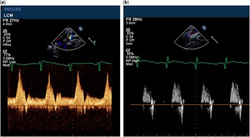
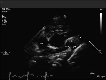
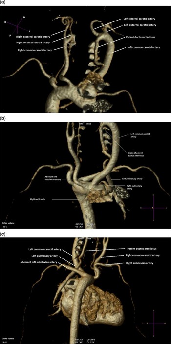
We present a neonate with echocardiographic diagnosis of dextrocardia, segmental anatomy {S,D,L}, tetralogy of Fallot, discontinuous left pulmonary artery with aortic origin from the left internal carotid artery via a patent ductus arteriosus, and right aortic arch with aberrant left subclavian artery. Doppler interrogation of the left common carotid artery demonstrated antegrade diastolic flow (Fig 1a), which was absent in the left subclavian artery (Fig 1b), confirming aberrancy of the left subclavian artery and indicating that the pulmonary artery origin was from the carotid. The left pulmonary artery was in close proximity to the right pulmonary artery at midline (Fig 2); it was tracked to the left internal carotid artery (Fig 3) by placing the transducer just below the patient’s left mandible. This finding was confirmed by CT scan (Fig 4). Intra-operative inspection confirmed all the above findings.
( a ) Doppler interrogation of the left common carotid artery demonstrates antegrade flow in diastole due to runoff into the left pulmonary artery. ( b ) Doppler interrogation of the left subclavian artery shows absence of diastolic flow, confirming aberrant origin of the left subclavian artery.

Suprasternal view shows the discontinuous branch pulmonary arteries in close proximity to each other. LCC=left common carotid artery; LPA=left pulmonary artery; RPA=right pulmonary artery; SVC=superior vena cava.

The probe is placed on the left neck with the notch just below the left mandible. The patent ductus ateriosus origin is noted along with the internal carotid artery.

Three-dimensional CT reconstruction confirmed ( a ) the origin of the patent ductus arteriosus from the left internal carotid artery. ( b ) The proximity of the right and left pulmonary arteries at their proximal course is delineated. ( c ) When visualised from a posterior aspect, the transition zone from patent ductus arteriosus to the discontinuous left pulmonary artery is better seen.

Our case highlights an unusual, not previously described, ductal origin of the left pulmonary artery from the left internal carotid artery. The diagnosis was made prospectively on transthoracic echocardiogram by tracking the left common carotid artery until the antegrade diastolic flow was not present and the origin of the ductus was visualised. This case highlights the importance of non-traditional echocardiographic views in conjunction with a thorough understanding of cardiac physiology in congenital heart disease to define the unusual anatomy. In addition, CT with three-dimensional reconstruction may assist in surgical planning.
Acknowledgements
None.
Financial Support
This research received no specific grant from any funding agency, commercial, or not-for-profit sectors.
Conflicts of Interest
None.