In a setting of single-ventricle physiology, some complications such as heart failure, hypoxia, arrhythmia, and hepatic fibrosis are well known. Reference Pundi, Johnson and Dearani1,Reference Gewillig2 Recently, a few reports have revealed a serious complication regarding coronary circulation, such as coronary arterial fistulae. Reference Finn, Walsh, Roberson and McMahon3,Reference Tannous, Ghelani, Marshall and Porras4 However, detailed clinical information about coronary arterial fistulae in patients with single-ventricle physiology is not fully understood, and its precise mechanism has not been established yet.
This study evaluated three patients who developed coronary arterial fistulae after right heart bypass surgery to clarify the clinical characteristics and mechanisms for coronary arterial fistulae in patients with single-ventricle physiology.
Materials and methods
We retrospectively investigated patients who underwent right heart bypass surgery and were followed up at our institute. Patients who underwent cardiac catheterisation and developed coronary arterial fistulae were included in the study.
The clinical records of these patients including their clinical findings and courses were reviewed. We excluded patients with sinusoidal communication that could develop in the setting of pulmonary atresia with intact ventricular septum and hypoplastic left heart syndrome. Reference Lowry, Olabiyi, Adachi, Moodie and Knudson5,Reference Anderson and Spicer6
The Institutional Review Board of the Osaka Medical College Hospital approved this study, and informed consent was obtained via opt-out due to the retrospective nature of this study.
Results
Three out of 21 cases (14.3%) developed coronary arterial fistulae after right heart bypass surgery at our institute. These coronary arterial fistulae were acquired and progressive in all three cases. In two of these patients, the coronary arterial fistulae contributed to their heart failure. All three cases had the right ventricle as the main chamber, suffered from hypoxia for a long duration of time, and have been administered pulmonary vasodilators such as phosphodiesterase type V inhibitors. The remaining 18 cases did not have these combinations of clinical features.
Case 1
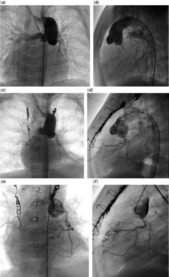
Case 1 was a 9-year-old boy. Cardiac anomalies were recognised during the foetal period. We diagnosed him with single right ventricle, pulmonary atresia, patent ductus arteriosus, and dextrocardia. He underwent a modified Blalock–Taussig shunt operation at 11 days of life. His post-operative clinical course was very stormy due to excessive pulmonary flow resulting in cardiogenic shock and fungal sepsis. This caused chronic lung disease. Therefore, long-term ventilator management was required. He underwent bidirectional Glenn shunt and Fontan completion at 8 months and 7 years of age, respectively. His oxygen saturation using pulse oximetry was 93% at follow-up in outpatient clinics due to chronic lung disease and recurrent respiratory tract infections. He had no symptoms of NYHA class I. No cardiac murmur was audible. His chest X-ray showed no cardiomegaly. We administered him frusemide, spironolactone, warfarin, aspirin, and tadalafil. The cardiac catheterisation performed at 4 years of age showed multiple small communications between the coronary artery and the right ventricle cavity. The cardiac catheterisation at 7 months of age showed no communication (Fig 1).
Serial angiograms and selective coronary angiograms of case 1. (a and b) Angiograms before right heart bypass surgery. The patient shows no coronary arterial fistulae. (a) AP view, (b) lateral view. (c and d) Angiograms at 6 years after Fontan completion, tortuous coronary arteries, and multiple and diffused coronary arterial fistulae were detected. (e and f) Selective coronary angiograms performed at 6 years old when the angiograms (c and d) were conducted. These show multiple and crackle communications between the tortuous coronary artery and ventricular chamber with detectable contrasts in a part of the right ventricle.

Case 2
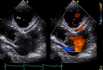
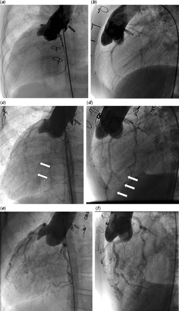
Case 2 was an 11-year-old girl. Her cardiac defects were right isomerism, dextrocardia, unbalanced atrioventricular septal defect, pulmonary atresia, supracardiac type total anomalous pulmonary venous connection, and major arterio-pulmonary collateral arteries. She underwent total anomalous pulmonary venous connection repair at the age of 11 months. This repair was followed by right and left unifocalisation with modified Blalock–Taussig–Bing shunt at 14 and 17 months of age, respectively. Subsequently, a bilateral bidirectional cava pulmonary shunt was performed at 20 months of age. Fontan completion was not indicated because the central pulmonary arteries did not cover more than two-thirds of the lung segments. She was followed up at the outpatient clinic and administered warfarin, aspirin, frusemide, spironolactone, carvedilol, enalapril, ambrisentan, and tadalafil. Her oxygen saturation using pulse oximetry was 85%. Finger clubbing and diastolic murmurs (Levine II/VI) were noted. Her chest X-ray showed mild cardiomegaly. She had symptoms of NYHA class II heart failure. Echocardiography showed a dilated coronary artery and multiple diastolic flow in the myocardium, which entered the cardiac chamber (Fig 2). Cardiac catheterisation, done at 11 years of age, revealed dilated coronary arteries that diffusely communicated with the cardiac chamber via multiple distal microvessels. Contrast medium from the coronary artery into the cardiac chamber enhanced the entire chamber. She underwent repeated catheterisation. However, her follow-up angiograms at 4 and 6 years of age showed no coronary arterial fistulae (Fig 3).
Echocardiography of case 2. Echocardiography shows a dilated coronary artery originating from the posterior cusp of the aorta. Ao=aorta.

Serial angiograms of case 2. (a and b) No coronary arterial fistulae before right heart bypass surgery at 14 months. Coronary arteries are dilated, and multiple small communications have developed in angiograms performed after the Glenn shunt in years. The white arrows indicate the contrast poured into the ventricular cavity through CAFs (c and d). Patient’s angiograms at 9 years old show dilated and generally tortuous coronaries with a progressive coronary arterial fistulae at the latest cardiac catheterisation (e and f).

Case 3
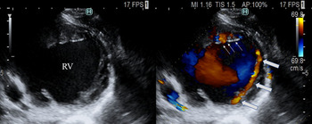
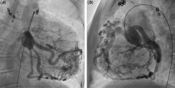
Case 3 was an 18-year-old man with hypoplastic left heart syndrome. At 2 years of age, fenestrated total cavopulmonary connection was performed. His oxygen saturation using pulse oximetry was 86% with moderate cyanosis and severe finger clubbing. Furthermore, he had NYHA class III heart failure. Diastolic murmur Levine (II/VI) was audible at the third left sternal border. Chest X-ray showed severe cardiomegaly and lung congestion. He was administered warfarin, aspirin, torasemide, eplerenone, trichlormethiazide, imidaprilat, carvedilol, macitentan, and tadalafil. Endothelin receptor blockade was administered for 4 years, and a phosphodiesterase V inhibitor was administered for 8 years. Diastolic murmur has been noted since he was 5 years old. His echocardiography showed a dilated coronary artery and multiple diastolic flows in the myocardium, which entered into the cardiac chamber (Fig 4). This finding was noted during the last 10 years and worsened. Cardiac catheterisation performed at 16 years of age revealed markedly dilated and tortuous coronary arteries. These coronary arteries had multiple communications with the distal end via microvessels such as the Thebesian veins. In addition, the right ventricle was enhanced entirely by contrast medium entered from these communications. His symptoms of heart failure worsened as echocardiographic findings of coronary arterial fistulae have deteriorated (Fig 5).
Echocardiography of case 3. Echocardiography revealed the diastolic blood flow through the dilated coronary arteries. The white arrows indicate the blood flow through the coronary artery, and the thin white arrows indicate multiple diastolic flows in the myocardium. RV=right ventricle.

Angiogram of case 3. Angiograms represent the dilated and tortuous coronary arteries that communicate into the right ventricle through diffuse and multiple coronary arterial fistulae. (a) AP view, (b) lateral view.

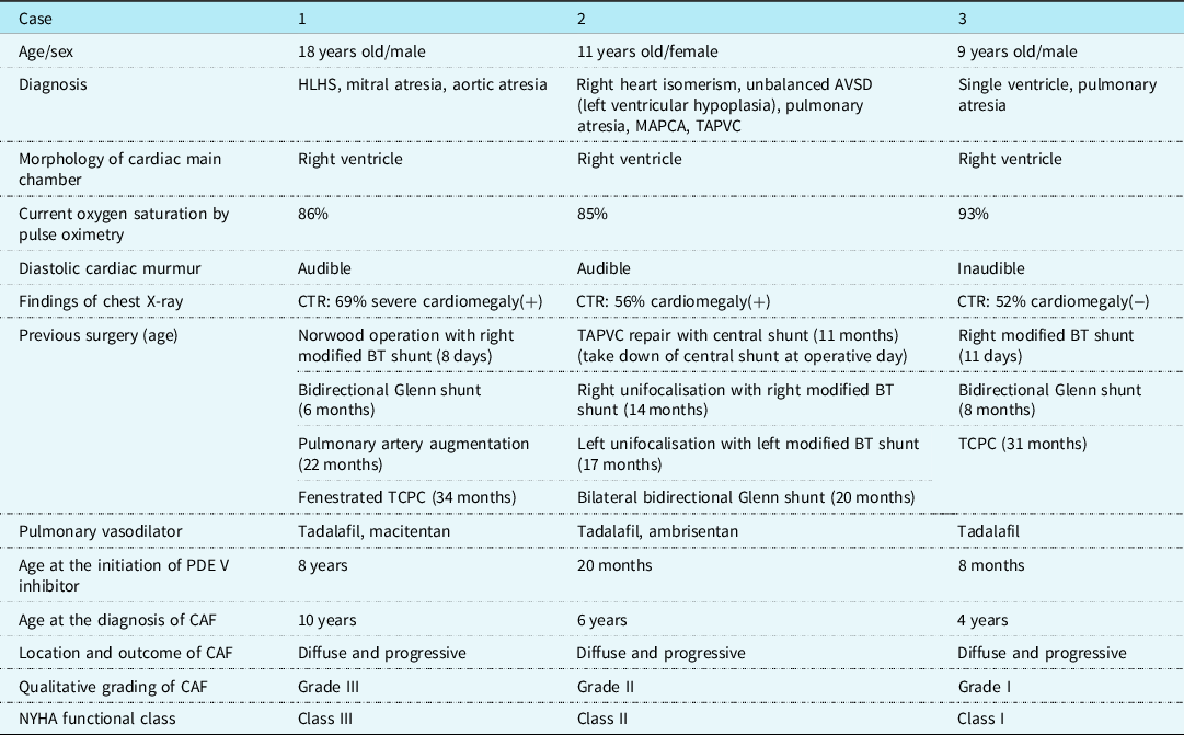
The clinical features of the three cases are listed in Table 1. They had some common features. First, the main ventricular chamber was the right ventricle. Second, they suffered from hypoxia for a long duration of time. Two of them had hypoxia even after Fontan completion. Third, all three cases were administered pulmonary vasodilators such as phosphodiesterase type V inhibitors. Fourth, coronary arterial fistulae were acquired and progressive over time. Furthermore, coronary arterial fistulae had multiple and diffuse communications via microvessels, such as the Thebesian vein, into the right ventricular chamber. Our three reported cases herein provide further evidence of this under-recognised phenomenon.
The demographics, clinical findings, surgical interventions, medical treatments, and clinical status of the three cases. We referred and modified the grading mentioned by Tannous et alReference Tannous, Ghelani, Marshall and Porras4 for qualitative grading of coronary arterial fistulae

AVSD=atrioventricular septal defect; BT shunt=Blalock–Taussig shunt; CAF=coronary arterial fistulae; CTR=cardio-thoracic ratio; HLHS=hypoplastic left heart syndrome; MAPCA=major aortopulmonary collateral arteries; PDE V=phosphodiesterase V; TAPVC=total anomalous of pulmonary venous connection; TCPC=total cavapulmonary connection.
Discussion
We could identify three cases of acquired and progressive coronary arterial fistulae among patients with single-ventricle physiology after right heart bypass surgery. They had common features such as dominant right ventricle, prolonged hypoxia, and treatment with phosphodiesterase type V inhibitors. The coronary arterial fistulae were acquired and progressive. The entering site was diffuse and multiple, similar to the Thebesian veins. The more prominent the shunt flows via coronary arterial fistulae, the worse symptoms of heart failure were through possible mechanisms of steal phenomenon and volume overload due to shunt flow. The incidence of complications of coronary arterial fistulae in the setting of single-ventricle physiology was higher in our cohort than was it in previous reports because we actively use pulmonary vasodilators in patients with pulmonary impairments after right heart bypass surgery. Reference Nemoto, Sasaki and Ozawa7 Occurrence and development of coronary arterial fistulae should be closely monitored during follow-up in patients after right heart bypass surgery. In addition, the indication for pulmonary vasodilators should be optimised in the setting of single-ventricle physiology after right heart bypass surgery.
There are some possible mechanisms for acquired and progressive coronary arterial fistulae established by common features among the three cases and other reported cases. First, the main cardiac chamber was the right ventricle. Tannous et al reported that their cases also had a dominant right ventricle chamber. Reference Tannous, Ghelani, Marshall and Porras4 The right ventricle essentially has rough trabeculation with space between the myocardia. Moreover, hypoxia can induce hypoxia-inducible factor 1, which can change matured cardiocytes into a foetal type that has not completed the compaction of the cardiac layer. Reference Sharma, Taegtmeyer and Adrogue8 Moreover, Ansari et al revealed in their study of sheep that the Thebesian veins were detected in the right ventricle more than in the left ventricle. Reference Ansari9 These histological features of the right ventricle are linked to the occurrence of diffuse and multiple coronary arterial fistulae. Second, hypoxia can lead to angiogenesis and vasculogenesis. Reference Semenza10,Reference McMahon, Coleman and Oslizlok11 Prolonged hypoxia can enhance the generation of hypoxia-inducible factor 1, which contributes to angiogenesis between the coronary arteries and cardiac chamber. Third, the phosphodiesterase type V inhibitors can also induce angiogenesis and dilatation of the coronary artery via the nitric oxide pathway. Regarding angiogenesis, Doganci et al performed a comparison between the three major pulmonary vasodilators: prostacyclins, phosphodiesterase type V inhibitors, and endothelin receptor antagonists using a chicken model. Reference Doganci, Yildirim and Yesildal12 They demonstrated that phosphodiesterase type V inhibitors significantly induced angiogenesis compared to the other two pulmonary vasodilators. A previous report regarding angiogenesis, especially in the setting of phosphodiesterase type V inhibitors and endothelin receptor antagonists, also suggested that phosphodiesterase type V inhibitors had angiogenetic effects rather than did endothelin receptor antagonists. Reference Zhu, Zhang and Alexeyev13–Reference Wanga, Li and Zhang17 McMahon et al and Finn et al have already described these mechanisms in their previous reports, especially regarding hypoxia and pulmonary vasodilators. Reference Finn, Walsh, Roberson and McMahon3,Reference McMahon, Coleman and Oslizlok11 Similarly, we could establish the same mechanisms based on our findings from our cohort and emphasise the potential contribution of phosphodiesterase type V inhibitors.
We referred to this rare complication as described in this article, coronary arterial fistulae. Finn et al also reported this complication as coronary arterial fistulae. Reference Finn, Walsh, Roberson and McMahon3 On the other hand, Tannous et al suggested these lesions as Thebesian veins in their article. However, the Thebesian veins cannot be detected by angiography and do not distribute diffusely with multiple entering. Reference Ansari9,Reference Wearn18 Therefore, we considered this lesion as a pathophysiological condition and decided to call it a coronary arterial fistulae.
The most important treatment of acquired and progressive coronary arterial fistulae is prevention. Coronary arterial fistulae can contribute to worsening symptoms of heart failure. Reference Snipelisky, Reddy, Lennon, Crusan, Rihal and Lapeyre19,Reference Khoury, Baydoun, Rafeh, McCord and Olkovky20 We need to avoid hypoxia and maintain oxygen saturation levels as high as possible using home oxygen therapy. We also need to evaluate the pulmonary circulation and keep in mind to make appropriate use of pulmonary vasodilators. We should avoid unnecessary prescription of pulmonary vasodilators to inhibit the progression of coronary arterial fistulae if possible. Hernández et al reported the efficacy of beta-blockers in reversing the coronary arterial fistulae. Reference Hernández, Carretero and Prada21
There are several limitations to this study. Due to its retrospective design performed in a single institute, the number of patients undergoing right heart bypass surgery included for analysis was small. We tended to use pulmonary vasodilators more frequently after right heart bypass surgery. A nation-wide survey is needed to investigate facts regarding coronary arterial fistulae in single-ventricle physiology.
The combination of the right ventricle as the main chamber, prolonged hypoxia, and phosphodiesterase type V inhibitors leads to these rarely acquired and progressive coronary arterial fistulae in patients with single-ventricle physiology, eventually resulting in heart failure. Patients should be followed up after right heart bypass surgery, noting this complication and evaluating it regularly by echocardiography and angiography. In addition, the progression of this complication should be avoided by optimising the use of pulmonary vasodilators.
Acknowledgements
The authors thank Shiro Ishikawa, from the Division of Cardiovascular Medicine, Department of Internal Medicine, Fukuoka-Kieikai Hospital, Koichi Sagawa, and Yasunori Nabeshima, from the Department of Cardiology, Fukuoka Children’s Hospital, for providing an angiogram of Case 3. We would like to thank Editage ( www.editage.com ) for the English language editing.
Financial support
This research received no specific grant from any funding agency, commercial, or not-for-profit sectors.
Conflicts of interest
None.
Ethical standards
The authors assert that all procedures contributing to this work comply with the ethical standards of the Ethical Guidelines for Medical and Health Research Involving Human Subjects and with the Helsinki Declaration of 1975, as revised in 2008, and have been approved by The Institutional Review Board of the Osaka Medical College.