Introduction
The introduction of molecular methods into the taxonomy of Pannariaceae has resulted in many surprising consequences. Ekman & Jørgensen (Reference Ekman and Jørgensen2002) showed that parts of the genera Moelleropsis, Degelia and Fuscopannaria did not belong to this family. Wedin et al. (Reference Wedin, Jørgensen and Wiklund2007, Reference Wedin, Jørgensen and Ekman2011) described the new families Massalongiaceae and Vahliellaceae with members previously placed within Pannariaceae, although Muggia et al. (Reference Muggia, Nelson, Wheeler, Yakovchenko, Tønsberg and Spribille2011) showed that Polychidium was heterogeneous, and they retransferred members of the reinstated genus Leptogidium from Massalongaceae to Pannariaceae. The new family Koerberiaceae described by Spribille & Muggia (Reference Spribille and Muggia2013) also includes ex-members of Pannariaceae. However, the family has also increased by the inclusion of genera previously belonging to Collemataceae. Wedin et al. (Reference Wedin, Wiklund, Jørgensen and Ekman2009) showed that the genera Leciophysma, Physma, Ramalodium and Staurolemma, all with non-septate spores, have a phylogenetic position in Pannariaceae in spite of a strong gross morphological resemblance to Collemataceae. This conclusion was also subsequently reached independently by Otálora et al. (Reference Otálora, Aragón, Molina and Martínez2010) in the case of Physma and Staurolemma. They also indicated that the genera Homothecium and Leightoniella might deserve a similar revised family affiliation; genera which still have not been studied genetically.
Two new phylogenetic studies on the family (Ekman et al. Reference Ekman, Wedin, Lindblom and Jørgensen2014; Magain & Sérusiaux Reference Magain and Sérusiaux2014) confirmed the affiliation of the ex-Collemataceae genera and presented topologies of the family, both with two major, well-supported clades. One of these, the ‘Parmeliella s. str. clade’, was well supported in both studies, and actually represents a still unrecognized family of its own, according to Spribille & Muggia (Reference Spribille and Muggia2013). The remaining samples of Pannariaceae were subdivided into three subgroups by both Ekman et al. (Reference Ekman, Wedin, Lindblom and Jørgensen2014) and Magain & Sérusiaux (Reference Magain and Sérusiaux2014), including a subgroup with Fuscopannaria and neighbouring genera, defined in a similar way in both studies. The other subgroups were classified differently, a fact which can be partly attributed to the sequencing of different genes and taxa and partly to different major topics defined for the studies. Psoroma is a key genus in the process of redefining generic limits within Pannariaceae. It is now defined as species resembling its generitype P. hypnorum (Vahl) S. F. Gray, but was previously interpreted in a wider sense, including most tripartite species within the family. Several squamulose species have been transferred to the new genera Joergensenia, Psorophorus, and Xanthopsoroma, described with molecular support (Passo et al. Reference Passo, Stenroos and Calvelo2008; Elvebakk et al. Reference Elvebakk, Robertsen, Park and Hong2010). The austral foliose species have been transferred to Pannaria, for example by Jørgensen (Reference Jørgensen2001) and Elvebakk & Galloway (Reference Elvebakk and Galloway2003). However, there are also foliose, tripartite species reported from many Paleotropical areas. Psoroma sphinctrinum (Mont.) Nyl. was reported from ‘Insula Borbonia’ (=Réunion), Mauritius and ‘Promontorio Bonæ Spei’ (in South Africa) by Nylander (Reference Nylander1859). Psoroma sphinctrinum has now been shown to be a panaustral Pannaria species, known as Pannaria sphinctrina (Nyl.) Tuck. ex Hue (Elvebakk Reference Elvebakk2007, Reference Elvebakk2011). The only tripartite and foliose Pannariaceae species described exclusively from Palaeotropical areas is Psoroma boninense Kurok. from the Bonin Islands (=Ogasawara Gunto Islands) by Kurokawa (Reference Kurokawa1969), in addition to Pannaria lobulifera Elvebakk, described from New Caledonia (Elvebakk Reference Elvebakk2007).
The first aim of the present study is to revise Paleotropical material of ‘Psoroma’, which instead represents an undescribed genus, very distinct from Pannaria, as already indicated by Elvebakk (Reference Elvebakk2007). During our initial studies of this group, we had the impression that this new tropical, ex-Psoroma genus included one or two widespread species, because the samples from widely separate geographical areas were found to be quite similar, except for cephalodium and spore morphology. However, we now conclude that the genus has a relatively high number of geographically differentiated species differing in several characters.
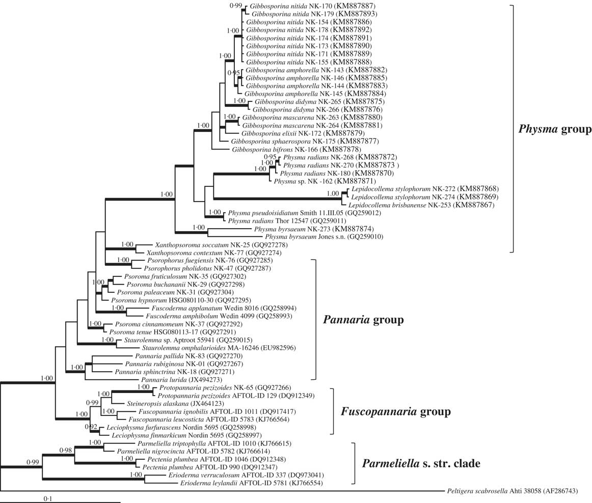
The previous transfer of tropical ex-Collemataceae genera to Pannariaceae by Wedin et al. (Reference Wedin, Wiklund, Jørgensen and Ekman2009) and Otálora et al. (Reference Otálora, Aragón, Molina and Martínez2010) has now been supported in the case of Physma by the new phylogenies presented by Magain & Sérusiaux (Reference Magain and Sérusiaux2014) and Ekman et al. (Reference Ekman, Wedin, Lindblom and Jørgensen2014). The former study presented a separate ‘Physma group’ represented by 20 sequences, 18 of these in a well-defined clade with species of Physma, Parmeliella s. lat. (now Lepidocollema) and a single ‘tripartite Pannaria’ sample from Réunion. The latter appeared to represent the undescribed genus which is the subject of the present study. Surprisingly, their ‘Physma group’ included, although with low support, two sequences of the very different genus Xanthopsoroma, which was a sister group to the remaining sequences. Ekman et al. (Reference Ekman, Wedin, Lindblom and Jørgensen2014) included four sequences of Physma and Parmeliella, the latter transferred to the redefined genus Lepidocollema, in a well-supported branch, nested within the Psoroma clade (‘Clade 2c’).
The second objective of the present study is to examine the new genus phylogenetically, both for possible molecular support for some of the newly described species, and for affiliation with other genera of Pannariaceae in the present phylogeny as well as in the recently published phylogenies of the family.
Material and Methods
Taxon sampling and identification
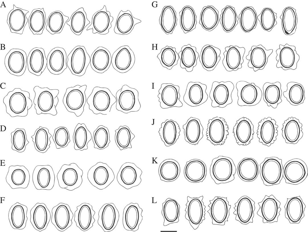
Herbarium material used in this study is housed in ABL, BG, BM, CBG, PC, E, H, IRD, L, O, REU, S, TNS, TROM, TUR and UPS. A total of 119 samples were examined. In microscope sections, iodine reactions were tested by adding IKI to mounts pretreated with KOH (Orange et al. Reference Orange, James and White2001). Perispore structures were studied in water mounts and restricted to spores liberated from the asci. Ascospore morphology was studied in detail by drawing detailed sketches of c. 880 ascospores from almost all collections, except for several samples of one species from the Solomon Islands housed at BM, where time did not permit this. The illustrations presented here aim to depict the variation in shape and size of ascospores within and between the species studied here (Fig. 7). Thin-layer chromatography of acetone extracts followed standard procedures and used solvents A and C (Culberson Reference Culberson1972; Orange et al. Reference Orange, James and White2001). Nomenclature of ascospore structure follows Nordin (Reference Nordin1997). Some of the type collections were also analyzed by HPLC following the procedure used by Bjerke et al. (Reference Bjerke, Lerfall and Elvebakk2002).
Specimens, DNA extraction, and sequencing
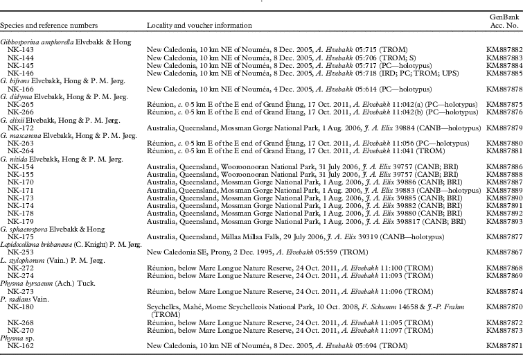
Twenty-seven specimens including 19 samples of seven species of Gibbosporina, five samples comprising three Physma species, and three samples representing two Lepidocollema species, were newly sequenced for this study (Table 1). Successful sequencing included seven holotypes. Complete Gibbosporina voucher information is found where these samples are cited in the present paper. Reference sequences were selected to represent the major phylogenetic lineages of Pannariaceae recently published by Magain & Sérusiaux (Reference Magain and Sérusiaux2014) and Ekman et al. (Reference Ekman, Wedin, Lindblom and Jørgensen2014), with clade names in the phylogram following those used by the former. Information on the previously published sequences included in Fig. 9 can be found in the tables of Elvebakk et al. (Reference Elvebakk, Robertsen, Park and Hong2010), Magain & Sérusiaux (Reference Magain and Sérusiaux2014) and Ekman et al. (Reference Ekman, Wedin, Lindblom and Jørgensen2014). Peltigera scabrosella was included to root the tree. The freeze-dried lichen materials were ground using TissueLyser (Qiagen, Hilden, Germany) after freezing in liquid nitrogen, and genomic DNAs were extracted using FastDNA™ SPIN Kit for Soil (MP Biomedical, Santa Ana, California) according to the manufacturer’s instructions. To determine phylogenetic relationships among lichenized fungi, amplification and sequencing of the ITS1, 5.8S, ITS2, and partial large subunit rDNA (LSU) were conducted using the ITS1F, ITS4, LR0R, and LR5 primers, following the procedures described in a previous study (Elvebakk et al. Reference Elvebakk, Robertsen, Park and Hong2010). Sequences were deposited in the GenBank database under the accession numbers KM 887867–887893.
Newly sequenced specimens in this phylogenetic study with locality information and GenBank accession number

Phylogenetic analyses
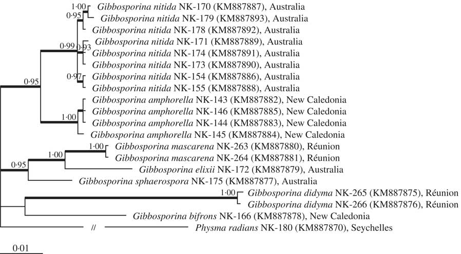
Sequence alignment of ITS1, 5.8S, ITS2 and partial LSU was conducted by the program ClustalX (Larkin et al. Reference Larkin, Blackshields, Brown, Chenna, McGettigan, McWilliam, Valentin, Wallace, Wilm and Lopez2007) and manually adjusted. Ambiguously aligned sites were excluded from the phylogenetic analyses. Phylogenetic trees were inferred from the datasets by neighbour-joining (NJ), maximum parsimony (MP), maximum likelihood (ML), and Bayesian analyses. The NJ tree was reconstructed using MEGA6 (Tamura et al. Reference Tamura, Stecher, Peterson, Filipski and Kumar2013) under Kimura’s 2-parameter model (Kimura Reference Kimura1980). The MP tree was obtained using the Tree-Bisection-Regrafting (TBR) algorithm of MEGA6 with search level 5, in which the initial trees were obtained by the random addition of sequences (1000 replicates). The ML tree was searched for by using PhyML ver 3.1 (Guindon & Gascuel Reference Guindon and Gascuel2003) with the GTR+I+G evolutionary model (Lanave et al. Reference Lanave, Preparata, Saccone and Serio1984) and the search options of best tree topology finding by branch swapping of NNIs and SPRs, random addition of sequences (100 replicates), and parameter estimation for proportion of invariant and transition/transversion ratio. The Bayesian tree was searched for by MrBayes ver. 3.2 (Ronquist et al. Reference Ronquist, Teslenko, van der Mark, Ayres, Darling, Höhna, Larget, Liu, Suchard and Huelsenbeck2012) with the GTR+I+G evolutionary model. Two parallel Markov chain Monte Carlo (MCMC) runs were performed, each with three heated chains and one cold chain, and the temperature parameter set to 0·1. Every 100th tree was sampled from 2 000 000 generations of analysis, and a consensus tree was calculated after discarding the first 25% trees as burn-in. The default search conditions were used for other options. The DNA evolutionary model was selected by AIC calculation implemented in jModelTest 2 (Darriba et al. Reference Darriba, Taboada, Doallo and Posada2012). Robustness of phylogenetic lineages was tested by posterior probability of Bayesian analysis and conservation in NJ, MP, and ML analyses. As the ITS sequences were hardly alignable among remotely related genera, phylogenetic relationships among genera of Pannariaceae were calculated based on LSU sequences only, and phylogenetic relationships among species of Gibbosporina were calculated based on combined sequences of ITS1, 5.8S, ITS2, and partial LSU.
Results
Taxonomy
Gibbosporina Elvebakk, Hong & P. M. Jørg. gen. nov.
MycoBank No.: MB 811978
Pannariae similis, sed sine acidis lichenosis, apotheciis amylodeis asci tholis et perisporis gibbis tumescentibus praeditis; photobionte maiore viridi et cephalodiis cyanobionticis bene evolutis instructo.
Typus generis: Gibbospora boninensis (Kurok.) Elvebakk & P. M. Jørg.
(Figs 1–8)
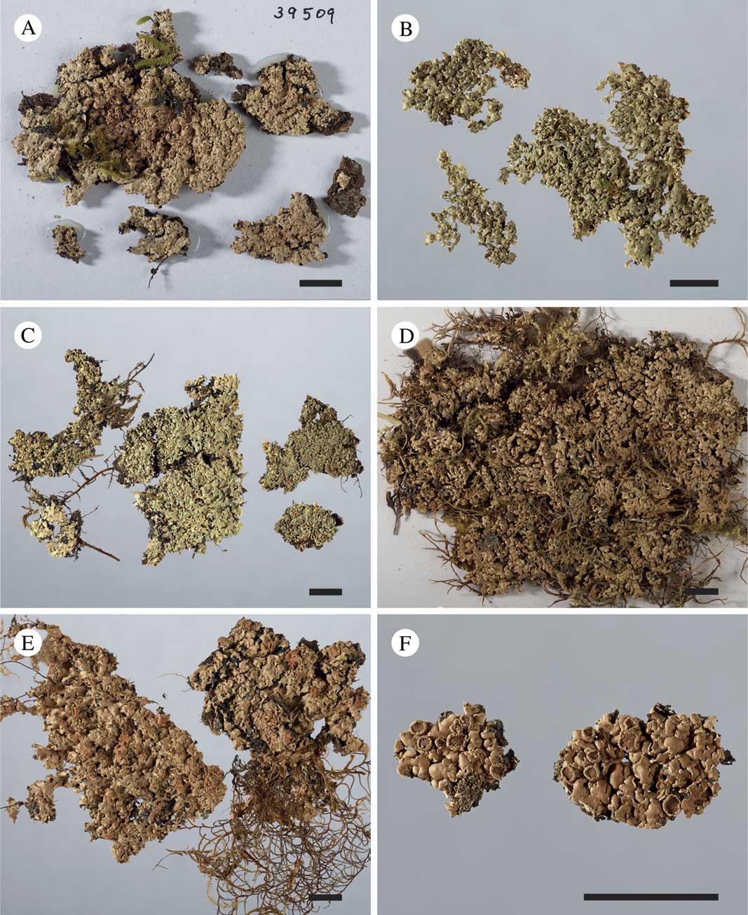
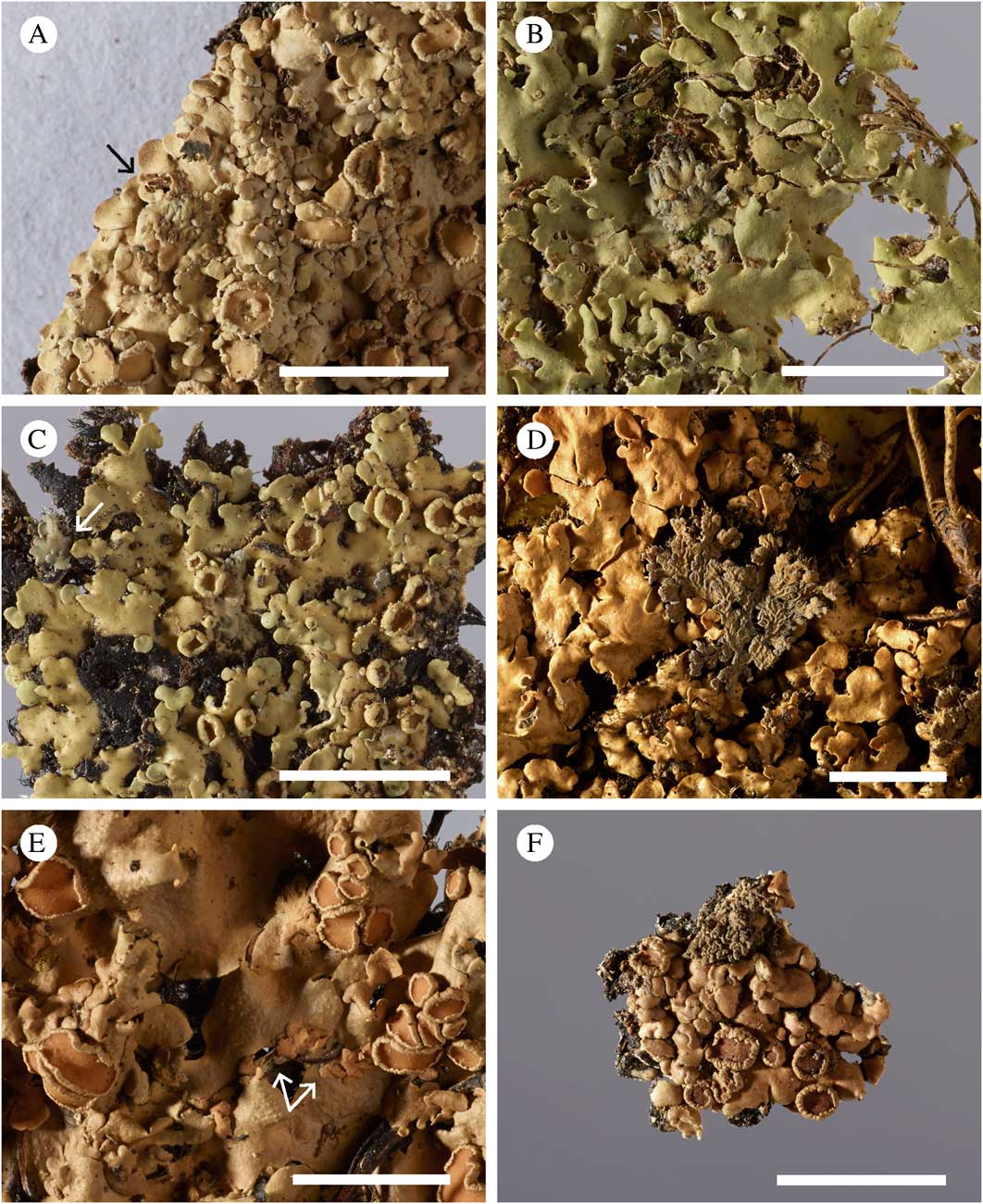
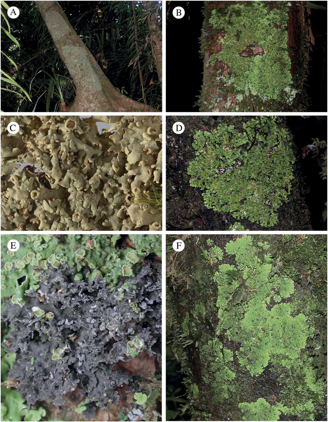
Gibbosporina species, habitus. A, G. acuminata (holotype); B, G. amphorella (holotype); C, G. bifrons (holotype centre), IRD isotype specimen top left, remaining specimens from isotype at TROM; D, G. bifrons (Coppins 5440 et al.) from Malaysia; E, G. bifrons (Hill 9744) from Solomon Islands; F, G. boninensis, (Knight, PC 0012753); G, G. didyma (holotype); H, G. elixii (holotype). Scales=1 cm.
Thallus foliose, pale greyish green when fresh, brown on old herbarium specimens, forming extensive, adpressed patches, partly developing distinct prothalli. Upper cortex distinct, forming a smooth, sometimes glossy upper surface; lower cortex lacking, but lower surface hyphae denser, running parallel to the lower surface.
Ascomata common, as large and substipitate apothecia with distinct, crenate thalline margins obscuring proper margins; discs reddish brown to orange-brown, dark brown on old, dry specimens. Hymenium colourless, IKI+ persistently deep blue, with paraphyses consisting of simple, septate hyphae which are apically thickened with external yellowish brown pigmentation. Asci clavate, apically with amyloid tube structures, 8-spored with simple, colourless, ellipsoid proper spores, surrounded by swelling, asymmetric and gibbose perispores.
Conidiomata rare, of protruding, black pycnidia of Sticta-type, producing bacilliform conidia 0·5–1·0×2–4 μm, laterally or terminally on short-celled conidiophores.
Major photobiont green, myrmecioid, but with Nostoc-containing laminal cephalodia which can be squamulose to nodulose, placodioid, mini-fruticose or mini-foliose, occasionally free-living with own rhizohyphae, but not yet observed completely independent with own cyanobiont apothecia.
Secondary chemistry. No acetone-soluble compounds detected by TLC and HPLC analysis.
Etymology. From Latin ‘gibbus’ (=‘with hump-like swellings’), which in combination with ‘spora’ refers to the unusual gibbose form of the perispores in most of the species.
Distribution and ecology. Corticolous in lowland and montane tropical and subtropical forests from central parts of the Pacific through SE Asia and NE Australia to Sri Lanka and Madagascar.
Gibbosporina acuminata Elvebakk sp. nov.
MycoBank No.: MB 811979
Gibbosporinae boninensi similis sed lobis impolitis vel debile nitidis, cephalodiis placodiodeis, adpressique, sporis angustioribus.
Typus: Australia, Queensland, Zillie Falls, 12 km by road NE of Millaa Millaa, 17°28'29''S, 145°39'22'E, 705 m elev., remnant rainforest near falls, on fallen tree, 29 July 2006, J. A. Elix 39509 (CANB 00783313—holotypus; BRI—isotypus).
Thallus of chloromorph 5–15 cm diam., foliose, corticolous or muscicolous on corticolous bryophytes, when corticolous with a distinct black hypothallus, often also prothallus; lobes subdichotomously divided, 160–200 μm thick, 0·8–1·5 mm broad, discrete and flat to weakly concave in peripheral parts, gradually becoming coalescent and convex, and often with small, geotropically oriented lobules in central parts of the thallus. Upper surface glabrous but weakly tomentose on young parts of lobes, matt to weakly glossy, fresh specimens bright green with contrasting blue-grey cephalodia when moist, light greenish grey when dry, old herbarium specimens either ochraceous or dark brown. Upper cortex 30–40 μm thick, plectenchymatous, lumina up to 15×10 μm, irregularly subellipsoid, arranged perpendicularly to the surface, surface weakly sclerenchymatous, lower cortex absent. Algal layer 30–35 μm thick; photobiont myrmecioid, cells 3–8 μm, globose to ellipsoid or slightly irregular. Medulla of loosely interwoven hyphae, 80–120 μm thick, lowermost part brownly pigmented; rhizines marginally white, otherwise black, 0·5–1·5 mm long, simple or in bundles, fibrillose.
Cyanomorph as cephalodia, pale grey when dry, formed on chlorobiont lobes, placodioid both when young and mature, very rarely subfoliose and weakly ascending and rhizinate, surface smooth and with radiating furrows, 1–2 mm diam., with subdichotomously and weakly branching lobes, 0·2–0·3 mm wide, weakly convex. Cortex like in the chloromorph. Cyanobiont Nostoc, as 3–7×4–8 μm large, globose to short-ellipsoid, dark brownish green to greyish violet cells, blue-green on some older samples, organized within 20–50 μm large glomeruli, without a discernible chain structure.
Apothecia subsessile, laminal, 0·5–4·0 mm diam.; disc orange-brown; thalline excipulum 0·15–0·30 mm thick, crenate-striate; epithecium pale brown, 10–20 μm thick, IKI−; hymenium colourless, but strongly IKI+ blue, 80–100 μm thick; hypothecium pale brown, IKI−, 20–30 μm thick; algal layer extending below the hypothecium; paraphyses simple to sparingly branched, with slightly swollen apices, septate; asci clavate, 8-spored, with pronounced, internal, apical, IKI+blue amyloid tube structures, 50–70×10–15 μm. Proper ascospores colourless, simple, ellipsoid, 11–17×7·5–11·0 μm; perispores 13–22×11–18 μm, walls 0·5–1·5 μm thick in addition to irregular gibbae, some bullate, others up to 6 μm tall, acuminate and sometimes asymmetrically oriented.
Pycnidia not seen.
Chemistry. No TLC - detectable compounds found.
Etymology. The specific epithet acuminate, from the Latin ‘acumen’ (=sharp point), refers to the distinctly spiked gibbae of the perispores.
Distribution and ecology. Known from tropical forests in Australia and the Philippines.
Major characteristics. A species with thick lobes, matt to weakly glossy, and without conspicuous pycnidia. Perispores are intermediately thick, but some have strongly acuminate gibbae, often slightly asymmetrical, resembling fins of ray fish in outline. Cephalodia are small and inconspicuous and remain placodioid.
Additional specimens examined (paratypes). Australia: Queensland: Millaa Millaa Falls, 4 km S of Millaa Millaa, 17°29'44''S, 145°36'41''E, 750 m, remnant rainforest, on fallen branches, 2006, J. A. Elix 39320 (CANB 00783312); Credition State Forest, 20 km SSW of Finch Hatton, 21°19'S, 148°33'E, 840 m, in rainforest dominated by Syzygium and Argyrodendron trifoliatum, on canopy of Argyrodendron, 1986, J. A. Elix 21050 & H. Streimann (H; B, not seen); Boonjie State Forest, 22 km SE of Yungaburra, 17°24'S, 145°45'E, 600 m, logged rainforest on flats, crown of medium-sized Endiandra sp. nov., 1983, H. Streimann 27603A (H; CANB & B, not seen).—The Philippines: Luzon: Province of Rizal, Antipolo, on tree, 1917, M. Ramos & G. Edano as Vainio 11990 (TUR 012395); Bataan Province, Mt. Mariveles, 1905, E. D. Merrill 3977(A) as Vainio 11987 (TUR 011310), 3968 as Vainio 11992 (TUR 12412), 3968 as Vainio 11992b (TUR 12416); 1000 m, trunks of trees, 1904, E. D. Merrill 3684 (S L31696; TUR 011317, as Vainio 11976 ); Province of Laguna, vi–viii 1915, R. C. McGregor as Vainio 11989 (TUR 012408); Island of Polillo, viii 1909, C. B. Robinson as Vainio 11991 (TUR 12418). Mindanao: Bukidnon Subprovince, Tangculan and vicinity, M. Ramos & G. Edaño, Bureau of Science no. 38378 (H).
Gibbosporina amphorella Elvebakk & Hong sp. nov.
MycoBank No.: MB 811980
Gibbosporinae boninensi similis sed lobis impolitis, pycnidiis conspicuis, urcelatisque, perisporis sporarum aequatis et gibbis leviter evolutis instructis.
Typus: New Caledonia, 10 km NE of Nouméa, W slope of Monts des Koghis, c. 0·8 km E of Auberge, along path to Les Sommets, 350 m after its bifurcation with the path to Belvedère, 22°10'S, 166°31'E, 685 m, 10–20 thalli on a 15 cm thick trunk of a smooth-barked palm in a semi-shaded forest, 8 December 2005, A. Elvebakk 05:717 (PC—holotypus, sequenced as KM887884; BM, IRD, TROM—isotypi).
(Figs 1B, 4B, 7B, 8A–C)
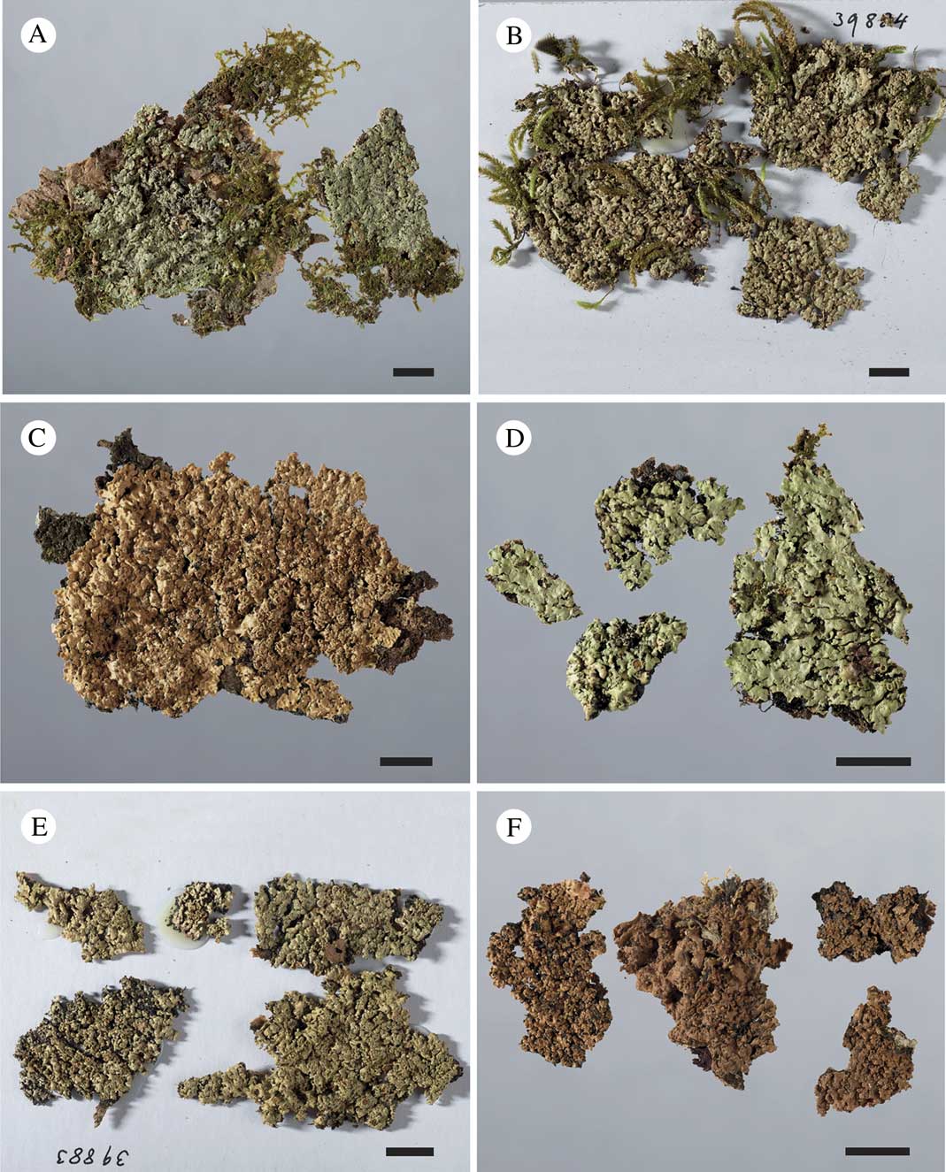
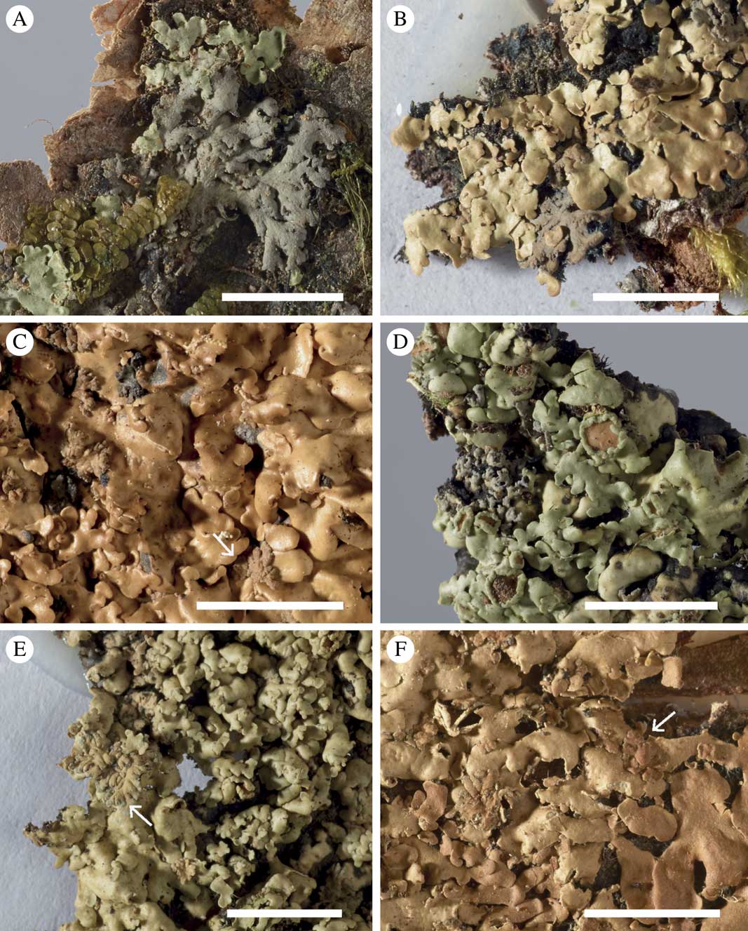
Gibbosporina species, habitus. A, G. didyma (holotype); B, G. elixii (holotype); C, G. leptospora (holotype); D, G. mascarena (holotype); E, G. nitida (holotype); F, G. nitida (Weber & McVean, S L-50422). Scales=1 cm.
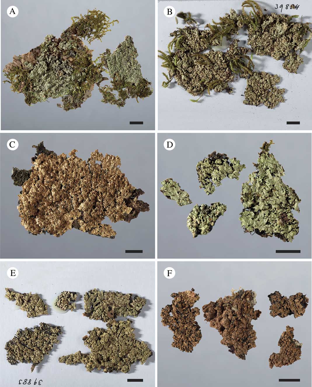
Thallus of chloromorph 5–30 cm diam., foliose, corticolous or muscicolous on corticolous bryophytes, when corticolous with a distinct black hypothallus, often also prothallus; lobes subdichotomously divided, 120–160 μm thick, 0·8–1·5 mm broad, discrete and flat in peripheral parts, gradually becoming coalescent and convex, centrally forming mats of ascending and geotropically oriented lobules. Upper surface glabrous and matt, fresh specimens bright green with contrasting blue-grey cephalodia when moist, light greenish grey when dry, 10 year-old herbarium specimens immediately become dark brown after application of water. Upper cortex 25–35 μm thick, plectenchymatous, lumina up to 15×10 μm, irregularly subellipsoid, arranged perpendicularly to the surface, surface weakly sclerenchymatous, lower cortex absent. Algal layer 25–35 μm thick; photobiont myrmecioid, cells small (3–7 μm) or large (6·0–12·5 μm), globose to ellipsoid or slightly irregular. Medulla of loosely interwoven hyphae, 60–90 μm thick, lowermost part brownly pigmented; rhizines marginally white, otherwise black, 0·5–1·5 mm long, simple or in bundles, fibrillose.
Cyanomorph as conspicuous cephalodia, pale grey when dry, formed on chlorobiont lobes, placodioid when young, becoming mini-foliose, often as weakly ascending fan-shaped lobe systems, 2–4 mm diam., lobes smooth, subdichotomously and densely branched, 0·2–0·3 mm wide, strongly convex, mostly erhizinate. Cortex like in the chloromorph. Cyanobiont Nostoc, as 3–6×4–7 μm large, globose to short-ellipsoid, dark brownish green to greyish violet cells, organized within 20–80 μm large glomeruli, without a discernible chain structure.
Apothecia subsessile, laminal, 0·5–5·0 mm diam., when large with irregular and wavy margins; disc orange-brown; thalline excipulum 0·15–0·25 mm thick, crenate-striate; epithecium pale brown, 10–20 μm thick, IKI−; hymenium colourless, but strongly IKI+ blue, 80–100 μm thick; hypothecium pale brown, IKI−, 50–80 μm thick; algal layer extending below the hypothecium and 50–80 μm thick; paraphyses simple to sparingly branched, with slightly swollen apices, septate; asci clavate, 8-spored, with pronounced, internal, apical, IKI+ blue amyloid tube structures, 50–80×15–20 μm. Proper ascospores colourless, simple, ellipsoid, 12·5–19·0×7·5–10·0 μm; perispores 13–23×12–16 μm, walls 2–3 μm thick, even, sometimes with a few low and wide gibbae.
Pycnidia very common, brown and conspicuous, swollen and urn-shaped, 0·2×0·2 mm, mostly marginal, sometimes laminal, occasionally even developed on cephalodia. conidia bacilliform, 2·5–4·0×1 μm.
Chemistry. No TLC-detectable compounds found.
Etymology. The specific epithet amphorella, from the Latin ‘amphora’ (=urn) and ‘-ella’ (a diminuitive suffix), refers to the small, urn-shaped pycnidia.
Distribution and ecology. Known only from a small area in a subtropical forest, around 700 m, in New Caledonia.
Major characteristics. A species with matt, rather thin lobes, and easily recognized by the numerous, conspicuous urn-shaped pycnidia, mainly along the margins, producing conidia larger than in most other species. Proper spores are narrow and short-ellipsoid, whereas perispores are thick and even, with few low and wide gibbae. Cephalodia mini-fruticose with narrow, strongly convex, erhizinate and fan-shaped lobes.
Additional specimens studied (paratypes). New Caledonia: 10 km NE of Nouméa, W slope of Monts des Koghis, c. 0·8 km E of Auberge, along path to Les Sommets, 250 m after its bifurcation with the path to Belvedère, 22°10'S, 166°31'E, 700 m, on unknown tree trunk in a semi-shaded forest, 2005, A. Elvebakk 05:706 (TROM; S); 400 m after its bifurcation with the path to Belvedère, 680 m, many 20–30 cm large specimens on the stooping trunk of a Montrouziera cauliflora tree, 3–4 m above the ground in semi-shaded forest, A. Elvebakk 05:718 (IRD; TROM; PC; UPS); 690 m, on Montrouzeria, A. Elvebakk 05:715 (TROM).
Gibbosporina bifrons Elvebakk, Hong & P. M. Jørg. sp. nov.
MycoBank No.: MB 811982
Gibbosporinae boninensi similis sed cephalodeis rhiziniphoris latiusque lobatis, perisporis crassissimis magisque gibbosis, plerumque cyanobiontibus distincte catenatis, pycnidiis parvis fuscis distinctisque.
Typus: New Caledonia, 10 km NE of Nouméa, W slope of Monts des Koghis, c. 0·8 km E of Auberge, near Belvedère, 22°10'S, 166°31'E, 750 m, two 10 cm-large thalli on the trunk of an unidentified small-leaved tree, 4 December 2005, A. Elvebakk 05:614 (PC—holotypus, sequenced as KM887878; IRD, TROM—isotypi) .
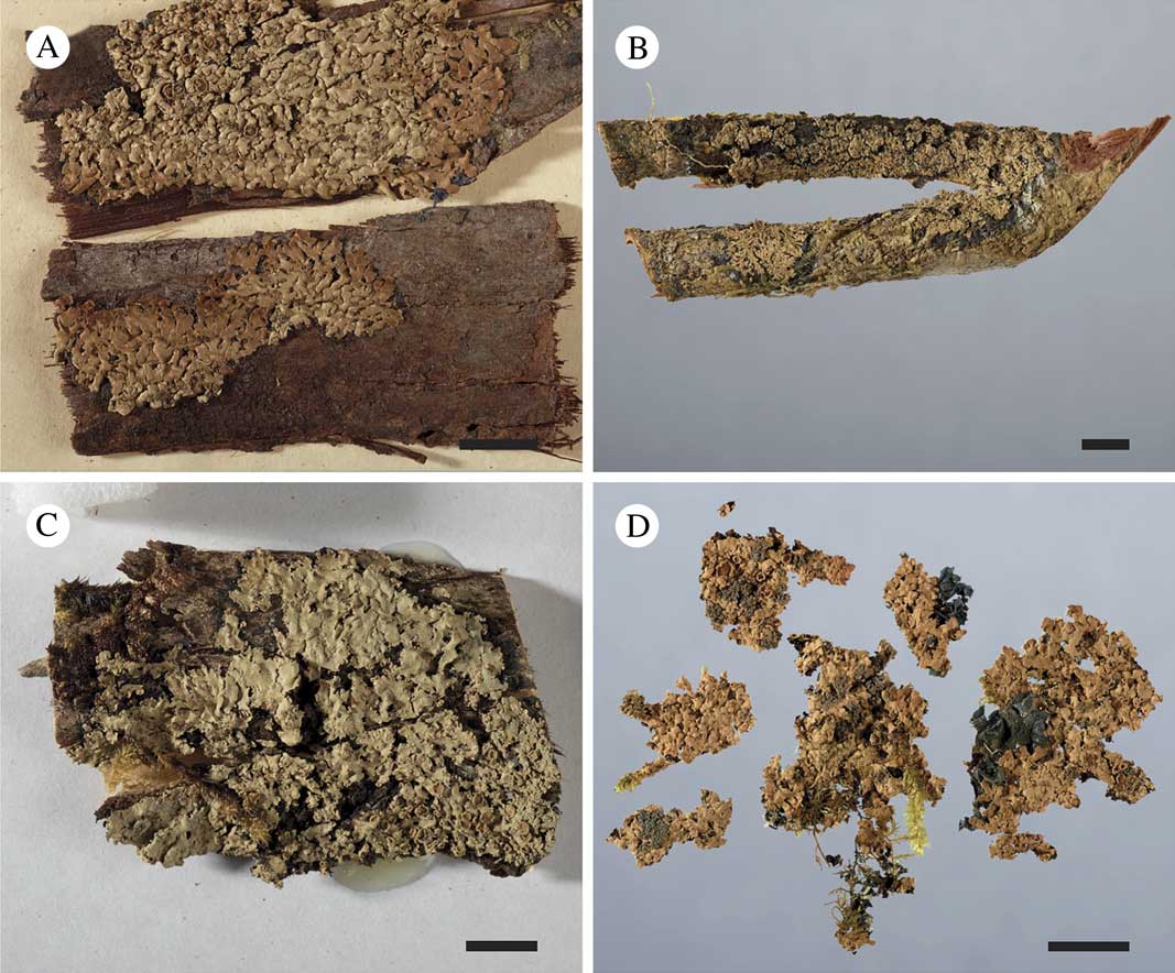
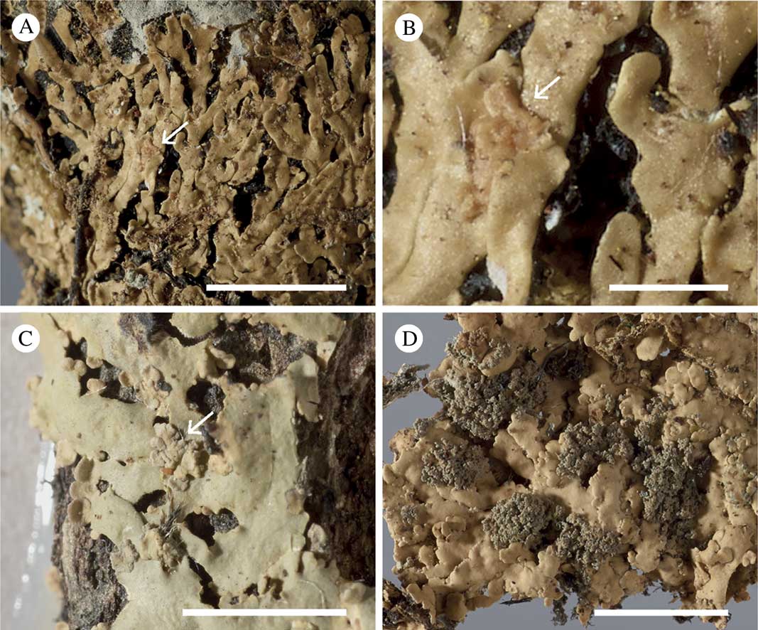
Gibbosporina species, habitus. A, G. papillospora (holotype); B, G. phyllidiata, (holotype); C, G. sphaerospora (holotype); D, G. thamnophora (holotype). Scales:=1 cm.
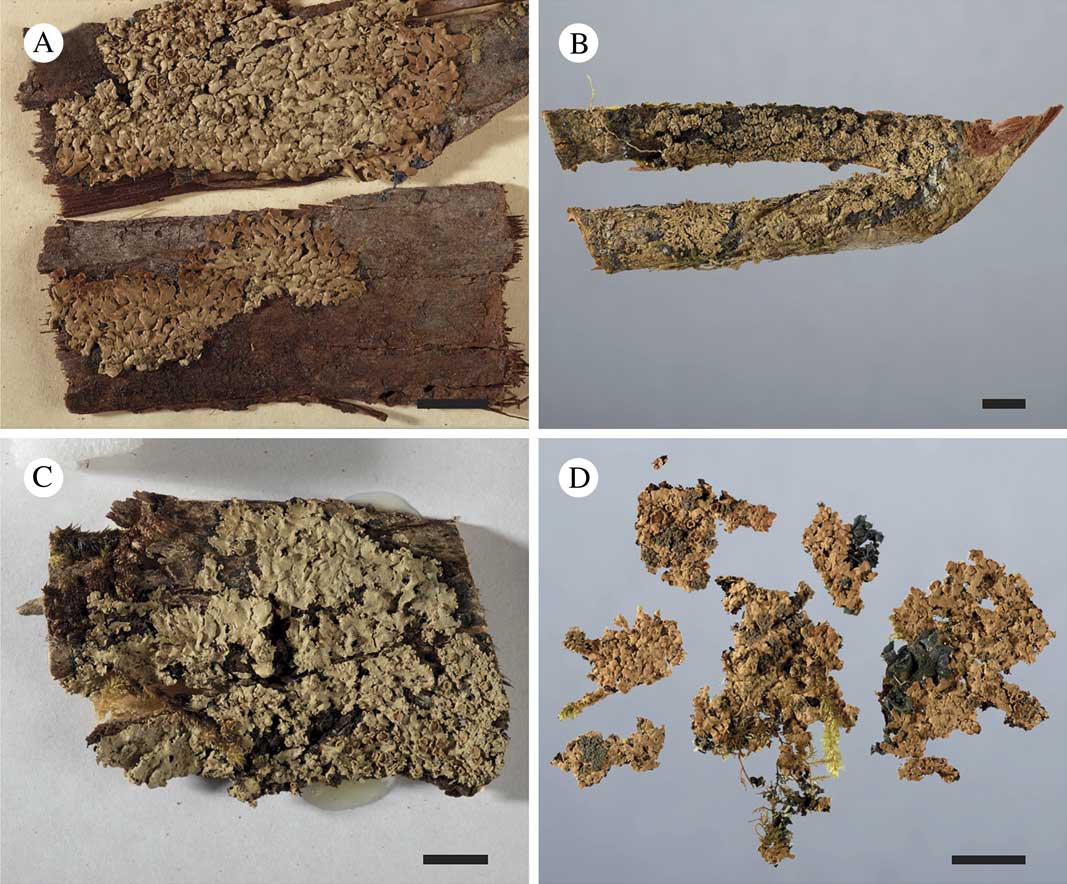
(Figs 1C–E, 4C–E, 7C, 8D)
Thallus of chlorobiont 5–20 cm diam., foliose, corticolous or muscicolous on corticolous bryophytes, when corticolous often with a distinct black prothallus; lobes subdichotomously divided, 120–150 μm thick, 0·8–2·0 mm broad, flat to convex, discrete and elongate when muscicolous, shorter and often imbricate when corticolous. Upper surface glabrous and glossy or weakly glossy, fresh specimens bright green with contrasting blue-green cephalodia when moist, light greenish grey when dry, old herbarium specimens ochraceous brown. Upper cortex 20–30 μm thick, plectenchymatous, lumina up to 10×15 μm, irregularly subellipsoid, arranged perpendicularly to the surface, surface weakly sclerenchymatous with ochraceous pigmentation on old herbarium specimens, no lower cortex present. Algal layer 15–25 μm thick; photobiont myrmecioid, cells 4·5–8·5 μm, globose to ellipsoid or slightly irregular. Medulla of loosely interwoven hyphae, 60–80 μm thick, lowermost part brownly pigmented; rhizines marginally white, otherwise blackish, 0·5–1·5 mm long, simple or in bundles .
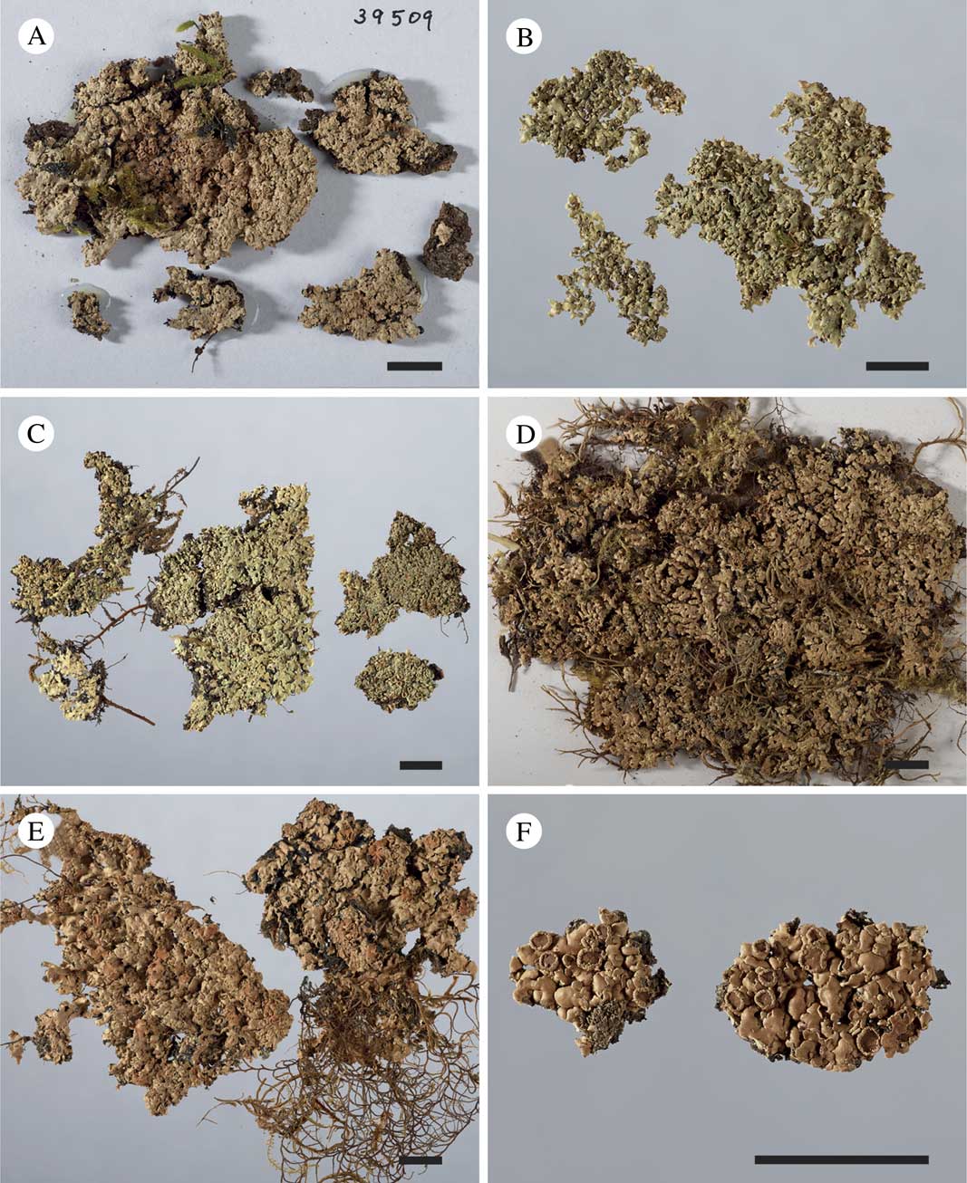
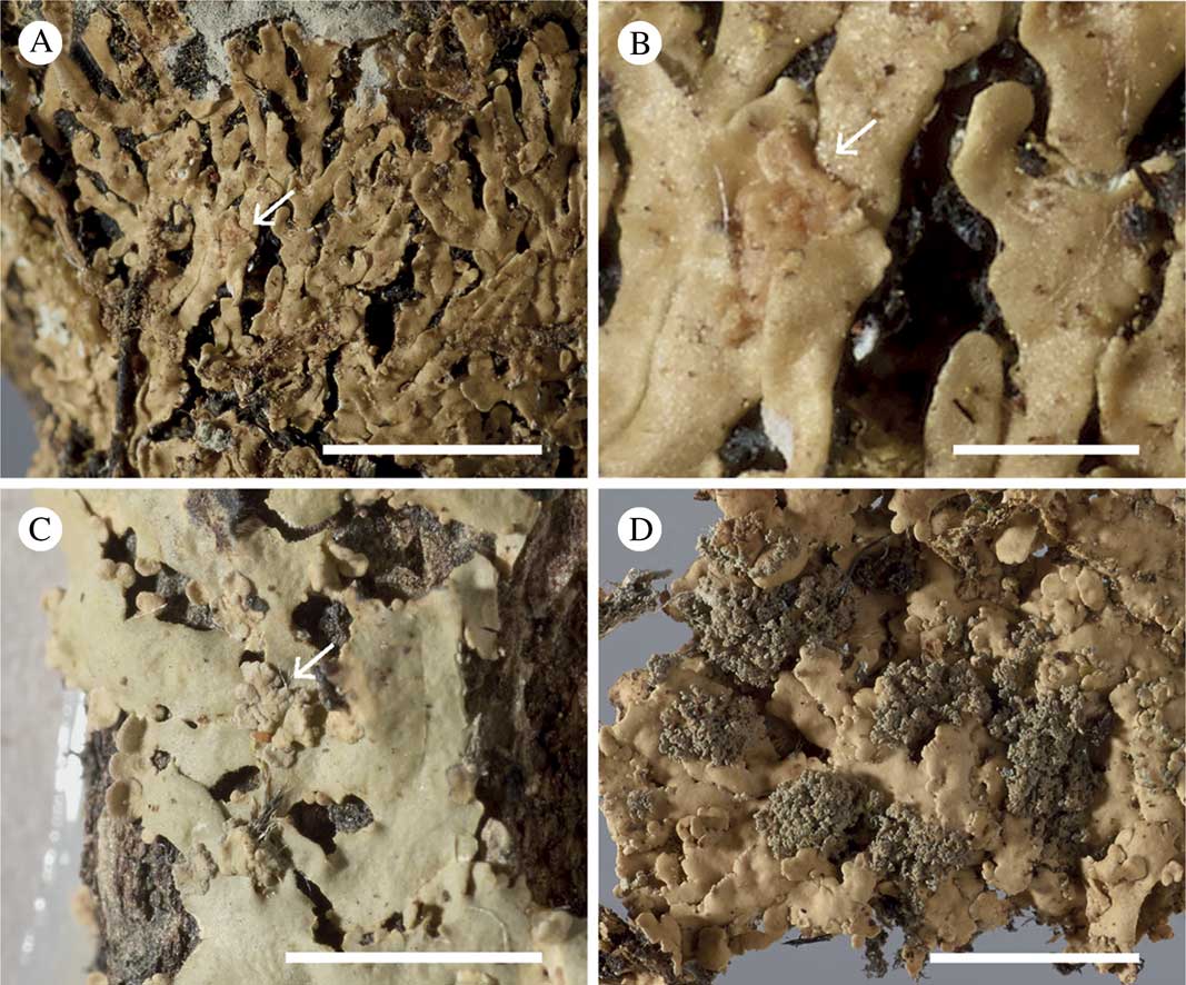
Gibbosporina species, cephalodia. A, Gibbosporina acuminata (holotype); B, G. amphorella (holotype); C, G. bifrons (isotype at IRD); D, G. bifrons (Coppins 5440 et al.) from Malaysia; E, G. bifrons (Hill 9744) from the Solomon Islands; F, G. boninensis (Knight, PC 0012753). Scales=0·5 cm.
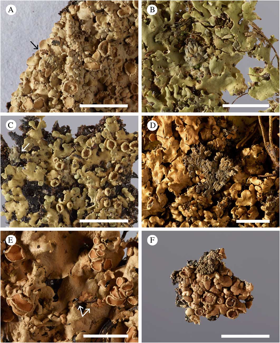
Cyanomorph as cephalodia, pale grey when dry, grey to dark brown on old herbarium specimens, formed on chlorobiont lobes or on the adjacent prothallus, placodioid when young, then becoming foliose, 2–12 mm diam., with 0·3–0·7 mm broad, subdichotomously branched lobes, becoming semi-erect and loosening from the chlorobiont substratum on its lower side, developing numerous rhizomorphs, in a few cases becoming apparently independent, although only immediately outside of the visible prothallus zone. In a few cases the cyanobiont has captured green algal cells and formed small chlorobiont lobules on the cephalodia. Upper surface smooth with radiating furrows to strongly areolate-ridged when dry, becoming smooth when moist. Cortex like in the chlorobiont, in some cases only 10–15 μm thick. Cyanobiont Nostoc, obviously of various strains, 3–6×4–7 μm diam., some with intensely violet-mauve cells in distinct chains, filling out most of the medulla within medullary compartments resulting in strongly gelatinous cephalodia, others greyish blue and small (2–4×3–5 μm diam.) or dark brownish violet and large (3–6×3–7 μm diam.), with cells in loose chain systems, sometimes the cyanobiont layer only occupying ¼ of the medulla, and occasionally as brownish violet cells in glomeruli without obvious visible chain structures .
Gibbosporina species, cephalodia. A, G. didyma (holotype). B, G. elixii (Elix 38759, CANB); C, G. leptospora (holotype); D, G. mascarena (holotype); E, G. nitida (holotype); F, G. papillospora (holotype). Scales:=0·5 cm.
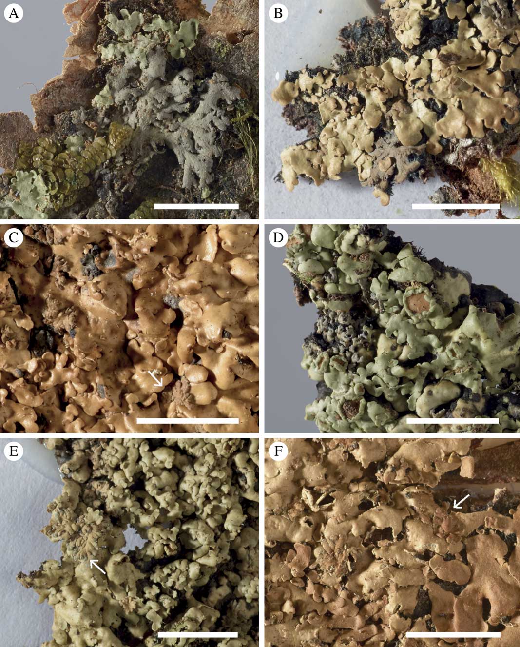
Apothecia sessile, laminal, 1–5 mm diam., when large with irregular and wavy margins; disc orange to reddish brown; thalline excipulum circular or strongly sinuous on old apothecia, 0·1–0·3 mm thick, crenate-striate; epithecium pale brown, 10–20 μm thick, IKI−; hymenium colourless, but strongly IKI+blue, 80–100 μm thick; hypothecium pale brown, IKI−, 30–50 μm thick; algal layer extending below the hypothecium; paraphyses simple to sparingly branched, with slightly swollen apices, septate; asci clavate, 8-spored, with internal, apical, amyloid tube structures, 50–70×10–15 μm in size. Proper ascospores colourless, simple, ellipsoid, 12–18×9–12 μm; perispores 17–23×13–20 μm, 1·0–5·5 μm thick, strongly and asymmetrically gibbose when mounted in water, gibbae up to 5 μm tall, obtuse .
Gibbosporina species, cephalodia. A & B, G. phyllidiata (holotype); C, G. sphaerospora (holotype); D, G. thamnophora (holotype). Scales: A, C & D=0·5 cm; B=0·1 cm.
Pycnidia common, small (0·05 mm), marginal, brown to blackish, bud-shaped, rarely swollen at base; conidia bacilliform, 2·5–3·0×0·5 μm.
Chemistry. No acetone-soluble compounds detected by TLC or HPLC.
Etymology. The epithet ‘bifrons’ means ‘two-faced’ and refers to the well-developed and large cephalodia occurring with the chlorobiont.
Distribution and ecology. Found scattered in montane tropical forests in the Philippines, Solomon Islands, Malaysia and New Caledonia. There are 18 collections from four of the islands of the Solomon Islands archipelago at BM, mostly collected by D. J. Hill in 1965. This represents a very thorough field study, showing that the species ranges from lowland tropical forest (‘trees overhanging beach’; ‘on fallen log in river bed’), to intermediate elevations (300–600 m), to 1600 m (‘on many tree trunks in montane rain forest’). The broad-lobed cephalodia are characteristic of all these collections; in a couple of cases, cephalodia were found on the bark outside of its probable mother thallus, although possibly within the prothallus zone.
Major characteristics. A robust species with glossy to moderately glossy lobes, which are rather thin, commonly with small marginal pycnidia. Perispores are thick with large irregular and obtuse gibbae. Cephalodia vary in size but have broad, rhizinate lobes. Several morphologically different cyanobionts occur, mostly, but not always, with cells in chain structures. In some cases cephalodia are strongly gelatinous, appearing apparently independent from the mother thallus.
Additional specimens examined (paratypes). Malaysia: Sarawak: Gunong Mulu National Park, 4th Division, Baram District, Long Pala, Limestone Hill, c. 2 km E of Base Camp, S side of Sungei Melinau Paku, 70–300 m alt., 20 iv 1978, G. Argent, B. Coppins (5439 & 5440), C. Jermy & P. Chai (E00153722; BG); Valley of Ulu Jerneh, on fallen tree, amongst upper canopy and lianas, 1978, C. Argent, B. Coppins 5310, C. Jermy & P. Chai (E).—The Philippines: Biliran: vi 1914, R. C. McGregor as Vainio 11988 (TUR 012365); Luzon: xi 1915, A. D. E. Elmer (S L31698); Province of Sorsogon, Irosin, Mt. Bulusan, 1915, A. D. E. Elmer 14959 (S L31697; BM); A. D. E. Elmer as Vainio 11986 (TUR 12411).—Solomon Islands: Guadalcanal Island: Mt. Popomansiu, on ridge on SE side of Sutakiki River (Vunuvalukama), 4400 ft, montane rainforest, 1965, D. J. Hill 9744 (TROM; BM 000732072), 9745 (BM); 5700 ft, on many tree trunks in montane rainforest, 1965, D. J. Hill 9361 (BM), 9653 (BM); 5800 ft, little knoll supporting an isolated area of ‘moss’ forest, 1965, D. J. Hill 9433 (BM); 4800–5400 ft, montane rainforest, 1965, D. J. Hill 9689 (BM); central part, Sutakiki River, on ridge on S side, c. 2 miles from its confluence with Balasuna River, 1200–1700 ft., 1965, D. J. Hill 9242 (BM); central part, Balasuna River, on ridge on N side of river, path from Nuhu Village to Parina Village, c. 2300 ft., 1965, D. J. Hill 9897 (BM); ultrabasic area c. 0·5 mile S of Nuhu Village, 1300 ft, 1965, D. J. Hill 9088 (BM); NW end, Mount Gallego, small peak to right of upper end of path down ridge on right of Hidden Valley, 1500–2500 ft, 1965, D. J. Hill 8370 (TROM; BM000732071), 8351 (BM). Santa Isabel Island: Thousand Ships Bay, Kockatoo Anchorage, trees overhanging beach on mainland, 1965, D. J. Hill 10999 (BM). San Cristobal Island: Wainoni Region, Warinito River, c. 8 miles inland, T. C. Whitmore 8820 (BM). Kolombangara Island: 2 miles up River Kolumbangara, lowland rainforest, 1965, D. J. Hill 10251 (BM); on fallen log in river bed, 1965, D. J. Hill 10437 (BM); trees along river bed, 1965, D. J. Hill 10345 (BM); ridge to W of River Kolumbangara, 1000–2000 ft., 1965, D. J. Hill 10743 (BM); 3000 ft., 1965, D. J. Hill 10574, 10579 (BM).
Gibbospora boninensis (Kurok.) Elvebakk & P. M. Jørg. comb. nov.
MycoBank No.: MB 812264
Basionym: Psoroma boninense Kurok. Bull. Nat. Sci. Mus. Tokyo 12: 688 (1969); typus: Bonin Islands, around mountain top (rugged area with many large outcrops of andesite), Mt. Tsutsuji, Chichijima Island, 25 Nov. 1968 Hiroshi Inoue 19066 (TNS—holotypus!).
Pannaria sphinctrina (Mont.) Tuck. ex Hue var. microphylla Hue. Nouv. Arch. Mus. Ser. 4, vol. VIII: p. 268 (1906); type: Japan, Bonin Island, US North Pacific exp. under command of Ringgold and Rodgers 1893–96, coll. C. Wright s. n., PC (PC0012754)! (lectotypus selected here), W (isolectotypus!), BM (isolectotypus!). ≡ Psoroma sphinctrinum (Mont.) Nyl. var. microphyllum (Hue) Zahlbr., Cat. Lich. Univ. 3: 276 (1925).
? Pannaria sphinctrina (Mont.) Tuck. ex Hue var. confusa Hue, nom. nud., Bull. Soc. Bot. France 48: 56 (1902).
(Figs 1F, 4F, 7D; other illustrations: Kurokawa Reference Kurokawa1969: Pl. I–II)
Thallus of chloromorph 5–15 cm diam., foliose, corticolous with a distinct black hypothallus, often also prothallus; lobes subdichotomously divided, 180–200 μm thick, 0·8–1·5 mm broad, discrete and flat to weakly concave in peripheral parts, gradually becoming coalescent and convex in central parts of the thallus. Upper surface glabrous, glossy or weakly glossy, fresh specimens not seen (probably bright green with contrasting blue-grey cephalodia when moist, light greenish grey when dry), old herbarium specimens dark brown. Upper cortex 30–40 μm thick, plectenchymatous, lumina up to 15×10 μm, irregularly subellipsoid, arranged perpendicularly to the surface, surface weakly sclerenchymatous, lower cortex absent. Algal layer 40–60 μm thick; photobiont myrmecioid, cells 3–8 μm, globose to ellipsoid or slightly irregular. Medulla of loosely interwoven hyphae, 60–100 μm thick, lowermost part brownly pigmented; rhizines marginally white, otherwise black, 0·5–1·5 mm long, simple or in bundles, fibrillose.
Cyanomorph as cephalodia, dark greyish brown on herbarium material, formed on chlorobiont lobes, placodioid when young, small-foliose when mature, densely branched and ascending, 1–3 mm diam.; lobes convex, weakly uneven above, very narrow (0·10–0·25 mm wide), erhizinate. Cortex like in the chloromorph. Cyanobiont Nostoc, as 3–6×3–7 μm large, globose to short-ellipsoid, dark greyish violet cells, organized within 20–50 μm large glomeruli filling up c. ¾ of the medulla, without a discernible chain structure.
Apothecia subsessile, laminal, 0·5–4·0 mm diam.; disc orange-brown when moist, dark brown when dry; thalline excipulum 0·15–0·30 mm thick, crenate-striate; epithecium pale brown, 10–20 μm thick, IKI−; hymenium colourless, but strongly IKI+ blue, 80–100 μm thick; hypothecium pale brown, IKI−, 20–30 μm thick; algal layer extending below the hypothecium; paraphyses simple to sparingly branched, with slightly swollen apices, septate; asci clavate, 8-spored, with pronounced, internal, apical, IKI+ blue amyloid tube structures, 50–70×10–15 μm. Proper ascospores colourless, simple, ellipsoid, 10–15×7·0–8·5 μm; perispores 14–20×9–13 μm, walls 0·5–1·5 μm thick in addition to irregular gibbae, some obtuse, others pointed, up to 5 μm tall.
Pycnidia rare and inconspicuous, bud-shaped; conidia bacilliform, 3–4×1 μm.
Chemistry. No TLC-detectable compounds found.
Note. The description above is in agreement with the original protologue presented by Kurokawa (Reference Kurokawa1969), although the latter did not include any reference to cephalodia. These had been described by Hue (Reference Hue1906, below Pannaria sphinctrina var. microphylla), who considered the photobiont to be Scytonema. Hue (Reference Hue1906) and Kurokawa (Reference Kurokawa1969) cited the perispore (called “exospore” and “gelatinous membrane”, respectively) as being 2–3 or 3 μm thick, and the perispores are described in more detail here. Conidiomata are described here for the first time.
Nomenclatural note. Hue (Reference Hue1902) listed the new name Pannaria sphinctrina var. confusa Hue without any accompanying description, but referred to his species number “Hue Lich. Exot. n. 1133”. This is not a specimen citation, but Hue’s own number of his accepted species, and collections of several of Hue’s varieties of his Pannaria sphinctrina carry this number. When Hue (Reference Hue1906) described var. microphyllum, he did not make any reference to his confusa name, but preferred the former cited with Tuckerman as author. However, this is an illegitimate author citation as he stated that Tucker’s microphylla is an invalid herbarium name, which apparently has not been published previously with Tuckerman’s authorship. There are three Wright collections from the same expedition to the Bonin Islands representing the same species in PC, all determined as var. microphylla, and one of them (PC0012753) additionally with the name var. confusa on a separate label. Thus, Pannaria sphinctrina var. confusa is considered a nomen nudum here, and PC0012754 is chosen as a lectotype of Pannaria sphinctrina var. microphylla.
Distribution and ecology. Known from the isolated subtropical Japanese Ogasawara Gunto Islands (a World Heritage Site c. 1000 km S of Tokyo).
Major characteristics. A species with thick and glossy lobes, indistinct pycnidia, rather thin-walled perispores with dramatic gibbae, and very narrow-lobed, often fan-shaped, erhizinate cephalodia.
Additional specimens examined. Japan: Bonin Island: US North Pacific exp. under command of Ringgold and Rodgers 1893–96, C. Wright s. n. (PC0012753), C. Wright s. n. (PC).
Gibbosporina didyma Elvebakk, Hong & P. M. Jørg. sp. nov.
MycoBank No.: MB 811983
Gibbosporinae amphorellae similis sed sine pycnidiis urniformibus conspicuisque; sporis brevioribus, cephalodiis maioribus latiusque lobatis et chlorobiontibus interdum praeditis.
Typus: Réunion, c. 0·5 km E of the E end of Grand Étang, 4–500 m W of the parking site, 21°05'45''S, 55°38'57''E, 525 m, on tree trunk near the path, 17 October 2011, A. Elvebakk 11:042 (PC—holotypus, sequenced as KM887875 and KM887876; BM, TROM, REU—isotypi).
(Figs 2A, 5A, 7E, 8E; additional illustration: Jørgensen & Tønsberg Reference Jørgensen and Tønsberg2012: 87)
Thallus of chloromorph 3–10 cm diam., foliose, corticolous or muscicolous on corticolous bryophytes, when corticolous often with a distinct black hypothallus, prothallus only developed occasionally; lobes subdichotomously divided, 120–180 μm thick, 0·5–1·3 mm broad, discrete and concave in distal parts, gradually becoming flat, before being coalescent and convex in central parts of the thallus. Upper surface glabrous and matt to weakly glossy, fresh specimens bright green with contrasting blue-green cephalodia when moist, light greenish grey when dry. Upper cortex 25–40 μm thick, plectenchymatous, lumina up to 12×18 μm, irregularly subellipsoid, arranged perpendicularly to the surface, surface weakly sclerenchymatous, lower cortex absent. Algal layer 15–30 μm thick; photobiont myrmecioid, cells 3–7 μm, globose to ellipsoid or slightly irregular. Medulla of loosely interwoven hyphae, 70–100 μm thick, lowermost part brownish; rhizines marginally white, otherwise black, 0·5–2·0 mm long, simple or in bundles, fibrillose.
Cyanomorph as cephalodia, pale grey when dry, formed on chlorobiont lobes on the hypothallus between them, or apparently as independent cyanobiont thalli on bryophytes or cortex close to the main thallus, placodioid when young, then becoming foliose or subfruticose, 2–15 mm diam., with subdichotomously branched lobes, mostly erhizinate, occasionally with scattered rhizines. Cephalodia either foliose, with lobes 0·3–0·7 mm wide, or subfruticose with 0·2–0·3 mm wide, convex lobes, densely branched into fan-shaped, lobe systems. Cyanobiont Nostoc, as 3–6×5–8 μm large ellipsoid, dark greenish brown to brownish violet cells, blue-green on older samples, organized within 20–80 μm large glomeruli, mostly without an obvious chain structure, short chains observed in single broad-lobed cephalodia. Small chloromorph apothecia and chloromorph thallus fragments have in several cases been developed on the cephalodia.
Apothecia subsessile, laminal, 0·5–3·0 mm diam.; disc orange-brown; thalline excipulum 0·15–0·30 mm thick, crenate-striate; epithecium pale brown, 10–20 μm thick, IKI−; hymenium colourless, but strongly IKI+ blue, 80–100 μm thick; hypothecium pale brown, IKI−, 20–30 μm thick; algal layer extending below the hypothecium; paraphyses simple to sparingly branched, with slightly swollen apices, septate; asci clavate, 8-spored, with pronounced, internal, apical, IKI+ blue amyloid tube structures, 50–70×10–15 μm. Proper ascospores colourless, simple, ellipsoid, 11–15×7·5–10·0 μm; perispores 15–20×12–16 μm, walls 2–5 μm thick, weakly gibbose when mounted in water.
Some possible pycnidia seen as poorly differentiated brown structures along margins; conidia not seen.
Chemistry. No acetone-soluble compounds detected by TLC analysis.
Etymology. The epithet ‘didyma’ means ‘in pairs’ and refers to the thallus being divided into a prominent cyanobiont in addition to the dominant chlorobiont. The name also refers to cephalodia occasionally being divided, by developing chlorobiont apothecia.
Distribution and ecology. A rare species known only from two localities, on Réunion and Mauritius, both in tropical forests at moderate altitudes of 5–600 m.
Major characteristics. A narrow-lobed species with matt lobes, without distinct pycnidia. Proper spores are narrow and short-ellipsoid, perispores very thick and even, with few, diffuse gibbae. Cephalodia are particularly well developed, large and foliose and broad-lobed, sometimes becoming fan-shaped and ascending. The species appears to be phytosymbiodemic, although cephalodia have so far been found only in the prothallus zone of the mother thallus.
Additional specimens examined (paratypes). Mauritius: Black River, along path from Plaine Champagne towards Piton de la Petite Rivière Noire, 20°25'S, 57°25'E, 600 m, corticolous, 1991, Krog, H. & Timdal, E. MAU09/62b (O – L 21227).—Réunion: Le Grand Étang along the trail from the road to the lake, 21°05'S, 55°39'E, on tree trunk, 520–540 m, 1996, Krog, H. & Timdal, E. RE48/12b (O).
Gibbosporina elixii Elvebakk, Hong & P. M. Jørg. sp. nov.
MycoBank No.: MB 811987
Gibbosporinae mascarenae similis sed cephalodiis maturis minus ramulosis distincteque foliosis et lobis latioribus instructis; apotheciorum thallorum marginibus distincte angustiis et subtiliter regulariterque crenulato-striatis; sporis sensu stricto angustioribus; parietibus perisporarum regulariter crassis vel leviter gibbosis.
Typus: Australia, Queensland, Mossman Gorge National Park, 6 km W of Mossman, 16°28'21''S, 145°19'54''E, 60 m, tropical rainforest along Mossman River, on base of tree, 1 August 2006, J. A. Elix 39884 (CANB 00783318—holotypus, sequenced as KM887879; BRI—isotypus).
(Figs 2B, 5B, 7F)
Thallus of chloromorph 3–7 cm diam., foliose, corticolous or muscicolous on corticolous bryophytes, when corticolous with a distinct black hypothallus, often also prothallus; lobes subdichotomously divided, 100–150 μm thick, 0·6–1·2 mm broad, discrete and flat in peripheral parts, gradually becoming coalescent and convex in central parts of the thallus. Upper surface glabrous and glossy, fresh specimens bright green with contrasting blue-grey cephalodia when moist, light greenish grey when dry. Upper cortex 20–30 μm thick, plectenchymatous, lumina up to 15×10 μm, globose or irregularly subellipsoid, and then arranged perpendicularly to the surface, surface weakly sclerenchymatous, lower cortex absent. Algal layer 30–45 μm thick; photobiont myrmecioid, cells 3–7 μm, globose to ellipsoid or slightly irregular. Medulla of loosely interwoven hyphae, 50–80 μm thick, lowermost part brownly pigmented; rhizines marginally white, otherwise black, 0·5–1·5 mm long, simple or in bundles, fibrillose.
Cyanomorph as cephalodia, pale grey when dry, formed on chlorobiont lobes, occasionally on the hypothallus, placodioid when young, then becoming foliose, surface slightly faveolate, 1–5 mm diam., with sparsely branched lobes, erhizinate or rarely with scattered rhizines, convex, lobes 0·2–0·3 mm wide, slightly detached from the substratum. Cortex like in the chloromorph. Cyanobiont Nostoc, as 3–6×4–8 μm large, ellipsoid, dark greenish brown to brownish violet cells, organized within 15–60 μm large glomeruli, without a discernible chain structure.
Apothecia subsessile, laminal, 0·5–1·5 mm diam.; disc orange-brown; thalline excipulum 0·1–0·2 mm thick, regularly and finely crenate-striate; epithecium pale brown, 10–20 μm thick, IKI−; hymenium colourless, but strongly IKI+ blue, 80–100 μm thick; hypothecium pale brown, IKI−, 20–30 μm thick; algal layer extending below the hypothecium; paraphyses simple to sparingly branched, with slightly swollen apices, septate; asci clavate, 8-spored, with pronounced, internal, apical, IKI+ blue amyloid tube structures, 50–70×10–15 μm. Proper ascospores colourless, simple, ellipsoid, 12–16×6·5–8·0 μm; perispores 15–19×10·0–12·5 μm, walls 1·5–2·5 μm thick and predominantly even or weakly gibbose when mounted in water.
Pycnidia not seen.
Chemistry. No TLC-detectable compounds found.
Etymology. The species is named in honour of the Australian lichenologist Jack Elix, who collected the type as well as many samples of other Gibbosporina species described here.
Distribution and ecology. Known only from two localities in lowland tropical forests of NE Australia.
Major characteristics. Lobes thin and glossy without visible pycnidia. The apothecia have narrow and regular margins, the proper spores are narrow and almost long-ellipsoid, whereas perispores are intermediately thick, rather even, with few and low gibbae. Cephalodia are mini-foliose with narrow, erhizinate lobes.
Additional samples examined (paratypes). Australia: Queensland: Wooroonooran National Park, Josephine Falls, 20 km NW of Innisfail, 17°26'16''S, 145°51'33''E, 80 m, lowland tropical rainforest, on tree trunk, 2006, J. A. Elix 38759 (CANB 00783310).
Gibbosporina leptospora Elvebakk sp. nov.
MycoBank No.: MB 811988
Gibbosporinae nitidae similis, sed sporis ellipsoidibus, parietibus perisporarum tenuibus et gibbis humilibus subconvexisque praeditis.
Typus: Australia, Queensland, Moses Creek, Rossville-Bloomfield River road, 35 km SSE of Cooktown, 15°47'S, 145°17'E, 240 m, lowland rainforest on flats beside creek, on shaded upper tree trunk, 21 October 1995, H. Streimann 57369 (H—holotypus; CANB—isotypus, not seen; B—isotypus, not seen).
(Figs 2C, 5C, 7G)
Thallus of chloromorph 3–10 cm diam., foliose, corticolous with a distinct black hypothallus, often also prothallus; lobes subdichotomously branched, 100–170 μm thick, 0·8–2·5 mm broad, discrete and flat to weakly concave in distal parts, gradually becoming coalescent in central parts of the thallus. Upper surface glabrous, except often with minute, erect, 10–15 μm long hairs on young lobe tips, strongly glossy and mostly even, fresh specimens not seen (probably bright green with contrasting blue-grey cephalodia when moist, light greenish grey when dry), old herbarium specimens ochraceous brown. Upper cortex 20–35 μm thick, plectenchymatous, lumina up to 15×10 μm, irregularly subellipsoid, arranged perpendicularly to the surface, surface weakly sclerenchymatous, lower cortex absent. Algal layer 25–40 μm thick; photobiont myrmecioid, cells 5–8 μm, globose to ellipsoid or slightly irregular. Medulla of loosely interwoven hyphae, 50–100 μm thick, lowermost part brownly pigmented; rhizines in marginal positions white, otherwise black, 0·5–1·5 mm long, simple or in bundles, fibrillose.
Cyanomorph as cephalodia, pale grey when dry, formed on chlorobiont lobes, occasionally on the hypothallus, mostly placodioid, occasionally becoming mini-foliose and loosely attached to the substratum, surface smooth with radiating furrows, 1–3 mm diam., commonly with a corona of whitish short rhizines as seen from above, lobes convex, 0·15–0·30 mm wide, moderately branched. Cortex like in the chlorobiont. Cyanobiont Nostoc, as 3–6×4–6 μm large globose, subglobose or short-ellipsoid, brownish green to brownish violet cells, organized within 20–80 μm large glomeruli, without a discernible chain structure.
Apothecia subsessile, laminal, 0·5–2·0 mm diam.; disc orange-brown, circular, often becoming strongly irregular in outline; thalline excipulum 0·2–0·3 mm thick, crenate-striate; epithecium pale brown, 10–20 μm thick, IKI−; hymenium colourless, but strongly IKI+ blue, 80–100 μm thick; hypothecium pale brown, IKI−, 20–30 μm thick; algal layer extending below the hypothecium; paraphyses simple to sparingly branched, with slightly swollen apices, septate; asci clavate, 8-spored, with pronounced, internal, apical, IKI+ blue amyloid tube structures, 50–70×10–15 μm. Proper ascospores colourless, simple, ellipsoid, 12–17×6·5–9·5 μm, many almost twice as long as broad; perispores 14–19×8–12 μm, with thin walls, 0·5–1·0 μm thick, with low and wide gibbae, rarely protruding more than 1 μm above the general surface of the perispore.
Pycnidia present, formed like low dark brown verrucae, 0·10×0·05 mm. A few conidia observed, rod-shaped, 2·0×0·5 μm.
Chemistry. No TLC-detectable compounds found.
Etymology. From ‘lepto-’ in Greek composite words, meaning ‘thin-’, referring to the thin perispores.
Distribution and ecology. Known only from two tropical lowland forest localities, one in NE Australia, one in Papua New Guinea.
Major characteristics. Strongly glossy and moderately thick lobes, with small marginal pycnidia. Proper spores are very narrow, and short- to long-ellipsoid. Perispores are very thin, with scattered low gibbae. Cephalodia are placodioid to mini-foliose, often with a conspicuous corona of white rhizines when seen from above.
Additional specimens examined (paratypes). Papua New Guinea: Milne Bay Province: Woodlark Island, Mt. Kabati-Kulumadau Road, 5 km E of Kulumadau, 9°04'S, 152°47'E, 100 m, lowland forest disturbed by roading, on Endospermum stem, 11×1984, R. Kumei (H; CANB, US, LAE, B – not seen).
Gibbosporina mascarena Elvebakk, Hong & P. M. Jørg. sp. nov.
MycoBank No.: MB 811989
Gibbosporinae boninensi similis, sed sporis latioribus, perisporis gibbis obtusioribus praeditis, cephalodiis latius lobatis.
Typus: Réunion, c. 0·5 km NE of E end of Grand Étang, at La Vue, 21°05'39''S, 55°38'43''E, 560 m, on a trunk of cf. Dracaena on the slope below the viewpoint, 17 October 2011, A. Elvebakk 11:056 (PC—holotypus, sequenced as KM887880; TROM, REU—isotypi).
(Figs 2D, 5D, 7H, 8F)
Thallus of chloromorph 3–10 cm diam., foliose, corticolous or muscicolous on corticolous bryophytes, when corticolous with a distinct black hypothallus, often also prothallus; lobes subdichotomously divided, 120–180 μm thick, 0·8–2·0 mm broad, discrete and flat in peripheral parts, gradually becoming coalescent and convex in central parts of the thallus. Upper surface glabrous and glossy, fresh specimens bright green with contrasting blue-grey cephalodia when moist, light greenish grey when dry. Upper cortex 25–40 μm thick, plectenchymatous, lumina up to 15×10 μm, irregularly subellipsoid, arranged perpendicularly to the surface, surface weakly sclerenchymatous, lower cortex absent. Algal layer 20–35 μm thick; photobiont myrmecioid, cells 3–7 μm, globose to ellipsoid or slightly irregular. Medulla of loosely interwoven hyphae, 70–100 μm thick, lowermost part brownly pigmented; rhizines marginally white, otherwise black, 0·5–1·5 mm long, simple or in bundles, fibrillose.
Cyanomorph as cephalodia, pale grey when dry, formed on chlorobiont lobes, occasionally on the hypothallus, placodioid when young, then becoming mini-foliose, occasionally subfruticose, surface smooth, 1–4 mm diam., with subdichotomously branched lobes, erhizinate or rarely with scattered rhizines, convex, lobes 0·2–0·3 mm wide, moderately to densely branched into fan-shaped lobe systems. Cortex like in the chloromorph. Cyanobiont Nostoc, as 3–6×5–8 μm large ellipsoid, dark greenish brown to brownish violet cells, blue-green on older samples, organized within 20–50 μm large glomeruli, without a discernible chain structure.
Apothecia subsessile, laminal, 0·5–3·0 mm diam.; disc orange-brown; thalline excipulum 0·2–0·3 mm thick, crenate-striate; epithecium pale brown, 10–20 μm thick, IKI−; hymenium colourless, but strongly IKI+ blue, 80–100 μm thick; hypothecium pale brown, IKI−, 20–30 μm thick; algal layer extending below the hypothecium; paraphyses simple to sparingly branched, with slightly swollen apices, septate; asci clavate, 8-spored, with pronounced, internal, apical, IKI+ blue amyloid tube structures, 50–70×10–15 μm. Proper ascospores colourless, simple, ellipsoid, 10–16×7·5–10·0 μm; perispores 15–20×10–16 μm, walls 0·5–3·0 μm thick and strongly and irregularly gibbose when mounted in water.
Pycnidia marginal, small, brown, bud-shaped, rarely common or well developed; conidia bacilliform 3·0×0·5 μm.
Chemistry. No TLC-detectable compounds found.
Etymology. The epithet means ‘from the Mascarenes’, a name referring to Réunion and Mauritius.
Distribution and ecology. Known from tree trunks in tropical forests at altitudes of 5–700 m in Réunion, Mauritius and Sri Lanka, where the species is probably common.
Major characteristics. A broad-lobed, robust and strongly glossy species with indistinct marginal pycnidia and apothecia with coarse and irregular margins. Proper spores are short-ellipsoid and perispores intermediately thick with abundant high and obtuse gibbae. Cephalodia have intermediately broad lobes, become mini-foliose, rarely rhizinate, occasionally also mini-fruticose.
Additional specimens examined (paratypes). Mauritius: Black River, along path from Plaine Champagne towards Piton de la Petite Rivière Noire, 20°25'S, 57°25'E, 620–825 m, 1991, Krog, H. & Timdal, E. MAU51/95 (O–L21928), MAU51/92 (O–L21295), MAU51/93 (O–L 21926), MAU51/94 (O–L21927); 600 m, corticolous, 1991, Krog, H. & Timdal, E. MAU09/62 (O–L21227); without further geographical information, Robillard (PC); Plaines Wilhelms, Macchabee Forest, at the divide of the roads leading to Macchabee Kiosk and Mt. Bris Fer, 20°24'S, 57°27'E, 630 m, 1991, Krog, H. & Timdal, E. MAU14/16 (O–L21364); 0·5–1 km ESE of Macchabee Kiosk, 20°24'S, 57°26'E, 1991, Krog, H. & Timdal, E. MAU13/11 (O–L21343); Savanne, Mt. Cocotte, SE of the peak, along the road towards Bassin Blanc, 20°26'S, 57°28'E, 640 m, 1991, Krog, H. & Timdal, E. MAU21/33 (O–L21443); Mt. Cocotte, along the path towards the peak, Krog, H. & Timdal, E. MAU32/75 (O–L21629).—Réunion: Grand Étang, along the trail from the road to the lake, 21°05'S, 55°39'E, 520–540 m, on tree trunk, 1996, Krog, H. & Timdal, E. RE48/12, RE48/12c (O–L107313); 0·5 km E of the E end of le Grand Étang, 4–500 m W of the parking site, 21°05'45''S, 55°38'57''E, 525 m, on tree trunk near the path, 2011, A. Elvebakk 11:041 (TROM); ‘Bourbon’, 1840, Lepervanche-Mézières (PC).—Sri Lanka: Twaites 52 (S–L31706); ‘Leighton 52’ (=‘Twaites 52’) (S–L31705).
Gibbosporina nitida Elvebakk, Hong & P. M. Jørg. sp. nov.
MycoBank No.: MB 811990
Gibbosporinae mascarenae similis, sed superficie loborum aspera et valde nitida, pycnidiis marginalibus saepe conspicuis, cephalodiis rosettiformis, perisporis crassioribus gibbisque maioribus praeditis.
Typus: Australia, Queensland, Mossman Gorge National Park, 6 km W of Mossman, 16°28'21''S, 145°19'54''E, 60 m, tropical rainforest along Mossman River, on base of tree, 1 August 2006, J. A. Elix 39883 (CANB 00783317—holotypus, sequenced as KM887889; BRI—isotypus).
(Figs 2E, 2F, 5E, 7I)
Thallus of chloromorph 3–10 cm diam., foliose, corticolous or muscicolous on corticolous bryophytes, when corticolous with a distinct black hypothallus, often also prothallus; lobes densely and irregularly divided, 120–180 μm thick, 0·8–2·5 mm broad, discrete and flat in distal parts, gradually becoming coalescent and convex in central parts of the thallus, where they form scalariform side lobules, apparently in vertically-oriented individuals. Upper surface glabrous, strongly glossy and uneven, fresh specimens bright green with contrasting blue-grey cephalodia when moist, light greenish grey when dry, becoming pale brown in old herbarium samples. Upper cortex 20–35 μm thick, plectenchymatous, lumina up to 15×10 μm, irregularly subellipsoid, arranged perpendicularly to the surface, surface weakly sclerenchymatous, lower cortex absent. Algal layer 25–40 μm thick; photobiont myrmecioid, cells 5–8 μm, globose to ellipsoid or slightly irregular. Medulla of loosely interwoven hyphae, 50–100 μm thick, lowermost part brownly pigmented; rhizines in marginal positions white, otherwise black, 0·5–1·5 mm long, simple or in bundles, fibrillose.
Cyanomorph as cephalodia, pale grey when dry, formed on chlorobiont lobes, occasionally on the hypothallus, placodioid when young, then becoming mini-foliose and loosely attached to the substratum, surface smooth, 1–4 mm diam., with densely branched lobes, erhizinate or rarely with scattered rhizines, convex, 0·15–0·30 mm wide, densely branched into rosette-shaped lobe systems. Cortex like in the chlorobiont. Cyanobiont Nostoc, as 3–6×3–7 μm large globose, subglobose or short-ellipsoid, dirty green cells, brownish violet on older samples, organized within 20–50 μm large glomeruli, without a discernible chain structure.
Apothecia subsessile, laminal, 0·5–4·0 mm diam.; disc orange-brown, circular, often becoming strongly irregular in outline; thalline excipulum 0·15–0·30 mm thick, crenate-striate; epithecium pale brown,10–20 μm thick, IKI−; hymenium colourless, but strongly IKI+ blue, 80–100 μm thick; hypothecium pale brown, IKI−, 20–30 μm thick; algal layer extending below the hypothecium; paraphyses simple to sparingly branched, with slightly swollen apices, septate; asci clavate, 8-spored, with pronounced, internal, apical, IKI+ blue amyloid tube structures, 50–70×10–15 μm. Proper ascospores colourless, simple, ellipsoid, 10–15×7·5–10·0 μm; perispores 15–20×10–16 μm, walls 0·5–3·0 μm thick and strongly and irregularly gibbose when mounted in water.
Conidiomata pycnidia, scattered to common, marginal on ascending lobes, verruciform 0·1×0·1; conidia bacilliform to cigar-shaped, 2·0×0·5 μm.
Chemistry. No TLC-detectable compounds found.
Etymology. From Latin ‘nitidus’ (= glossy), referring to the structure of the upper lobe surfaces.
Distribution and ecology. Found in several places in NE Australia, Papua New Guinea and the Philippines, and probably widespread in tropical rainforest from lowlands to c. 1000 m.
Major characteristics. Lobes moderately thick, but upper surfaces strongly glossy and with regular depressions in well-developed parts of thalli, and with abundant and conspicuous marginal pycnidia. Apothecia have regular margins, short-ellipsoid proper spores, and particularly large and obtuse gibbae. The cephalodia are conspicuous, but erhizinate, narrow-lobed and form appressed rosettes.
Additional specimens examined (paratypes). Australia: Queensland: Mossman Gorge National Park, 6 km W of Mossman, 16°28'21''S, 145°19'54''E, 60 m, tropical rainforest along Mossman River, on base of tree, 2006, J. A. Elix 39880 (CANB 00783314; BRI), 39881 (CANB 00783315), 39882 (CANB 00783316; BRI), 39885 (CANB 00783319; BRI), 39886 (CANB 00783320; BRI); Josephine Falls, Wooroonooran National Park, 20 km NW of Innisfail, 17°26'16''S, 145°51'33''E, 80 m, lowland tropical rainforest, on base of tree, 2006, J. E. Elix 38756 (CANB 00783308), 38757 (CANB 00783309; BRI).—Papua New Guinea: Central Province: near Dabamura on Ower’s Corner Road, 40 km NE of Port Moresby, 9°23'S, 147°27'E, 580 m, Castanopsis dominated forest on gentle slopes, on tree trunk, 1981, H. Streimann & E. K. Naoni 14893 (H; CANB - not seen; B - not seen). Eastern Highlands: Kassem Pass, east end of Bismarck Ranges between Kaiapit and Kainantu, c. 3000 ft, clay banks on summit of pass, and on scattered rainforest trees on steep slope of ravine, on buttress, 21 vi 1968, W. A. Weber & D. McVean (UPS L-50422).—The Philippines: Luzon: Province of Sorsogon, Irosin, Mt. Bulusan, 1915, A. D. E. Elmer 14904 (S L31961); Province of Tayabas, Quinatacutan, 1911, F. W. Foxworthy & M. Ramos as Vainio 11983 (TUR 12415); Province of Pampanga, Mt. Arayat, iii 1910, H. M. Curran as Vainio 11981 (TUR 012406); Province of Rizal, on tree, M. Ramos as Vainio 11974 (TUR 012320). Mindanao: Lake Lanao, Camp Keithly, 1907, M. S. Clemens 1325 as Vainio 11977 (TUR 011308).
Gibbosporina papillospora Elvebakk sp. nov.
MycoBank No.: MB 811991
Gibbosporinae elixii similis, sed lobis latioribus, crassioribus minusque nitidis, pycnidiis conspicuis et abundantibus, perisporis papillis numerosis bulliformibusque praeditis.
Typus: The Philippines, Luzon, Prov. Bataan, Mount Mariveles, December 1908, E. D. Merrill 6260 (as Vainio 11987) (TUR 012322—holotypus).
(Figs 3A, 5F, 7J)
Thallus of chloromorph 3–7 cm diam., foliose, corticolous with a distinct black hypothallus/prothallus; lobes subdichotomously divided, 130–200 μm thick, 0·8–1·5 mm broad, discrete and flat in peripheral parts, gradually becoming coalescent and convex in central parts of the thallus. Upper surface glabrous and matt to weakly glossy, fresh specimens not seen (probably bright green with contrasting blue-grey cephalodia when moist, light greenish grey when dry), old herbarium specimens pale brown. Upper cortex 30–40 μm thick, plectenchymatous, lumina up to 15×10 μm, globose or irregularly subellipsoid, and then arranged perpendicularly to the surface, surface weakly sclerenchymatous, lower cortex absent. Algal layer 40–50 μm thick; photobiont myrmecioid, cells 4–9 μm, globose to ellipsoid or slightly irregular. Medulla of loosely interwoven hyphae, 70–120 μm thick, lowermost part brownly pigmented; rhizines marginally white, otherwise black, 0·5–1·5 mm long, simple or in bundles, fibrillose.
Cyanomorph as cephalodia, pale grey when dry, formed on chlorobiont lobes, placodioid when young, then becoming mini-foliose, surface even, 1·0–2·5 mm diam., with sparsely branched lobes, erhizinate or rarely with scattered rhizines, flat to weakly convex, lobes 0·2–0·3 mm wide, becoming loosely attached to the substratum. Cortex like in the chloromorph, 30–40 μm thick. Cyanobiont Nostoc, as 3–6×4–7 μm large ellipsoid, bluish to brownish violet cells, organized within 15–60 μm large glomeruli, filling ¾ of the medulla; without a discernible chain structure.
Apothecia subsessile, laminal, 0·5–2·0 mm diam.; disc orange-brown; thalline excipulum c. 0·2 mm thick, regularly and densely crenate-striate; epithecium pale brown,10–20 μm thick, IKI−; hymenium colourless, but strongly IKI+ blue, 80–100 μm thick; hypothecium pale brown, IKI−, 30–40 μm thick; algal layer extending below the hypothecium; paraphyses simple to sparingly branched, with slightly swollen apices, septate; asci clavate, 8-spored, with pronounced, internal, apical, IKI+ blue amyloid tube structures, 50–70×10–15 μm. Proper ascospores colourless, simple, ellipsoid, 11·5–17·0×7–10 μm; perispores 14–20×11–15 μm, walls c. 1·5 μm thick, almost even except for bubble-like small gibbae and/or papillae-like outgrowths on most spores.
Pycnidia common, dark brown, 0·05–0·10 mm diam., bud-like, marginal or laminal on strongly convex coaslescent lobes in central parts of thalli; conidia bacilliform, 2·5×0·5 μm.
Chemistry. No TLC-detectable compounds found.
Etymology. From Latin ‘papilla’ (= small wart, nipple), referring to the irregular surface of the perispores.
Distribution and ecology. Known only as two collections from the Philippines.
Major characteristics. A species with rather broad and thick lobes, matt to weakly glossy, and commonly with marginal conspicuous pycnidia. The apothecia have regular, thin margins, proper spores are short-ellipsoid, perispores are intermediately thick and almost even, except small bubble-like gibbae and/or small papillae-like outgrowths on most spores. Cephalodia are small, narrow-lobed, placodioid to mini-foliose, forming loosely appressed rosettes, rarely with rhizines visible from above.
Additional specimen examined (paratype). The Philippines: Luzon: Province of Rizal, on tree, vii 1911, M. Ramos as Vainio 11985 (TUR 012321).
Gibbosporina phyllidiata Elvebakk sp. nov.
MycoBank No.: MB 811992
Gibbosporinae boninensi similis, sed lobis angustioribus, phyllidiis lateralibus praesentibus et apotheciis deficientibus, cyanobionte in cephalodiis minutissimis indistinctisque incolente.
Typus: Solomon Islands, Guadalcanal Island, Mt. Popomansiu, on ridge SE of Sutakiki River (Vunuvalukama), c. 4400 ft, montane rainforest, 9 November 1965, D. Jackson Hill 9729 (BM 000731914 —holotypus).
(Figs 3B, 6A & B)
Thallus foliose, dominated by the chlorobiont, corticolous, forming rosettes 5–10 cm diam.; lobes dichotomously to subdichotomously divided, 120–170 μm thick, c. 0·5 mm broad, flat, adpressed, discrete in peripheral parts, imbricate centrally, resting on a distinct black hypothallus forming a peripheral 2–4 mm wide prothallus of thin byssoid hyphae as a film over the cortex and bryophytes growing on the cortex, with small, secondary chlorobiont thalli commonly developing on the prothallus; upper surface glabrous and glossy, fresh specimens supposedly light greenish grey when dry, old herbarium specimens pale brown; phyllidia common, 0·1–0·4 ×0·1mm, mostly developed along lobe margins, ascending to subascending, upper side corticate, ecorticate on the lower side. Upper cortex 30–50 μm thick, upper 10 μm sclerenchymatous with ochraceous pigmentation on old herbarium specimens, plectenchymatous below, with cell lumina 10–15×5–7 μm in size, arranged perpendicularly to the surface with walls c. 5 μm thick. Photobiont layer 15–25 μm thick, with globose to semiglobose cf. Myrmecia cells, 4–7 μm diam. Medulla pale, 60–80 μm thick, lowermost 15–10 μm with brown pigmentation, with scattered simple to sparingly branched rhizines; no lower cortex present.
Cephalodia scattered and inconspicuous, laminal on the upper surface, 0·3–1·0 mm diam., placodioid, with short, nodulose lobes, c. 0·2 mm broad, upper surface smooth, upper cortex as in the chlorobiont. Cyanobiont Nostoc, cells blue-green, ellipsoid to irregularly shaped, 2·5–3·0×4–6 μm, in clusters without any obvious visible chain or glomerulae structures.
Apothecia and conidiomata not seen.
Chemistry. No TLC-detectable secondary substances were found.
Etymology. Named after its phyllidia, as it is the only Gibbosporina species known at present where the chlorobiont has distinctly differentiated vegetative propagules.
Distribution and ecology. Known only as the holotype specimen from montane rainforest in the Solomon Islands.
Major characteristics. A narrow-lobed species, primarily characterized by the presence of phyllidia, and the only known species in the genus which is not primarily fertile. It has not been found with apothecia, and the cephalodia are minute and inconspicuous with distinct and blue-green Nostoc cells.
Gibbosporina sphaerospora Elvebakk & Hong sp. nov.
MycoBank No.: MB 811993
Gibbosporinae bifronti similis, sed cephalodiis minoribus, pycnidiis maioribus, sporis humilibus et plerumque gibbosis, parietibus perisporarum tenuioribus et plerumque aequatis gibbisque tantum dispersis praeditis.
Typus: Australia, Queensland, Millaa Millaa Falls, 4 km S of Millaa Millaa, 17°29'44''S, 145°36'41''E, 750 m, remnant rainforest near falls, on fallen branches, 29 July 2006, J. A. Elix 39319 (CANB 00783311—holotypus, sequenced as KM887877).
Psoroma sphinctrinum var. endoxanthellum Zahlbr. in Rechinger, Denkschr. Mat. Nat. Kl. K. K. Akad. Wiss. Wien 81: 258 (1907); typus: Samoa, Upolu, Launtoo, 700 m, 1–4 August 1905, K & L. Rechinger (W—holotypus!).
(Figs 3C, 6C, 7K)
Thallus of chloromorph 3–15 cm diam., foliose, corticolous or muscicolous on corticolous bryophytes, when corticolous with a distinct black hypothallus, often also prothallus; lobes subdichotomously divided, 150–200 μm thick, 1·0–2·5 mm broad, discrete and flat in peripheral parts, gradually becoming coalescent and convex in central parts of the thallus. Upper surface matt, moderately glossy on peripheral lobes, glabrous, except weakly tomentose on young lobe tips, light greenish grey when young and dry, old herbarium specimens dark brown. Upper cortex 25–40 μm thick, plectenchymatous, lumina up to 10×15 μm, irregularly subellipsoid, arranged perpendicularly to the surface, surface weakly sclerenchymatous, lower cortex absent. Algal layer 15–25 μm thick; photobiont myrmecioid, cells 4–8 μm, globose to ellipsoid or slightly irregular. Medulla of loosely interwoven hyphae, 70–120 μm thick, lowermost part brownly pigmented; rhizines marginally white, otherwise black, 0·5–1·5 mm long, simple or in bundles, fibrillose.
Cyanomorph as cephalodia, pale grey when dry, brown on old herbarium specimens, 0·5–3·0 mm diam., formed on chlorobiont lobes, occasionally on the hypothallus, placodioid when young, then becoming foliose, with moderately branched, short, 0·3–0·4 mm wide, moderately faveolate and weakly ascending lobes, rhizines common on the lower side, even on very small cephalodia. Cortex as in the chloromorph. Cyanobiont Nostoc, as 3–7×4–8 μm large globose to short-ellipsoid, dark greenish brown to brownish violet cells, bluish on older samples, organized within 20–50 μm large glomeruli, without obvious chain structures.
Apothecia subsessile, laminal, 0·5–3·0 mm diam.; disc orange-brown on young specimens, dark brown on old herbarium specimens; thalline excipulum 0·15–0·20 mm thick, crenate-striate, sometimes with 0·1–0·2 mm long bundles of hyphae, resembling white rhizinomorphs, but mostly radiating from central parts of the excipulum; epithecium pale brown, 10–20 μm thick, IKI−; hymenium colourless, but strongly IKI+ blue, 80–100 μm thick; hypothecium pale brown, IKI−, 20–30 μm thick; algal layer extending below the hypothecium; paraphyses simple to sparingly branched, with slightly swollen apices, septate; asci clavate, 8-spored, with pronounced, internal, apical, IKI+ blue amyloid tube structures, 50–70×10–15 μm. Proper ascospores colourless, simple, globose to short-ellipsoid, 9–14×9·0–12·5 μm; perispores of similar shape, 13–18×12·5–15·0 μm, walls 0·5–2·5 μm thick, even and with scattered low gibbae.
Conidiomata pycnidia, common, blackish, 0·1–0·2 mm wide, button-like, formed apically on swollen, short erect lobules developed from laminal or marginal parts of main lobes; conidia bacilliform with obtuse ends, 0·5×2·0–2·5 μm.
Chemistry. No TLC-detectable compounds found.
Etymology. Named from ‘sphaero-’ in Greek composites (= globose), referring to the shape of the spores.
Notes. The type of P. sphinctrinum var. endoxanthellum has spores which match exactly the distinct ones of the present taxon, in addition to the presence of pycnidia, and relatively broad cephalodium lobes, although the thallus lobes are more glossy than in normal G. sphaerospora. The yellow pigment referred to by Zahlbruckner (Reference Zahlbruckner1907) is invisible and no compound could be detected by TLC. Still, dried herbarium specimens of tripartite Pannariaceae species remain greyish green for c. 10 years and gradually take on a brownish colour. However, as soon as the chlorobiont cells have died, which normally takes place after a little more than one month during dry storage, the thallus turns rapidly brownish when exposed to water. This process, which is not well understood but is probably due to degradation of the chlorobiont [see discussion in Elvebakk (Reference Elvebakk2007)], may explain Zahlbruckner’s choice of epithet. For this reason, the name endoxanthellum was avoided as an alternative epithet for this taxon when raised to species level. Psoroma sphinctrinum var. endoxanthellum has previously been placed into synonomy with Psoroma boninense by Jørgensen (Reference Jørgensen2003).
Distribution and ecology. Widely distributed in SE Asia and NE Australia in lowland and montane tropical rainforests.
Major characteristics. A matt to weakly glossy species, having cephalodia with short and broad lobes with well-developed rhizines. Pycnidia are common and relatively large. The spores are very distinct, as this is the only known Gibbosporina species where both the proper spores and the perispores are predominantly globose. Some proper spores are subglobose to short-ellipsoid, but are broader than those of other species. The perispore walls are mostly even, although scattered gibbae occur. Most perispore walls are thin, 0·5–1·0 μm thick, but some are occasionally thicker.
Additional specimens examined (paratypes). Australia: Queensland: 39 km WSW of Ingham, Lannercost State Forest, along Blue Water Creek close to Old Mill Road, 18°42'S, 145°50'E, c. 600 m, dense montane rainforest, 1985, G. Thor 6709 (S L31986); Boonjie State Forest, 22 km SE of Yungaburra, 17°24'S, 145°45'E, 600 m, logged rainforest on flats, crown of medium-sized Endiandra sp. nov., 1983, H. Streimann 27603B (H); Eungella National Park, 66 km W of Mackay, 21°11'S, 148°20'E, close to the campsite along Broken River, fringe of rainforest, on bark, 1983, L. Tibell 14691 (UPS 170715).—Indonesia: Java: Jungh (PC); Supra Tjibodas, V. Schiffner (O–L106899).—Malaysia: Sarawak: Gunong Mulu National Park, 4th Division, Baram District, Bukit Long Pala, 70–100 m, on trunk of old tree on small limestone hill, 1978, G. Argent, B. Coppins 5021, C. Jermy & P. Chai (E 00153721).—The Philippines: Luzon: Benguet Subprovince, v 1911, M. Ramos as Vainio 11984 (TUR 012407); Bataan Prov., Mt. Mariveles, iii 1905, E. D. Merrill as Vainio 11996 (TUR 011320); Bataan Province, Mt. Mariveles, 1905, E. D. Merrill 3977(B) as Vainio 11978 (TUR 011310); Sorsogon, Irosin, on Radermachera, E. D. Merrill as Vainio 11973 (TUR 012314); Elmer as Vainio 11980 (TUR 12414); Prov. of Pampanga, Mt. Arayat, iii 1910, H. M. Curran as Vainio 11979 (TUR 012404); District of Lepanto, Mt. Data, on trees at 1000 ft., 1905, E. D. Merrill 4886 as Vainio 11982 (TUR 12413). Mindanao: District of Zamboanga, 3900 ft., on Agathis philippinensis, iv 1905, E. B. Copeland as Vainio 11975 (TUR 011340); on Agathis, 3900 ft., iv 1905, E. B. Copeland as Vainio 11995 (TUR 012417); Bukidnon Subprovince, Mt. Dumalucpihan, vi–vii 1920, M. Ramos & G. Edaño, Bureau of Science Hb. No. 38392 (H); Butuan Subprovince, alt. 129·5 m, on bark of trees, 1911, C. M. Weber 1372 as Vainio 11994 (TUR 012377).
Gibbosporina thamnophora Elvebakk & P. M. Jørg. sp. nov.
MycoBank No.: MB 811994
Gibbosporinae acuminatae similis, sed apotheciis rarioribus, cephalodiis structuras dense ramulatas coralloideasque ut organa vegetativo-dispersalia formantibus.
Typus: Australia, Queensland, Eungella National Park c. 70 km W of Mackay, close to Broken River 1–2 km ESE of Broken River camping area, tropical rainforest, 21°02'S, 148°20'E, 11 November 1985, G. Thor 4983 (S L31957—holotypus; UPS 36646— isotypus).
(Figs 3D, 6D, 7L)
Thallus of chloromorph 5–10 cm diam., foliose, corticolous with a distinct black hypothallus/prothallus; lobes subdichotomously divided, 120–180 μm thick, 0·5–1·5 mm broad, discrete and flat to weakly concave in peripheral parts, gradually becoming coalescent and convex, and often with small geotropically oriented lobules in central parts of the thallus. Upper surface glabrous, matt to glossy, fresh specimens not seen (probably bright green with contrasting blue-grey cephalodia when moist, light greenish grey when dry), old herbarium specimens pale to dark brown. Upper cortex 30–40 μm thick, plectenchymatous, lumina up to 15×10 μm, irregularly subellipsoid, arranged perpendicularly to the surface, surface weakly sclerenchymatous, lower cortex absent. Algal layer 30–35 μm thick; photobiont myrmecioid, cells 3–8 μm, globose to ellipsoid or slightly irregular. Medulla of loosely interwoven hyphae, 60–100 μm thick, lowermost part brownly pigmented; rhizines marginally white, otherwise black, 0·5–1·5 mm long, simple or in bundles, fibrillose.
Cyanomorph as cephalodia, very abundant and conspicuous, bluish grey to dark brown, 2–6 mm diam., on the chlorobiont lobes, occasionally in the prothallus zone, pulvinate when very young, then becoming umbilicate before starting to branch densely into coralloid mini-fruticose mature cephalodia, branchlets 0·05–0·10 mm wide, breaking off and acting as vegetative propagules. Cortex like in the chloromorph. Cyanobiont Nostoc, as 3–5×4–6 μm large, globose to short-ellipsoid, dark brownish green to greyish violet cells, blue-green on some older samples, organized within 20–50 μm large glomeruli, without obvious chain structure.
Apothecia subsessile, laminal, 0·5–2·0 mm diam.; disc orange-brown when wet; thalline excipulum 0·2–0·3 mm thick, crenate-striate; epithecium pale brown, 10–20 μm thick, IKI−; hymenium colourless, but strongly IKI+ blue, 80–100 μm thick; hypothecium pale brown, IKI−, 20–30 μm thick; algal layer extending below the hypothecium; paraphyses simple to sparingly branched, with slightly swollen apices, septate; asci clavate, 8-spored, with pronounced, internal, apical, IKI+ blue amyloid tube structures, 50–70×10–15 μm. Proper ascospores colourless, simple, ellipsoid, 10–14×7·0–9·5 μm; perispores 14–20×9–13 μm, walls 0·5–1·5 μm thick in addition to irregular gibbae, some bullate, others up to 4 μm tall, acuminate and sometimes asymmetrically oriented.
Pycnidia marginal, dark brown, bud-shaped; conidia bacilliform, 2·5×0·5 μm.
Chemistry. No TLC-detectable compounds found.
Etymology. The specific epithet is derived from ‘thamnos’ (=‘shrub’ in Greek) and ‘-phora’ (‘carrier’ in Greek composite words) and refers to the finely branched, mini-fruticose cephalodia ‘carried’ by the chlorobiont.
Distribution and ecology. Known from lowland and montane tropical forests in Australia and Papua New Guinea, but all known Australian collections are from Eungella National Park.
Major characteristics. The species has ellipsoid proper spores and thin-walled perispores with irregular and acuminate gibbae, and are of the same type as those of G. acuminata, whereas the lobes and thallus surfaces are quite variable. However, the cephalodia are unique in being umbilicate when juvenile, but then becoming densely fine-branched and mini-fruticose, obviously serving as vegetative dispersal units through fragmentation. This is consistent with the fact that apothecia are less common here than in other Gibbosporina species and have only been observed on the type material.
Additional specimens examined (paratypes). Australia: Queensland: Eungella National Park, 66 km W of Mackay, 21°11'S, 148°20'E, close to the campsite along Broken River, fringe of rainforest, on bark, 1983, L. Tibell 14691 (UPS 170715), 14683 (UPS); c. 70 km W of Mackay, along the Discovery Walk W of Broken River camping area, 21°10'S, 148°20'E, tropical rainforest, 1985, G. Thor 4919 (S: L31988); Sky Window Lookout, c. 2·5 km NW of Broken River camping area, 21°09'S, 148°20'E, tropical rainforest, 1985, G. Thor 5098 (S: L31992).—Papua New Guinea: Morobe Province: Herzog Mountains, 15 km WSW of Lae, 6°45'S, 146°51'E, 760 m, on Castanopsis and Dipterocarpaceae dominated ridge, large tree trunk, 1981, H. Streimann 10990 & T. Umba (H; CANB – not seen, B – not seen).
Phylogenetic analyses
The genus Gibbosporina was found to be monophyletic in all phylogenetic analyses performed, and it is statistically supported in the Bayesian analyses (Figs 9 & 10). The genus was closely related to Lepidocollema and Physma, and the monophyletic grouping of these genera was strongly supported in the Bayesian analysis and maintained by all phylogenetic analyses, including NJ, MP, ML, and Bayesian analyses. However, relationships among these genera were not clearly resolved, and the genus Physma formed polyphyletic or paraphyletic relationships with Lepidocollema within the clade, depending on the phylogenetic algorithms used. In all cases, the genus Gibbosporina was clearly separated from the Lepidocollema and Physma clades. The major phylogenetic Pannariaceae clades defined by Magain & Sérusiaux (Reference Magain and Sérusiaux2014) and Ekman et al. (Reference Ekman, Wedin, Lindblom and Jørgensen2014) were generally well maintained in the present study. The position of the genus Xanthopsoroma in Fig. 9 was more similar to the phylogeny based on 5.8S, mtSSU, LSU, and RPB1 (Magain & Sérusiaux Reference Magain and Sérusiaux2014) than the phylogeny based on ITS, mtSSU, and RPB1 (Ekman et al. Reference Ekman, Wedin, Lindblom and Jørgensen2014). Relationships among Gibbosporina species were calculated based on 1593 aligned nucleotide positions spanning ITS1, 5.8S, ITS2 and LSU sequences (Fig. 10), and the relationships among species and specimens were maintained as in the LSU only phylogeny (Fig. 9). Support for monophyly of each species was, however, stronger. The clear distinction among species was also supported by the multiple insertion/deletion fragments in the ITS1 and ITS2 domains, which were not included in the phylogenetic analyses (data not shown).
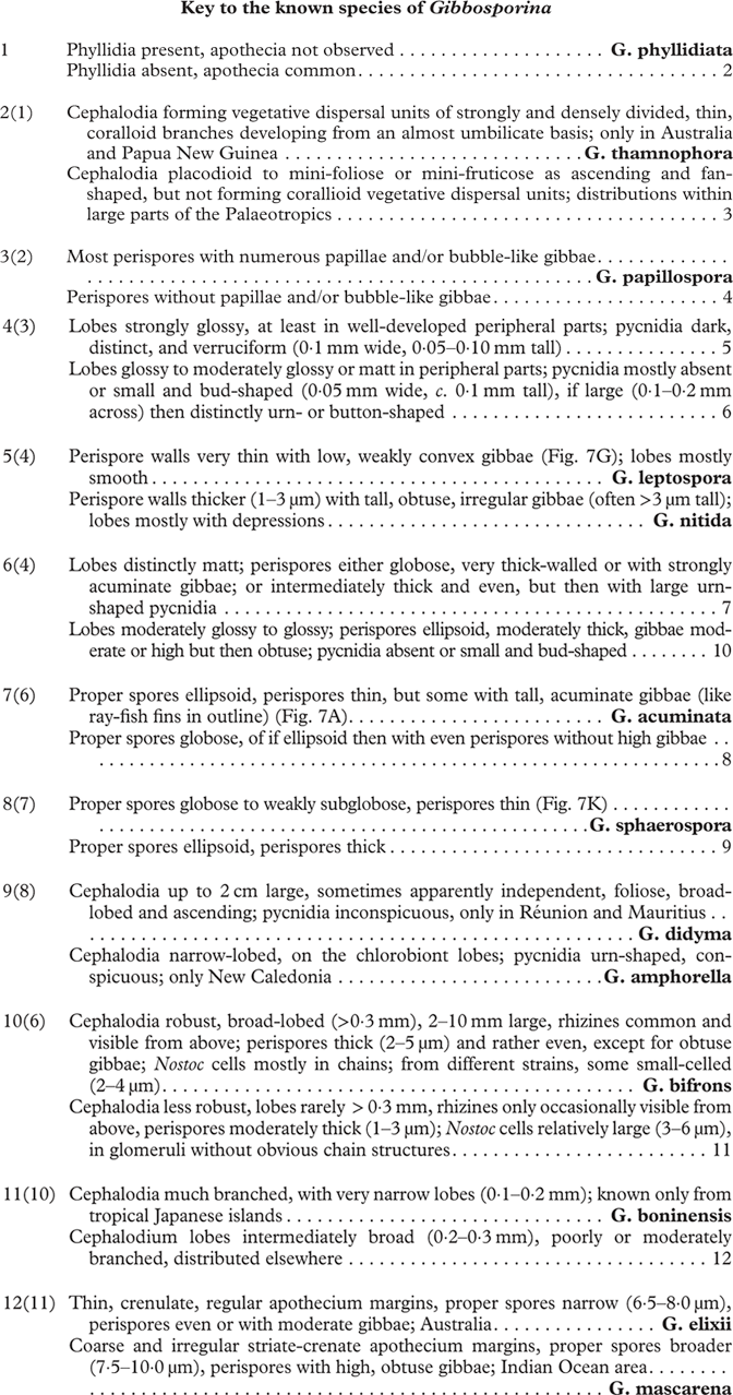
Gibbosporina species, ascospores. A, G. acuminata; B, G. amphorella; C, G. bifrons; D, G. boninensis; E, G. didyma; F, G. elixii; G, G. leptospora; H, G. mascarena; I, G. nitida; J, G. papillospora; K, G. sphaerospora; L, G. thamnophora. Scales:=10 µm.
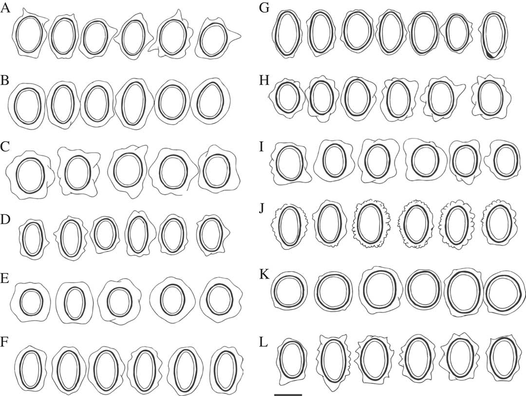
Gibbosporina amphorella (holotype, Elvebakk 05:718). A and B in situ New Caledonia; C, pycnidia; D Gibbosporina bifrons holotype, in situ New Caledonia; E, Gibbosporina didyma (holotype) cephalodia carrying chlorobiont apothecia. Photographed when hydrated immediately after collection in Réunion; F, Gibbosporina mascarena in situ Réunion (Elvebakk 11:041).
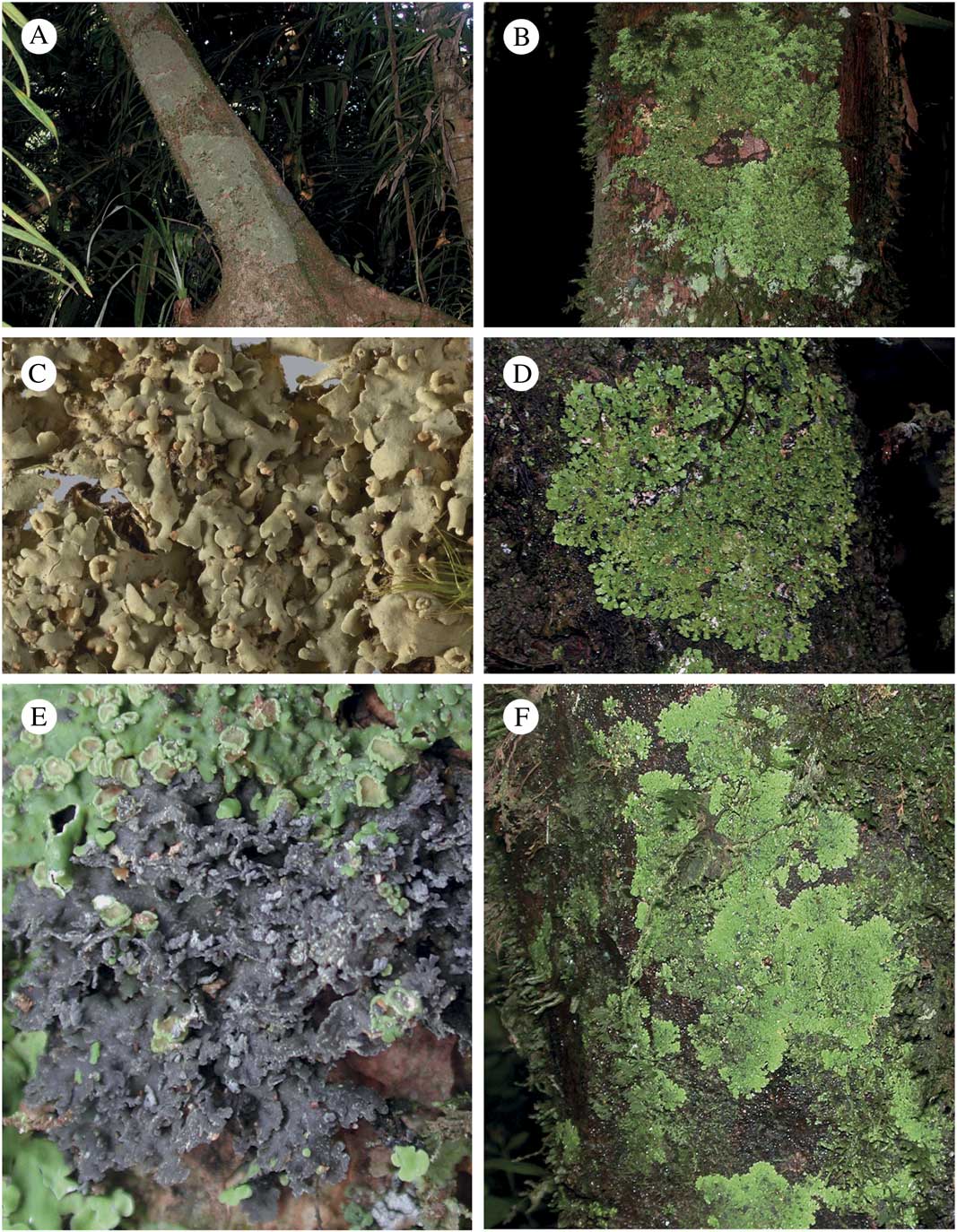
Discussion
According to our current knowledge, the genus Gibbosporina comprises 13 species which have a total distribution ranging from Samoa and Ogasawara (Bonin) Islands in the Pacific Ocean, through South-East Asia and northern Queensland in Australia to the islands Réunion, Mauritius and Sri Lanka in the Indian Ocean. Material obviously belonging to this genus has also been published from Taiwan (Zahlbruckner Reference Zahlbruckner1933; Kurokawa Reference Kurokawa1969), and we have also seen material from Fiji and Madagascar, which will be restudied for a future publication. Material representing reports of ‘Psoroma sphinctrinum’ from South Africa (Doidge Reference Doidge1950) has been searched for in vain in South African herbaria. Three immediate questions arise: is there good support for such an unexpectedly large number of species in a previously unrecognized genus, what patterns can be found in its species diversity, and what can we hypothesize about its evolutionary history?
Evaluation of characters
Spores
To evaluate whether the many new species are well supported, characters are discussed and compared with phylogenetic data when available. Spore attributes, including proper spore and perispore shape and size, perispore wall thickness and surface structure, represent the most important set of characters found here for separating the species. A thorough examination of c. 10 spores in one sample alone will normally show the specific traits and their variation, and make it possible to determine the sample to species level by comparing it with the illustrations in Fig. 7. The exceptions are G. acuminata and G. thamnophora which have similar spores (both with characteristic acuminate gibbae), G. bifrons and G. nitida which also have similar spores, and G. phyllidiata which has not been found fertile.
The most striking spores are those with very thick perispore walls and those with high gibbae. The former type appears like fried eggs in outline. It should be noticed that the sequences of the two species with the thickest perispore walls, G. didyma from Réunion and G. bifrons from New Caledonia, have basal and neighbouring positions in the phylograms. The sister species in the phylograms, G. amphorella and G. nitida, have perispores with intermediate thickness, whereas the three remaining and associated sequenced species and the two remaining non-sequenced species have thin to moderately thin perispores. Only two collections have been determined as G. papillospora; however, a thorough examination of many spores indicated that the presence of papillae and small bubble-like gibbae is a general characteristic and not a casual variation.
Cephalodia
The morphology of the cephalodia also helps in definition and identification of the species. Those of G. thamnophora are very distinct and develop into conspicuous mini-fruticose vegetative reproductive structures (Fig. 6D). This species is comparable, both in general appearance and function to Pannaria durietzii (Henssen & James) Elvebakk & D. J. Galloway, a very conspicuous pan-austral species (James & Henssen Reference James and Henssen1975). The cephalodia of G. phyllidiata are so small (Fig. 6B) that they were first overlooked, whereas G. bifrons and G. didyma can have very large cephalodia (Fig. 4C, 4D & 5A), apparently capable of existing independently, although they have not yet been observed with certainty outside of the prothallus zone. In a species like G. sphaerospora, the cephalodia are less than 3 mm large but relatively broad-lobed and with rhizomorphs clearly visible from above, even in several small cephalodia (Fig. 6C). The remaining species have relatively narrow-lobed cephalodia. In two species, G. mascarena and G. nitida, the cephalodia become rather prominent and mini-foliose or even subfruticose, up to 4 mm across, with lobes loosening from the substratum and becoming suberect, but still erhizinate. These two species also have spores which are somewhat similar, and were morphologically the most closely related species, even though they have very different distributions. Their close relationship was confirmed by the phylogenetic analyses, where they are in a sister species position (Fig. 10). Genetic distances, morphological differences and distribution patterns support their status as different species.
Photobiont diversity
Although photobionts have not been sequenced here, all samples have been studied microscopically, and it is possible to discuss general patterns from these studies, and form hypotheses for future phylogenetic studies. The chlorobiont appears in all cases to be myrmecioid, but varies greatly in cell size. Even two specimens of Gibbosporina amphorella which grew quite close to each other in New Caledonia had large (6·0–12·5 μm) versus very small (3–7 μm) chlorobiont cells, obviously representing different strains.
There is a high morphological diversity of cyanobionts. One extreme case is the Coppins et al. 5440 sample of G. bifrons from Malaysia. The Nostoc cells are intensely mauve-violet, strictly arranged in chains within medullary compartments, with a photobiont layer 150 μm thick, filling out almost the whole thallus, except a reduced 20 μm thick upper cortex and a similarly sized lower medulla. This pattern comes close to a homoiomerous thallus, and is strongly gelatinous. The dominance of the gelatinous Nostoc layer in this specimen is easily observed macroscopically when cephalodia are dry, due to their strongly wrinkled upper surface.
When transferring four gelatinous ex-Collemataceae genera to Pannariaceae, Wedin et al. (Reference Wedin, Wiklund, Jørgensen and Ekman2009) provided a review of gelatinous representatives already included in Pannariaceae, both as bipartite lichens and cyanobionts in tripartite ones. Later, Magain & Sérusiaux (Reference Magain and Sérusiaux2014) studied the phylogeny of cyanobionts in lichens, with a focus on Pannariaceae. They introduced the terms ‘collematoid’ and ‘pannarioid’, to separate two types of cyanobacterial bipartite thalli. The former has a homoiomerous anatomy and the latter is heteromerous. Magain & Sérusiaux (Reference Magain and Sérusiaux2014) described a tripartite species from Réunion named as ‘Pannaria tripartite R969’. We have not examined the sample itself, however its sequence is identical to G. mascarena except for a single base pair in the ITS domain, and can therefore be determined as this species. Its cyanobiont was analyzed phylogenetically by Magain & Sérusiaux (Reference Magain and Sérusiaux2014), and was classified as a distinct ‘Phylotype E’, shared by four samples from Physma byrsaeum. They concluded that the cephalodia of R969 (=Gibbosporina mascarena) is comparable to the collematoid thallus of Physma byrsaeum. In their mycobiont tree, R969 is in a basal position in their Physma clade, and their hypothesis is that the cyanolichen genera dominating this clade arose through emancipation of cephalodia in ancestral tripartite lichens. With this hypothesis in mind, a better knowledge of the cyanobionts of Gibbosporina would be highly desirable. This is illustrated by the cyanobiont phylogram of Magain & Sérusiaux (Reference Magain and Sérusiaux2014), where three phylotypes exclusively consisted of Nostoc strains of the three collematoid Pannariaceae genera Physma, Kroswia, and Gibbosporina (R969), partly associated with cyanobionts of Leptogium.
Our material of G. mascarena and G. didyma have Nostoc cells arranged in glomeruli, and only occasionally with short chain structures clearly discernible. The pattern agrees very well with Nostoc of our own Physma radians material from the same island, where there is a transition from cells in glomeruli with indistinct chain structures above, through short chains and then longer ones in the lower part of a rather thin cyanobiont layer. Physma byrsaeum from Réunion, on the other hand, has a typical homoiomerous anatomy, dominated by the cyanobiont layer with the filamentous morphology of Nostoc clearly discernible. It should be added that several Physma determinations are unreliable, as illustrated by strongly different phylogenetic positions of the same mycobiont species in the phylograms where several samples of the genus are represented (Magain & Sérusiaux Reference Magain and Sérusiaux2014; the present study).
However, the anatomy of the G. bifrons cyanobiont of Coppins et al. 5440, which is similar to the homoiomerous one of Physma byrsaeum, is neither representative of Gibbosporina, nor of G. bifrons. Even a sample from the same area (Coppins et al. 5310) has a very different Nostoc strain, with small greyish blue cells, a distinct morphotype which is also present in a sample from the Philippines. Apart from these two extreme cases, most samples of G. bifrons have rather large, dark brownish violet Nostoc cells. In most cases these have discernible chain structures, although these are weak, and apparently represent a transition state to strains where chain structures are lacking. Sometimes the cyanobiont layer occupies only c. ¼ of the medulla, and in such cases the cephalodium is only moderately gelatinous. These cephalodia are heteromerous and represent a parallel to pannarioid thalli of bipartite cyanolichens. Their upper surface is smooth when dry, indicating moderate swelling in the moist state.
Two of the cyanobionts of G. bifrons were not found to have any discernible chain structures, and Nostoc cells were arranged in glomeruli. This is also the dominant state in Gibbosporina, involving greenish-brownish violet Nostoc cells, organized in glomeruli and with no discernible chain structures. Even within this morphological type, there is obviously a strong diversity, revealed as differences in both Nostoc cell shapes, colours and sizes. The cells of the tiny cephalodia of G. phyllidiata have a distinct blue-green colour not seen in any other Gibbosporina sample.
The conclusion, even without a phylogenetic analysis, is that there is a surprisingly high cyanobiont diversity in Gibbosporina. Our hypothesis for a future extended phylogenetic study of Gibbosporina cyanobionts, based on the morphological pattern presented here, is that they would be expected to show a pattern similar to the one presented by Magain & Sérusiaux (Reference Magain and Sérusiaux2014) for the major collematoid family Collemataceae. This family has Leptogium and Collema cyanobionts distributed widely within the species represented in their phylogram.
Surface structure, lobe thickness and pycnidia
The chlorobiont thalli are quite similar in most species, with coalescent short convex lobes centrally, often with short lobules, and with more discrete lobes peripherally, the latter more appressed on smooth bark than when growing over a mat of epiphytic bryophytes. The lobes of G. didyma are clearly broader than those of the sympatric G. mascarena. Some species are more robust than others, an impression explained by thicker lobes. The most useful thallus character, however, is the upper surface structure. The species G. leptospora and G. nitida have strongly glossy lobes, whereas they are matt in G. acuminata, G. amphorella, G. didyma, and G. sphaerospora. The remaining species are intermediate, and upper surface structure is therefore used in combination with other characters in the key.
The apothecia are substipitate and similar in most species, with thalline excipuli more finely crenate in some species than in others. Pycnidia, on the other hand, which are known in 70% of the species, differ both in external morphology and in their pycnoconidia. The most distinct ones are the urn-shaped pycnidia of G. amphiphorella, even visible in Fig. 4B. In G. sphaerospora, the pycnidia are wide and conspicuous, whereas they are verruciform or bud-shaped in the remaining pycnidiate species. When abundant, they are conspicuous, even when they only measure 0·05 mm across. In two of the species (G. amphorella and G. boninensis), the pycnoconidia are significantly larger (1×2·5–4·0 μm) than in the other species (0·5×2–3 μm).
The main conclusion is that there is variation in quite a number of characters which makes it possible to define the species based on morphology and anatomy. In several cases the species which are most similar according to some given characters are also most closely positioned in the phylograms. The phylograms provide good support for the seven species which have been sequenced. The genus is common in some areas where fieldwork has focused on it, and there are already quite a number of herbarium specimens which could not be included in the present study. We have also treated some species in a wide sense, for example included the Sri Lankan material in G. mascarena, and the single Kumei s. n. specimen at H from Papua New Guinea in G. leptospora. When G. leptospora is compared with the holotype from Australia, it has some lobe depressions, larger pycnidia, and has slightly broader proper spores, which are not only long-ellipsoid, but also can be short-ellipsoid and in some cases even subglobose. The tiny erect hairs on young lobes of the holotype are replaced by more diffuse tomentum in the Kumei s. n. specimen, but the distinctly thin perispore is shared with the holotype. Future studies of additional material will decide whether these species will be split up or not. Nevertheless, these are many reasons to believe that the genus contains more than 13 species and that it has a long evolutionary history.
Species and diversity pattern in the Indian Ocean area
Gibbosporina mascarena appears to be the most common species in Réunion and Mauritius. A total of 16 specimens have been examined from these islands, while the endemic G. didyma is known only from three collections. The holotypes of both these species were collected SE of the lake Grand Étang in east Réunion, in a closed valley opening towards the cyclones from the east. The amount of rainfall in this area is extremely high, and van den Boom et al. (Reference van den Boom, Brand, Ertz, Kalb, Magain, Masson, Schiefelbein, Sipman and Sérusiaux2011) reported no less than 10–12 000 mm annually at Hauts-de-Sainte-Rose, slightly further to the east.
The holotype of G. mascarena was collected among 15–20 specimens growing on a single trunk, with a population of G. didyma present only 10 m away. The obvious difference noted immediately in the field was that most cepholodia in G. didyma were much more prominent than those of G. mascarena. In addition, those of G. didyma were partly foliose with relatively broad, matt lobes, and several cephalodia were growing apparently independently on bryophytes close to the major thalli. There may also be a third species in the Mascarenes, which is not yet sufficiently understood.
Two old collections from Sri Lanka have been studied and found to match G. mascarena. The Leighton 52 collection is heterogeneous, with one deviating individual where the perispores are much more even than those of G. mascarena. The cephalodia are strongly branched with thin and delicate, appressed lobes and the cephalodia are frequently positioned directly on a strongly developed prothallus. Its Nostoc cells appear visually identical to those of G. mascarena from Réunion/Mauritius. The neighbouring individual in this collection has spores which are identical to Mascarene material, but the cephalodia are more broad-lobed, semi-erect and partly rhizinate, and the Nostoc symbiont has smaller cells with short chain structures. Both Sri Lankan collections are placed within G. mascarena here, although future studies might well discover more than one taxon from this island.
The reports from Mauritius and Réunion by Nylander (Reference Nylander1859) of ‘Psoroma sphinctrinum’ and from the ‘Central province’ of Sri Lanka by Leighton (Reference Leighton1869) of ‘Pannaria pholidota’ are based on the old collections of G. mascarena at PC and S studied here, although Leighton (Reference Leighton1869) may have studied a duplicate of the latter.
Species and diversity patterns in Australia, SE Asia and Pacific Islands
Four of the species (G. acuminata, G. bifrons, G. nitida, and G. sphaerospora) are now known from 13–26 collections originating from two to five countries, and G. sphaerospora (19 collections from Australia, Indonesia, Malaysia, the Philippines, and Samoa) and G. bifrons (26 collections from the Philippines, Malaysia, Solomon Islands and New Caledonia) in particular appear to be common and widespread. The remaining species are rare, and with the exception of G. leptospora (known from Australia and Papua New Guinea), endemic: G. amphorella from New Caledonia, G. boninensis from the Japanese Ogasawara Islands, G. elixii and G. thamnophora from Australia, G. papillospora from the Philippines and G. phyllidiata from the Solomon Islands. This pattern can of course change with future studies.
A centre of diversity is Queensland in Australia, where six species are known, and many additional samples are awaiting determination. None of the c. 11 austral tripartite Pannaria species in Australia are known with certainty as far north as Queensland, except P. phyllidiata close to its southern boundary (Lumbsch et al. Reference Lumbsch, Ahti, Altermann, Amo de Paz, Aptroot, Arup, Bárcenas Peña, Bawingan, Benatti and Betancourt2011). However, they resemble Gibbosporina species, and a specimen at BM from the Atherton Tableland in northern Queensland, published as Psoroma sphinctrinum (Jørgensen & Galloway Reference Jørgensen and Galloway1992: 288), Psoroma contortum (Passo et al. Reference Passo, Calvelo and Stocker-Wörgötter2004: 364) and Pannaria contorta (Passo & Calvelo Reference Passo and Calvelo2006: 554), represents a Gibbosporina, but has not been studied sufficiently yet.
In Australia, most of the collections are from the forests of the mountain range between the latitudes of Cairns and Ingham (c. 16°30'−18°40'S). The single Australian collection of G. leptospora was found somewhat further north (15°47'S), while all six Australian collections of G. thamnophora are from Eungella National Park, quite a bit further south (c. 21°S). However, these two species have also been found in Papua New Guinea, and it is not reasonable to compare the distribution patterns of the Australian species from the present data. Nevertheless, it should be emphasized that two different species have been found to co-occur in six of the areas (mostly national parks and state forests) where Gibbosporina species have been collected and studied.
A rather large number of specimens were collected in the Philippines in the early 20th century by persons associated with the US Bureau of Science established in Manila. Among the samples studied here, some have been deposited at S and H, but most of the material had been sent to Vainio, who published c. 25 specimens as Psoroma sphinctrinum (Vainio Reference Vainio1920). However, the material is diverse and represents five species of Gibbosporina. Four of these appear to be widespread, G. acuminata, G. nitida and G. sphaerospora also well represented in Australia, with G. bifrons appearing to have a more northern distribution. The most abundant species are G. sphaerospora, represented by 11 specimens, and G. acuminata with 9. Two collections from two different regions in southern Luzon near 14°N have strange papillose to small-bullate persipores and have been described as the endemic species G. papillospora.
Only eight samples among the widely distributed species have been seen from the large territory represented by the countries Indonesia, Malaysia and Papua New Guinea. However, the genus is probably widespread there, as illustrated by the 17 collections from Papua New Guinea present at CANB, but not yet studied. The Pacific area houses both widespread species and species at present known as endemics. The former category includes the isolated occurrence of G. sphaerospora in Samoa and a very large collection of G. bifrons from the Solomon Islands, in addition to a collection from its easternmost locality in New Caledonia. The endemics are G. amphorella from New Caledonia, G. boninensis from Ogasawara (Bonin) Islands and G. phyllidiata from the Solomon Islands. Gibbosporina boninensis resembles G. acuminata, as indicated in the diagnosis of the latter, whereas the two others are very distinctive species.
Evolution
Transoceanic dispersal has certainly played an important role in the distribution of Gibbosporina species. The distribution coincides well with the cyclone belt, at least as it is now, and there are no alternative explanations other than long-distance dispersal for the occurrences on isolated islands such as Samoa and Fiji. Also the phylogram shown in Fig. 10 does not show any geographical trend, and indicates that several migrations of groups within Gibbosporina have taken place. However, the specimens from the islands in the Indian Ocean are all from different species to those from further east, indicating that transoceanic dispersal across the Indian Ocean is a very rare event, even with the aid of easterly winds in the cyclone belt. The evolution of the sister species G. elixii and G. mascarena, as they are known today, requires a postulated, rather young, but rare transoceanic long-distance dispersal event between Réunion and Australia to have taken place. Co-occurrences of different and distantly related species in Réunion, New Caledonia and Australia, and two mixed samples from the Philippines and Mauritius, as well as an undetermined admixture to G. boninense, testify to an evolution involving both migration and vicariance.
The well-known Réunion endemic Acacia heterophylla is closely related to Australian Acacia s. str. species, a group with a newly proposed position in Austroacacia (Miller et al. Reference Miller, Seigler and Mischler2014), and poses a similar biogeographical challenge to Gibbosporina. A most extraordinary migration event was recently presented by Le Roux et al. (Reference Le Roux, Strasberg, Rouget, Morden, Koordom and Richardson2014), who showed that Acacia heterophylla is derived from long-distance dispersal, potentially by means of petrels, to Réunion 1·4 million years ago, not directly from its ancestral stock in Australia, but from its more closely related species, A. koa from the Hawaiian Islands. Dispersal over very long distances can obviously occur as very rare events. However, there is no evidence that Gibbosporina ever made it to tropical America, nor mainland Africa, except for unconfirmed reports from South Africa. Its centre is in South-East Asia and NE Australia, as is the case for species in several other genera (see e.g. Jørgensen Reference Jørgensen1983).
As compared with other lichen genera, a strikingly high proportion of Gibbosporina species are primarily fertile. The phyllidiate and sterile G. phyllidiata sample from the Solomon Islands is an exception. Its tiny cephalodia and morphological dissimilarity to typical Gibbosporina samples may explain why this and possibly similar species with vegetative propagules have probably been overlooked. On the other hand, the very finely branched cephalodia of G. thamnophora obviously disperse the cyanobiont, in an adaptation shared as a convergence with Pannaria durietzii. These two examples indicate that future studies on Gibbosporina should involve a search for both bipartite chlorolichens and cyanolichens, the latter from truly emancipated cephalodia, which are not yet known. Gibbosporina bifrons and G. didyma have some large rhizinate cephalodia which appear to have developed physiological independence. However, they cannot be confirmed at present to be fully photosymbiodemic, as cephalodia have not been found with certainty outside of the prothallus zone. In addition, cephalodiate cyanomorphs appear to be sexually independent, because when cephalodia were collected as fertile for the first time, the apothecia were unexpectedly found to belong to the chloromorph (Fig. 8E).
Magain & Sérusiaux (Reference Magain and Sérusiaux2014) indicated, in their hypothesis on the evolution of the whole Physma group, that a tripartite thallus is the most likely ancestral form, based on the positions of Xanthopsoroma and Gibbosporina in their phylogram. Interestingly, the two most ancestral species of Gibbosporina in our phylograms are G. didyma and G. bifrons, with entirely different distributions, but sharing a particularly thick-walled perispore and large cephalodia, the latter being almost independent. Thus the evolution within Gibbosporina seems to be the opposite, from an almost photosymbiodemic state to a more specialized tripartite thallus type, with smaller and independent cyanomorphs as distinctly epiphytic cephalodia in the latter.
Gibbosporina species are superficially similar to austral tripartite Pannaria species. Old herbarium samples of both groups take on a similar dark brown colour, which is probably an effect of decomposition of the chlorobiont (see Elvebakk Reference Elvebakk2007). Small cephalodia tend to acquire the same colours, while large ones maintain a greyish colour. However, Pannaria is very remotely related to Gibbosporina, as shown in Fig. 9, and differs by lacking internal ascus amyloid structures, by the different perispores and by the presence of diverse and characteristic sets of TLC-detectable secondary compounds.
Bayesian phylogenetic tree of Gibbosporina and representative clades of Pannariaceae inferred by MrBayes ver. 3.2 under a GTR+I+G model based on LSU sequences. Branches that were maintained in NJ, MP, and ML trees are indicated by thick lines. Bayesian posterior probabilities are indicated when the values were greater than 0·9. Clades are labelled as indicated in Magain & Sérusiaux (2014).
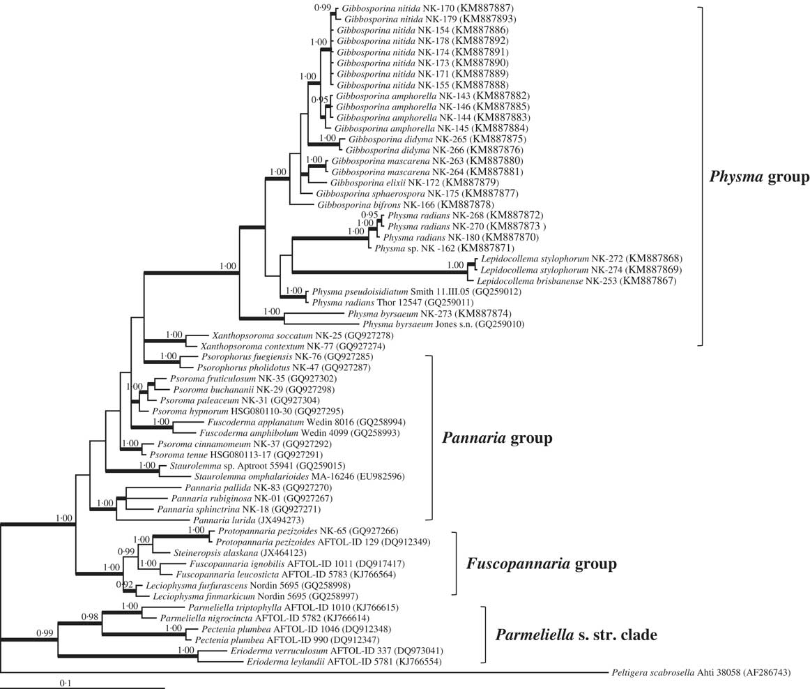
Bayesian phylogenetic tree of the genus Gibbosporina with Physma radians as outgroup taxon. The tree was calculated by MrBayes ver. 3.2 under a GTR+I+G model based on ITS1, 5.8S, ITS2, and LSU sequences. Branches that were maintained in NJ, MP, and ML trees are indicated by thick lines. Bayesian posterior probabilities are indicated when the values were greater than 0·9.
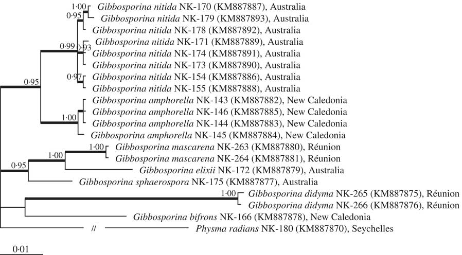
A discussion of the potential evolutionary history of Gibbosporina should therefore focus instead on the ‘Physma group’, where Gibbosporina forms a well-supported clade associated with Physma and Lepidocollema (Fig. 9). There are no modern taxonomic studies on Physma, except for a study from Australia (Verdon & Elix Reference Verdon and Elix1994). However, it should be noted that Physma was shown to be paraphyletic by Magain & Sérusiaux (Reference Magain and Sérusiaux2014), and appears even polyphyletic in Fig. 9. Also, Lepidocollema was paraphyletic according to Magain & Sérusiaux (Reference Magain and Sérusiaux2014, as Parmeliella pro parte). Recent phylogenetic studies of Pannariaceae (Ekman et al. Reference Ekman, Wedin, Lindblom and Jørgensen2014; Magain & Sérusiaux Reference Magain and Sérusiaux2014; the present study) all have constraints, in terms of numbers of loci analyzed, selections of loci, numbers of samples in each clade, and incompatability of loci analyzed for different sequences compared. Thus, many remaining challenges represented by Physma and related genera should be left for future studies.
The inclusion of Gibbosporina in the ‘Physma group’ adds to its general gross morphological heterogeneity, a feature strongly enhanced if Xanthopsoroma is also added. Nonetheless, Physma, Lepidocollema and Gibbosporina share tropical distribution patterns, a lack of TLC-detectable compounds, and the presence of distinct ring-like thalline excipuli and amyloid internal ascus structures, as pointed out in previous studies. However, we propose here that perispore structure (gibbose surfaces and/or presence of apical extensions) could be an additional synapomorphy of the ‘Physma group’, even including Xanthopsoroma. All specimens in these four genera studied by us have perispores corresponding to this pattern. It can even be presented here that the Jones s. n. sample of ‘Physma byrsaeum’ from Tahiti (an unreliable species identification like several other sequenced Physma samples), included in the phylograms by Wedin et al. (Reference Wedin, Wiklund, Jørgensen and Ekman2009), Muggia et al. (Reference Muggia, Nelson, Wheeler, Yakovchenko, Tønsberg and Spribille2011), Magain & Sérusiaux (Reference Magain and Sérusiaux2014), Ekman et al. (Reference Ekman, Wedin, Lindblom and Jørgensen2014) and by the present study, has perispores which are neatly intermediate between those of Gibbosporina and Xanthopsoroma; gibbose, but with wide nodulose apical extensions.
The old evolutionary history of both Lecanoromycetes and Peltigerales (Prieto & Wedin Reference Prieto and Wedin2013; Beimforde et al. Reference Beimforde, Feldberg, Nylinder, Rikkinen, Tuovila, Dorfelt, Gube, Jackson, Reitner and Seyfullah2014), and early diversification of Pannariaceae versus Collemataceae (Wedin et al. Reference Wedin, Wiklund, Jørgensen and Ekman2009; Spribille & Muggia Reference Spribille and Muggia2013), would indicate that the ‘Physma group’ as a basal diversification within Pannariaceae is also of considerable age. We have made a tentative secondary calibration (only conclusion referred to here) following the procedures of a previous molecular dating study involving nucleotide differences of ITS in Erysiphales (Takamatsu & Matsuda Reference Takamatsu and Matsuda2004), where nucleotide substitution rates of the ITS region were found to be 2·52×10−9 per site per year. The divergence between Physma radians (NK-180) and Gibbosporina elixii corresponds to 75 Ma. Although not incompatible with the datings by Beimforde et al. (Reference Beimforde, Feldberg, Nylinder, Rikkinen, Tuovila, Dorfelt, Gube, Jackson, Reitner and Seyfullah2014), we should treat this estimate with care.
In the northern part of the Great Dividing Range in east Australia, the Pannariaceae flora is dominated by ‘Physma group’ genera and members of the Pannaria lurida group (Jørgensen Reference Jørgensen2015), whereas a very different flora of austral Pannariaceae genera and groups instead dominate in temperate areas further south along the same mountain range. It could be tempting to postulate an evolutionary connection between the ‘Physma group’ and representatives among the numerous austral Pannariaceae genera in this area, coupled with migrations into South-East Asia. The high-latitude cooling during the Eocene associated with the opening of the Drake Passage at 33·7 Ma (e.g. Pagani et al. Reference Pagani, Zachos, Freeman, Tipple and Bohaty2005), as well as New Guinean orogeny, initiated important northern migrations. The classic example is the genus Nothofagus, with its subgenus Brassospora; now the genus Trisyngyne according to Heenan & Smissen (Reference Heenan and Smissen2013). A recent study on the liverwort family Schistochilaceae is another example. Yu et al. (Reference Yu, Xiaolan and Glenny2014) showed that the group migrated into New Guinea at c. 19 Ma, shortly after the orogeny had been initiated there.
However, all phylogenies presented so far indicate that the separation of the ‘Physma group’ from remaining clades in Pannariaceae is far older than the austral biogeographical events taking place after the Eocene cooling. Most austral Pannariaceae genera have now been represented in Pannariaceae phylogenies, but they are all well nested within the four clades presented by Magain & Sérusiaux (Reference Magain and Sérusiaux2014), Ekman et al. (Reference Ekman, Wedin, Lindblom and Jørgensen2014) and the present study. The only exception is Xanthopsoroma, the only Pannariaceae genus with usnic acid (Elvebakk et al. Reference Elvebakk, Robertsen, Park and Hong2010), which had deviating and surprising positions in all these studies. Its basal position in the Physma group of Magain & Sérusiaux (Reference Magain and Sérusiaux2014) was not well supported, and was not confirmed by Ekman et al. (Reference Ekman, Wedin, Lindblom and Jørgensen2014), where instead a clade of two samples was a sister group to their whole ‘Clade 2’, comprising no less than 16 Pannariaceae genera. In our LSU-based phylogram (Fig. 9), Xanthopsoroma forms, together with Psorophorus, two unresolved sister groups to the Physma clade. The topic obviously needs to be restudied with better multi-locus sequence data.
The conclusion is that the evolutionary history of the ‘Physma clade’ remains unresolved and with open gaps, although Gibbosporina obviously belongs there. At present, the studied members of the clade should be considered to represent a small, remaining part of a diverse and probably large group with an old evolutionary history.
We are indebted to the curators and directors of the cited herbaria for sending material on loan, to Direction des Ressources Naturelles, Nouméa, for permission to collect in New Caledonia, to Parc National de La Réunion and Office National des Forêts, St. Denis, Réunion for permission to collect there, Prof. J. A. Elix, CANB, for sending invaluable Gibbosporina material from Australia, F. Schumm, Wangen, Germany, for sending Physma material, M. Karlstad, Tromsø University Museum, for taking all photographs in Figures 1 to 6, to her colleague E. Høgtun for preparing the plates, P. P. Aspaas, University Library, Universiy of Tromsø, for help with the Latin diagnoses, and N. Magain, University of Liège, Belgium, for interesting discussions on the topic and comments on the manuscript. Comments on the manuscript by an anonymous referee were also appreciated. Tina Dahl, then Department of Biology, University of Tromsø, carried out the HPLC analyses. This study was partly supported by the Korea Polar Research Institute (Grant PE14020).