I. INTRODUCTION
Bhadeshia and Caballero have reported a medium transformation product composed of bainite ferrite laths separated by retained austenite films in the high carbon high silicon steel, and it has excellent mechanical properties of great engineering application value, such as high tensile strength, good ductility, and fracture toughness etc.Reference Garcia-Mateo, Caballero and Bhadeshia1,Reference Caballero and Bhadeshia2 This mixture is so-called low temperature bainite, regarding to the fact that it is generated at the temperature a little bit higher than M S for a long time.Reference Garcia-Mateo, Caballero and Bhadeshia1 It can also be named as nanoscale bainite, because the bainite ferrite laths characterized as sheaves in a parallel arrangement have nanoscale size in width. These newly discovered characteristics contain useful information to reveal the essence of bainite transformation. Bainite transformation mechanism has been in debate for several decades. Investigation on nanoscale bainite could provide new experimental evidences and could be conducive to building a new transformation theory on nanoscale bainite transformation mechanism. This will make contributions to the development of novel steels with high mechanical properties. Investigators have been studying its formation mechanism in aspects of transformation driving force, subunit structure, solid solution of carbon atom, growth kinetics etc.Reference Garcia-Mateo and Bhadeshia3–Reference Gong, Tomota, Harjo, Sua and Aizawa11 Bhadeshia considered that formation of nanoscale bainite is achieved by a displacive mechanism.Reference Garcia-Mateo and Bhadeshia3,Reference Bhadeshia4 However, the formed bainite structure is distinctively different from the acicular martensite in general high-carbon steel in microscopical morphology, and this should be conducive to understand the phase transition by the diffusion mechanism. Other high carbon steel with similar composition had been studied,Reference Kong, Liu and Yuan12 and nanoscale bainite was obtained by isothermal transformation at 280 °C (above M S) after austenitization at 950 °C. These phenomena suggest that the structure of austenite grains formed by austenitization at high temperatures has a substantial impact on bainite transformation. It is worthy of attention to that, what condition does the high-temperature austenitization provide to form the nanoscale bainite? Investigations that focus on this point could properly reveal the nanoscale bainite formation mechanism.
II. EXPERIMENT MATERIALS AND PROCEDURES
The chemical composition of the experimental steel used in this study was similar to the high carbon steels studied by Bhadeshia.Reference Caballero and Bhadeshia2 Fe–0.88C–1.35Si–1.03Cr–0.43Mn (wt%) was melted in a ZG-25 vacuum medium frequency (Shenyang Ace Care Technology Co., Shenyang, China) induction melting furnace. The homogenizing treatment for the ingot was carried out in an argon protective atmosphere at 1200 °C for 24 hours. A c1 and A cm temperatures of this steel were measured as 770 °C and 830 °C, respectively, by differential thermal analysis (DTA) method using a TG/DTA6300 analyser (Netzsch, German). A Formastor-Digit 2 automatic measuring instrument was used to obtain phase transition points of a series of samples, and then connect these points into a curve. The experiment samples were heated to the austenitizing temperature at a rate of about 25 K/min and maintain for 20 min, then reduce the temperature to a medium isothermal temperature at a rate of about 45 K/s, and at last cool the samples down to room temperature after heat preservation of a certain amount of time. The M S temperature of this steel was measured as 210 °C using a Formastor-Digit 2 dilatometer (Rigaku, Japan) when austenitized at 880 °C. It has been proved by the measured results that the M S only changed a little with increasing the austenitization temperatures from 880 °C to 950 °C.
The austenitizing process for annealing and austempering of the steel samples was carried out in a vacuum heat treatment furnace. The austenitized samples were isothermally treated in dimethyl silicone oil, and then cooled to room temperature by water. To reveal the microstructure, the samples were twice etched samples by nitric acid alcohol and picric acid solution, respectively. The metallographic structure was observed by an Axio Scope A1 optical microscope (Zeiss, German). A D/Max-2500V diffractometer (Rigaku, Japan) was used to carry out x-ray diffraction with Cu Kα radiation at 40 kV voltage and at 100 mA current. The results of x-ray diffraction were analyzed by Analysis Software Jade 6.0. The fine structure of the samples was observed by JEM-2100 transmission electron microscope (JEOL, Japan). The transmission electron microscopy (TEM) films with Ф3 × 0.3 mm2 were cut from the heat treated samples and electrolytically polished in a twin jet electro-polishing apparatus using a solution of 5% perchloric acid and 95% methanol.
To know the austenitizing effect on nanoscale bainite nucleation mechanism, we used thermodynamic calculation formula to calculate the concentration of point defects (vacancy). And we calculated the formation energy of different vacancy combinations by analytical embedded atom method (AEAM).
III. RESULTS AND DISCUSSIONS
A. Transformation start temperature
Figure 1(a) shows the curve of the bainite transformation start temperature corresponding to the austenitization temperature at 880 °C. The shape of the bainite transformation start curve is characterized as a flat “C” (the curve is so-called “C’’ curve). There is an interval of ∼50 °C between the nose temperature and the martensitic transformation starts (M S) temperature. The temperature range of low-temperature bainite transformation is small, and is close to that of the martensitic transformation. Figure 1 illustrates the relation between the austenitization temperature and the bainite transformation temperature. Compared the curves in Fig. 1(a)–1(c), it is observed that the transformations curve moves to the lower left with increasing the austenitization temperature in the range of 880–950 °C, this implies that the incubation period of bainite transformation is shorter, and the bainite transformation temperature range is nearer to that of low temperature phase transformation. In the case of austenitization temperature at 950 °C, the bainite start transformation temperature drops to ∼235 °C and the incubation period decreases to ∼100 s. This probably to some extent means that the characteristics of bainite transformation become gradually similar with that of low temperature transformation as transformation temperature increases.
Curve of the bainite transformation start temperature after austenitization at temperatures (a) 880 °C, (b) 900 °C, (c) 930 °C, and (d) 950 °C.

Figure 2 shows the x-ray diffraction analysis (XRD) patterns of samples annealed from austenitization at different heating temperatures. It is calculated that, the structure of annealed samples mainly contains pearlitic ferrite and cementite. The detailed result of (200)α diffraction peak is shown in the top right corner of Fig. 2. Scrutinizing the shapes of (200)α diffraction peak corresponding to the samples annealed at austenitization temperatures 880, 900, 930, and 950 °C, the 2θ-coordinate position of (200)α diffraction peak has little change, while its integral breadth reduces gradually.
XRD patterns of the samples annealed from different austenitization temperatures (a) 880 °C, (b) 900 °C, (c) 930 °C, and (d) 950 °C.
Figure 3 demonstrates the integral breadth of the (200)α diffraction peak of the annealed samples. The curve is relatively flat where austenitization temperature is below 880 °C while the curve descends obviously where austenitization temperature is above 880 °C. However, the curve becomes flat again where austenitization temperature is higher than 950 °C. The α phase (i.e., pearlitic ferrite) is the product of high temperature diffusion phase transformation, and its structure is extremely close to equilibrium state. Therefore, the changes of the integral breadth of the (200)α diffraction peak directly reflect the variations of pearlite field in size. The smaller integral breadth corresponds to larger pearlite field, and the larger integral breadth to the smaller pearlite field. The size of pearlite field formed by high temperature diffusion phase transformation is determined by the grain size of parent phase (austenite). The pearlite with larger field size is formed in the austenite with larger grain size. Therefore, the results demonstrate that, to some extent, in the interval of 880–950 °C, austenite is likely to grow into obviously various grain sizes in the experimental steel.
Integral breadth of (200)α diffraction peak of samples annealed at different temperatures.
Figure 4 shows the optical microstructures of the annealed samples at different austenitization temperatures. Obvious grain boundary could be observed in the picture (such as indicated by red arrows). To some extent, the grain size of prior austenite could be characterized by measuring the pearlite grain size. The austenite grain size of the sample austenitized at 880 °C was larger than that at 850 °C, but the increase the amplitude of grain size was small. However, when austenitized at 900 °C, the austenite grain size of the sample was much larger, and the austenite grain size reached nearly the maximum when austenitized at 950 °C. This is in accordance with the deduction from Fig. 4.
Optical micrographs of the prior austenite grain boundary in annealed samples at different austenitization temperatures (a) 850 °C, (b) 880 °C, (c) 900 °C, and (d) 950 °C.
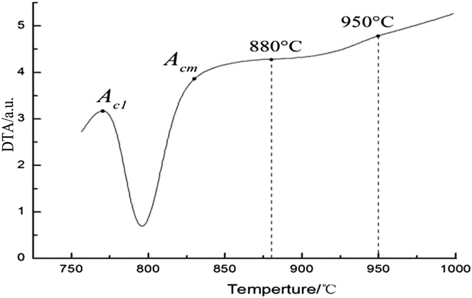
Figure 5 illustrates the DTA curves of the heat treated samples. There is an obvious endothermic peak between A c1 (770 °C) and A cm (830 °C), at which the cementite dissolves into austenite and austenite forms from ferrite. Nevertheless, the DTA curve presents a straight line in the temperature range from A cm to 880 °C, which means that the structure of austenite basically has little change. However, there is a steady endothermic peak between 880 °C and 950 °C, where the composition homogenization in austenite and the further growth of austenite grain takes place. This is also identical to the results of optical observation and XRD analyses.
The DTA curve of samples.
When heated in the range of 880–950 °C, the composition homogenization (especially the carbon atoms) takes place in austenite. Grains grow up, grain boundaries reduce, and crystal defects decrease. Meantime, the ability of heterogeneous nucleation of medium temperature transformation becomes lower. Therefore, the bainite transformation start temperature turns lower to meet the need for driving force of γ → α phase transformation.
B. Microstructure of bainite transformation products
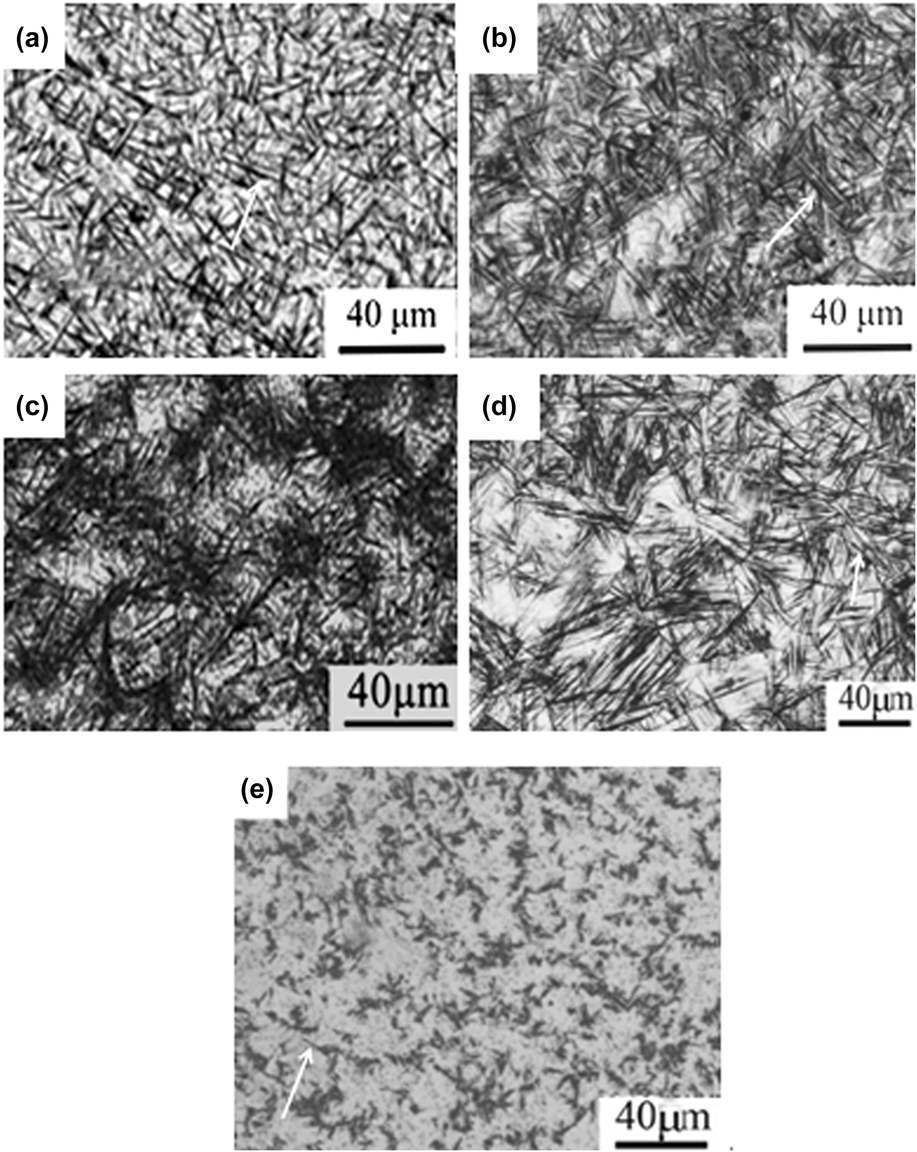
Figures 6(a)–6(d) show the microstructure of samples austenitized at different temperatures and isothermally treated at 240 °C for 30 min. Acicular structure in two forms can be seen in the pictures: independent bainite needles and bainite sheaves consisting of dark needles in a parallel arrangement. With austenitization temperatures increasing, the amount of the sheaves increases, and the size of sheave develops with increasing the aspect ratio (length/width) of the needle. Figure 6(e) depicts the dark needles intersected with each other in the sample quenched by water after austenitized at 880 °C. Evidently, the dark needles are product of the martensitic transformation at even lower temperature. The product of medium temperature transformation is named as sheaf-like bainite in this paper, which is distinct from that of acicular martensite-the product of low temperature transformation. The sheaf-like bainite has different morphology features even obtained at a same medium temperature after austenitized at different temperatures. The difference can be characterized as variation in the aspect ratio (length/width) of units constituting bainite sheave.
Optical micrographs of samples isothermally transformed at 240 °C for 30 min (a) 880 °C, (b) 900 °C, (c) 930 °C, (d) 950 °C, and sample quenched from (e) 880 °C.
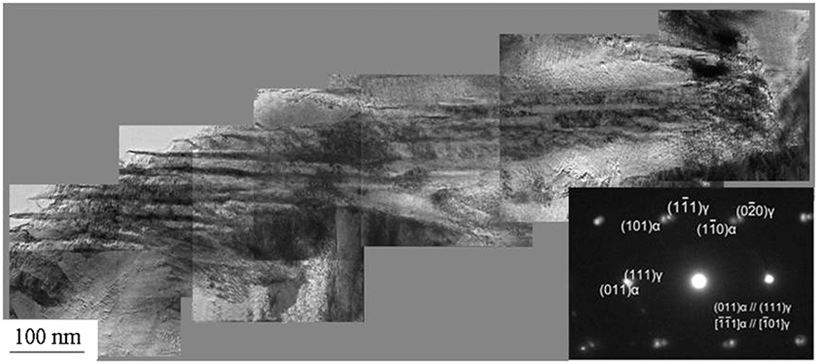
Figure 7 is joined with a set of pictures to display the morphology of fine microstructure of bainite isothermally transformed at 240 °C for 30 min after austenitization at 880 °C. The obtained bainite consists of a row of parallel laths, the length in micrometers and the width about 20 nanometers. Therefore, we also call it nanoscale bainite. Considering the analysis of dark-field and bright-field images and electron diffraction measurement of bainite sheave, the products of bainite transformation are composed of α-phase laths without carbide separated by γ-phase retained austenite films.
TEM morphology of the sheave-like bainite in the sample isothermally transformed at 240 °C after austenitization at 880 °C.
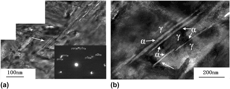
Figure 8(a) shows the TEM morphology of fine structure isothermally transformed at 240 °C after austenitization at 950 °C. However, the structure also consists of several parallel laths. The electron diffraction pattern declares that the laths are α phase among γ-phase. The length of the laths is also in micron-size while the width becomes smaller. The bainite lath has a higher length/width ratio. Figure 8(b) magnifies the arrowed area in Fig. 8(a), and shows the morphology of leading tip of the sheave-like bainite ferrite lath. The sublath structure of bainite laths is only several nanometers in width, extremely like a needle in geometry. Sublaths line up end to end, and develop into a lath which could be observed with optical microscopy.
TEM morphology of the sheave-like bainite in a sample isothermally transformed at 240 °C for 30 min after austenitization at 950 °C dimensional combination.
Due to the curves in Fig. 1, in the case of austenitization at 880 °C, 240 °C is located below the nose temperature within the lower bainite transformation range; on the other hand, in the case of austenitization at 950 °C, 240 °C is located within the upper bainite transformation range. According to the conventional viewpoints on medium transformation, the former could generate the needle-like lower bainite, and the latter could generate the feather-like upper bainite. However, as shown in Figs. 7 and 8, the products have quite similar structure and are composed of parallel α phase laths only with variation of width of a lath and size of sublath. This implies that the nanoscale bainite in Figs. 7 and 8 are generated via a similar formation mechanism.
With increasing austenization temperature in the range of 880–950 °C, it is seemingly difficult for nonhomogeneous nucleation of bainite ferrite to take place. However, it does not mean that the nucleation of bainite transformation is restrained because at higher temperature carbon atoms can be divorced from the equilibrium position easily and the vacancies form, and the impact of the vacancy defects also benefit the bainite transformation. The vacancy concentration with the lowest free energy of the system at a certain temperature is named the equilibrium vacancy concentration as a function of temperature expressed by the formula (1).Reference Leibfried and Breuer13
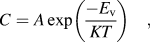 $$C = A\,\exp \left( {{{ - {E_{\rm{v}}}} \over {KT}}} \right)\quad ,$$
where A is material constant, usually equal to 1; K is Boltzmann's constant, 1.38 × 10⁻²³ J/K; and E v is the formation energy for 1 mole vacancies, J/mol.
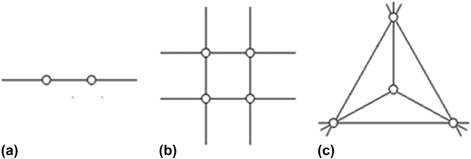
The measured activation energy of a Fe vacancy is ∼1.80 eV in face-centered cubic crystal structure.Reference Wang14 Accordingly the equilibrium concentration of Fe vacancy can be calculated: C 880 °C = 2.06 × 10⁻⁹, C 900 °C = 1.860 × 10⁻⁸, and C 950 °C = 4.139 × 10⁻⁸. The vacancy concentration increases distinctly as austenitization temperature increases. For instance, the vacancy concentration at 950 °C is ∼20 times as much as that at 880 °C. When the austenite is cooled to the bainite transformation range at a rapid speed, most of vacancies are retained at low temperature. To lower the energy of system, the vacancies start to combine and migrate.Reference Kuramoto, Ohsawa and Tsutsumi15,Reference Kulikov and Hou16 There are three types of vacancy combinations: linear combination (such as di-vacancies), planar combination, and dimensional combination, shown as Fig. 9.Reference Croeker, Doneghan and Ingle17
Three types of vacancy combination (a) linear combination, (b) planar combination, (c) dimensional combination.
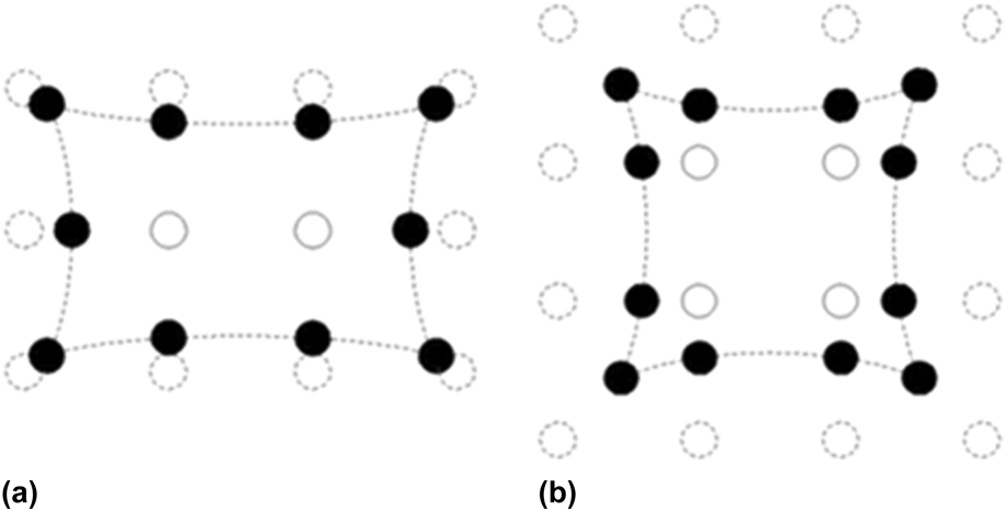
The lattice distortion shown in Fig. 10 caused by vacancy combinations is simulated by computer. The lattice distortion extent caused by the planar combination of four vacancies [as shown in Fig. 10(b)] is much bigger than that of di-vacancies [showed in Fig. 10(a)]. The more vacancies in one combination, the more complex the combination structure is; the bigger extent of lattice distortion, the more activation energy is needed. How the vacancies' structure impact on free energy of the system has aroused the researchers' attention. In this paper, the formation energy of different vacancy combinations is calculated by AEAM reported by Johnson,Reference Johnson18,Reference Johnson19 and the results show that di-vacancies are most possibly to exist in face-centered cubic austenite rather than other combinations.
Lattice deformation caused by different vacancy combination: (a) di-vacancies and (b) planar combination of four vacancies.
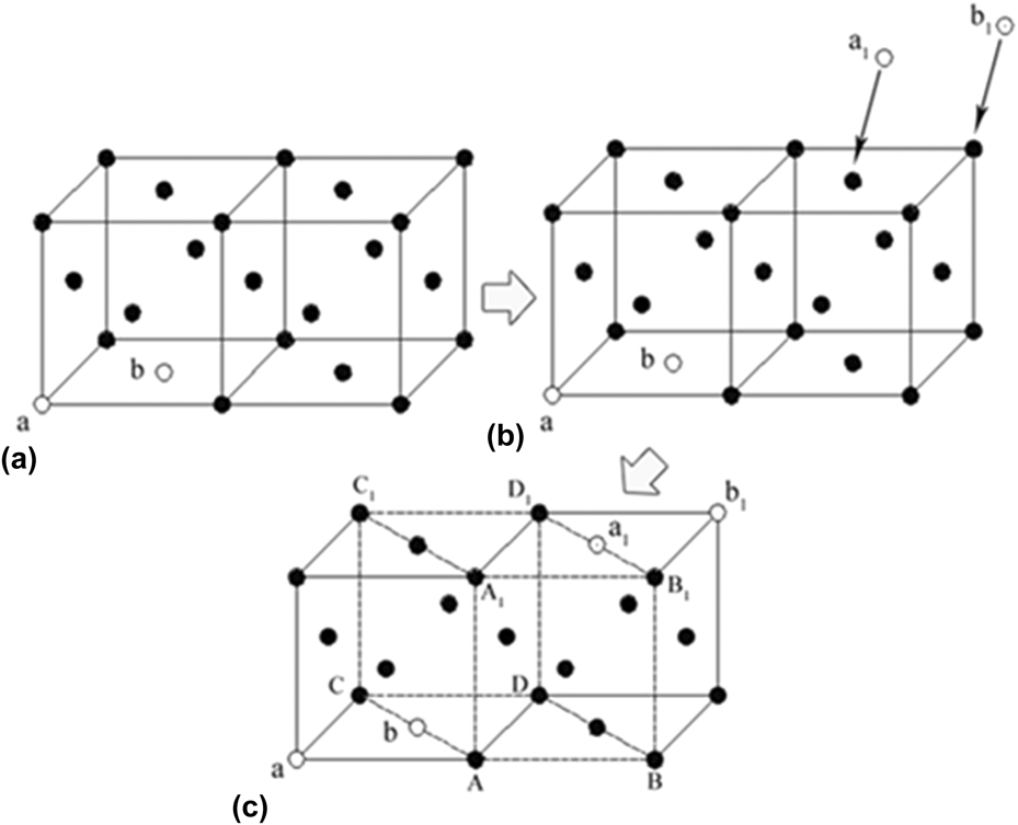
Figure 11(a) describes the face-centered cubic unit cell of normal austenite in balance, and a vacancy combination [indicated by ab in the Fig. 11(a)] is supposed there. When a di-vacancy [indicated by a1b1 in the Fig. 11(b)] migrates into the unit cell [Fig. 11(b)], the compactness of the unit cell becomes lower and lattice distortion happens partially. The change of atomic arrangement results in the adjustment of the lattice constant. Therefore, puny areas are generated where crystal structure is deviated from the normal unit cell of austenite. With the help of structure fluctuation and energy fluctuation of system, these puny areas turn into embryo nucleus of phase transformation: face-centered cubic γ → body-centered cubic α [hexahedral unit ABCD–A1B1C1D1 in Fig. 11(c)]. Due to the fact that the vacancy concentration increases distinctly as austenitization temperature increases, puny areas suitable for the embryo nucleus of γ → α increase overtly, which can benefit the nucleation of bainite transformation. This mechanism is in accordance with the experimental results (as shown in Fig. 1) that the transformations curve moves to the lower left with increasing austenitization temperature.
Di-vacancy string migration caused lattice diagram adjustment.
Besides the di-vacancies, influence of the interstitial atom carbon on the unit cell of austenite shall be also considered. Recently, Caballero et al. characterized the distribution of carbon atoms in bainite on the atomic scale, confirming the migration of carbon atoms with the formation of bainite.Reference Caballero, Miller, Garcia-Mateo and Cornide7,Reference Caballero, Miller, Bahu and Garcia-Mateo20 The interstitial atoms can cause local expansion-type lattice distortion of the unit cell, which weakens the shrinkage-type lattice distortion by effect of vacancies. Therefore, the interstitial carbon atom plays a role to restrain the formation of the embryo nucleus of γ → α.
When austenitized at lower temperature such as 880 °C, the homogenization level of the interstitial atom carbon is lower, and there are local microzones in austenite crystal with lower concentration of carbon atom, where the interstitial atom carbon has a weaker ability to hinder the effect of vacancies on embryo nucleus. Embryo nucleus of bainite ferrite is in advantageous condition to form in these local microzones, and then grows into bainite lath with hundreds of nanometer of width. While austenitized at 950 °C, the homogenization level of the interstitial atom carbon is higher. As a result, these local microzones become smaller in size where the embryo nucleus of γ → α can be generated. Accordingly, the bainite ferrite grows into more slender laths with several tens nanometers in width.
IV. CONCLUSIONS
Nanoscale bainite can be generated at low temperature (∼30 °C above M S) in the Fe–0.88C–1.35Si–1.03Cr–0.43Mn (wt%). The austenitization temperature has a strong effect on the formation of the γ → α embryo nucleus, as a consequence of comprehensive influence given by migration of the vacancies and the interstitial carbon atoms. A bainite lath, as the component of bainite, grows into different microscopic geometrical morphologies: very slender lath with a width of several tens nanometers because of the higher vacancy concentration from austenitized at higher temperatures (e.g., 950 °C), and lath with a larger width because of the lower vacancy concentration from austenitized at lower temperatures (e.g., 880 °C).
ACKNOWLEDGMENT
The study was supported by Tianjin Momentous Technology Supporting Program Foundation of China (No. 11ZCKFGX20500).