Introduction
Amyloid plaque, neurofibrillary tangle (NFT) and neuronal tissue loss are typical pathological hallmarks of Alzheimer's disease (AD). Deposition of amyloid beta (Aβ), which is the major component of amyloid plaque, plays a critical role in the etiology of AD.
Aβ is derived from β-amyloid precursor protein (β-APP) and appears to be degraded mainly by neprilysin (NEP). Neprilysin, known as a neural endopeptidase, is a 97-KD glycoprotein widely expressed in many tissues including the central nervous system (CNS), fibroblasts and plasma (Haouas et al., Reference Haouas, Morello, Lavenu, Billard, Jasmin and Boucheix1995). Increasing evidence suggests that NEP may be the major degrading enzyme of Aβ in the brain and the NEP gene has been proposed as a candidate gene for AD. The neprilysin (NEP) gene is located within Chromosome 3 (region 3q25.1–q25.2) which has also been reported to be associated with AD (Tanzi et al., Reference Tanzi, Kovacs, Kim, Moir, Guenette and Wasco1996; Poduslo et al., Reference Poduslo1999; Jiajun et al., Reference Jiajun2005). In-vivo experiments in NEP-deficient mice with decreased NEP activity or exposure to Aβ peptide have increased Aβ levels and induced Aβ deposition in brain (Iwata et al., Reference Iwata2001; Mohajeri et al., Reference Mohajeri, Kuehnle, Li, Poirier, Tracy and Nitsch2004). Both NEP mRNA and protein levels have been shown to be reduced in AD brains, especially in those areas typically affected by plaques (Yasojima et al., Reference Yasojima, Akiyama, McGeer and McGeer2001; Reilly, Reference Reilly2001; Carpentier et al., Reference Carpentier, Robitaille, DesGroseillers, Boileau and Marcinkiewicz2002). Although the association results were still preliminary, recent reports have found that genetic variances within the NEP gene were associated with AD (Clarimon et al., Reference Clarimon2003; Helisalmi et al., Reference Helisalmi2004; Sakai et al., Reference Sakai2004).
In the present study, we chose two polymorphisms of −204G/C and 159C/T in the NEP gene and explored their effects on the risk of AD. The −204G/C polymorphism, a newly-identified single nucleotide polymorphism (SNP) by denaturing high-performance liquid chromatography (dHPLC) and sequencing methods in our previous research (Jiajun et al., Reference Jiajun2005), was located within the putative transcription repressor factor Pax-4 binding region and therefore might result in subtle alterations in the NEP structure or expression. The 159C/T polymorphism consists of a C to T substitution at position 159 downstream of the gene stop codon and has been found to associate with AD in an age-dependent manner in a Spanish population (Clarimon et al., Reference Clarimon2003). We aim to evaluate whether these two SNPs in the NEP gene confer susceptibility to the development of AD in the Chinese population.
Method
Participants
Patients with sporadic late-onset AD were recruited from the Guangzhou Psychiatric Hospital in southern China. The subjects were diagnosed with possible or probable AD using National Institute of Neurological and Communicative Disorder and Stroke and the Alzheimer's Disease and Related Disorders Association (NINCDS-ADRDA) criteria. Healthy elderly controls were enrolled from three old people's homes in suburban Guangzhou. The subjects comprised 236 sporadic AD patients (169 female and 67 male; age range 56–103 years; mean age 81.9 ± 8.6 years) and 332 controls (203 female and 129 male; age range 55–97 years; mean age 74.3 ± 9.5 years). The AD group was older than controls (t-test, t = −9.503, p < 0.001), and there were more females in the AD group (Pearsonχ2 = 6.685, p = 0.010). Informed consent was obtained from all subjects or their caregivers and the project was approved by the ethics committee of Guangzhou Psychiatric Hospital.
Genotyping
Genomic DNA was extracted from peripheral blood lymphocytes by using the standard phenol-chloroform method. Genotyping for NEP −204G/C alleles was performed by polymerase chair reaction (PCR) amplification using the forward primers 5’-ACGGGGAGGGAGAGTAAGAA-3’ and backward primers 5’-GCAATGAAAATGTCCCATCC-3’. The amplified product of 315bp was digested with 5 units of MboI and then electrophoresed on 12% polyacrylamide non-denaturing gel with silver staining. Similarly, NEP159C/T alleles were amplified by PCR with the following primers: 5’-GTCACTGTACTGACTTGAGGG-3’ and 5’-ACATCAGCAAACTGGTAGACC-3’, then the PCR product of 212bp was subject to 5 units of XbaI and followed by electrophoresis on 12% polyacrylamide gel with silver staining. ApoE was genotyped as previously described (Hixson et al., Reference Hixson and Vernier1990).
Analysis
Allele frequencies were determined by counting alleles and calculating sample proportions. The Hardy-Weinberg equilibrium (HWE) was confirmed by using the χ2 test. Differences in genotypic and allelic distribution between groups were analyzed by using the χ2 or log-likelihood ratio test (as appropriate). The t-test was performed to compare mean age between groups while the χ2 test was used to assess differences relating to sex. The data were analyzed with the Statistical Package for Social Sciences for Windows, version11.0.
Results
Association between NEP genotypes and AD
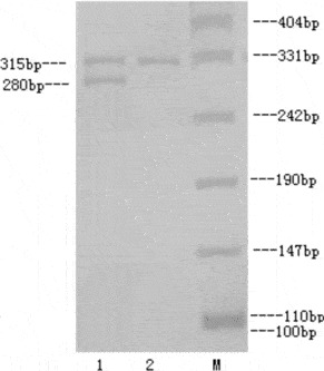
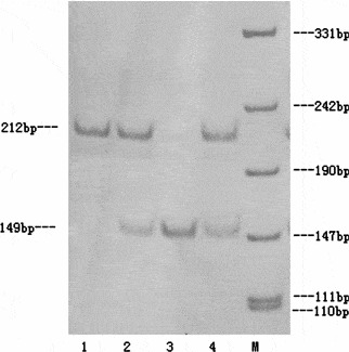
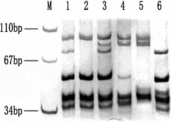
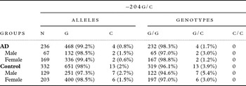
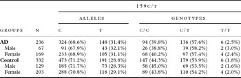
The NEP −204G/C, 159C/T and ApoE genotypes were determined by using PCR-RFLP (Figures 1–3). The distribution of genotype and allelic frequencies for −204G/C and 159C/T in AD group and controls are shown in Table 1. As for NEP −204G/C polymorphism, allele frequencies followed HWE (for the AD group: χ2 = 0.02, p = 0.89; for the control group: χ2 = 0.13, p = 0.72). However, both groups deviated from HWE for 159C/T polymorphism (for the AD group: χ2 = 27.07, p < 0.001; for the control group: χ2 = 33.07, p < 0.001).
NEP −204G/C genotype as determined by PCR-RFLP. M: PUC19/Msp DNA Marker; lane1: G/C genotype; lane2: G/G genotype PCR-RFLP = polymerase chain reactions–restriction fragment length polymorphisms.

NEP 159C/T genotype as determined by PCR-RFLP. M: PUC19/Msp DNA marker; lane 1: C/C genotype; lane 2, 4: C/T genotype; lane 3:T/T genotype.

ApoE genotype as determined by PCR-RFLP. M: pUC19/Msp DNA Marker; lane1: 3/4 genotype; lane2: 3/3 genotype; lane3: 2/4 genotype; lane4: 2/3 genotype; lane5: 2/2 genotype; lane6: 4/4 genotype.

The genotype and allele distribution of NEP −204G/C gene polymorphism in AD and control groups

We did not find any difference in the frequencies of genotype and allele in −204G/C or 159C/T polymorphisms between AD and control group (for −204G/C genotype: χ2 = 2.34, p > 0.05; for allele: χ2 = 2.31, p > 0.05; for 159 C/T genotype: χ2 = 1.34, p > 0.05; for allele: χ2 = 0.88, p > 0.05). Nor were any differences observed when stratified by sex or age (Tables 1 and 2).
The genotype and allele distribution of NEP 159C/T gene polymorphism in AD and control groups

Interaction between NEP genotype and ApoE4
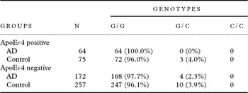
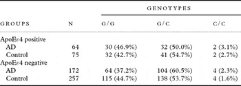
In order to analyze the interaction effects of the ApoE ε4 allele on the association of the NEP genotype with AD, we stratified AD cases and controls according to ApoE genotypes. The results are shown in Tables 3 and 4.
The genotype and allele distribution of NEP −204G/C gene polymorphism in ApoEɛ4 positive and negative carriers

The genotype and allele distribution of NEP 159C/T gene polymorphism in ApoEɛ4 positive and negative carriers

Discussion
Aβ aggregation is of primary importance in the pathogenesis of AD. It was found to trigger a cascade of physical changes such as inflammation by cell activation, oxidative stress, the formation of tau protein, synaptic loss and cognitive decline. However, recent research showed that synaptic dysfunction precedes the formation of plaques and tangles and also that there is weak correlation between the degree of dementia and the distribution of Aβ. Therefore, it may not be a causative factor but rather an associated one in AD.
Previous results have confirmed that mutations in the APP, PS1 and PS2 genes, increasing the production of Aβ, were associated with familial AD (Goate et al., Reference Goate1991; Sherrington et al., Reference Sherrington1995; Rogaev et al., Reference Rogaev1995). Recent attention has been focused on searching for other candidate genes involved in affecting Aβ degradation. NEP, as the major degrading enzyme of Aβ in the brain, and its gene have been investigated with AD across different ethnic groups. Among them, two SNPs, rs989692 and rs3736187 in the NEP gene have been reported to increase risk for AD in the Finnish population (Helisalmi et al., Reference Helisalmi2004). Additionally, NEP159C/T polymorphism has been found to associate with AD among patients younger than 75 years old in a Spanish population (Clarimon et al., Reference Clarimon2003). However, the results have been inconsistent. Notably, a Japanese team has reported lack of association between NEP polymorphisms and AD in Japanese subjects (Sodeyama et al., Reference Sodeyama, Mizusawa, Yamada, Itoh, Otomo and Matsushita2001).
In the present study, we failed to find any association between −204G/C polymorphism and AD in Chinese subjects. This differed from our previous result in a smaller sample, which showed that not only did the frequency of the −204C allele decrease significantly in the patients, but the GC genotype was also associated with decreased risk of AD (Jiajun et al., Reference Jiajun2005). Several reasons might account for such a discrepancy. First, a small sample might decrease the statistical power, which would lead to a false positive result. Second, although it has been shown that the −204G/C polymorphism was located within the putative transcription repressor factor Pax−4 binding region, it remains uncertain whether it has a functional effect. Furthermore, −204G/C polymorphism may be in linkage disequilibrium with other functional variant(s) within the gene or a locus nearby.
We could not replicate the positive association result between NEP159C/T and AD either in subjects younger or older than 75 years, or stratified by gender and ApoE genotype. The plausible explanations might be between-race difference in terms of the frequency of polymorphisms or, alternatively, this polymorphism may be in linkage disequilibrium with a nearby regulatory sequence. Therefore, additional studies with a larger sample size ruling out possible population differences will be required to confirm our results, which parallel those of the Japanese study (Sodeyama et al., Reference Sodeyama, Mizusawa, Yamada, Itoh, Otomo and Matsushita2001).
In conclusion, our results indicate that the two SNPs analyzed here in the NEP gene did not contribute to genetic risk factors for sporadic AD. Exhaustive analyses of other known or still unknown variants and replication of the findings in much larger independent samples and diverse ethnic populations will be necessary to clarify the exact role of the NEP gene with regard to risk for AD.
Conflict of interest
None.
Description of authors' roles
Yan Fu and Jia Jun Shi were responsible for the genotyping, statistical analysis and writing the paper. Mou Ni Tang, Yang Bo Guo, Ai Feng Li and Zhen Huan Zhao were responsible for the subject recruitment. Nelson Leung Sang Tang assisted in the preparation of the paper.
Acknowledgments
This work was supported by the Medical Sci-Tech Research Foundation of Guangdong Province (20041201510107691015217) and grant (JB02-2000202601) from the Science and Technology Committee of Guangzhou. Professor Edmond Chiu, Professor Linda Lam and Professor Nelson Tang provided advice in the revision of this paper.