Introduction
Septoplasty is one of the most common otolaryngological operations. It is often dismissed as a simple procedure, despite the wide range of potential complications.Reference D'Ascanio and Manzini1 Complications that may arise from septal surgery include excessive bleeding, cerebrospinal fluid rhinorrhoea, extra-ocular muscle damage, wound infection, septal abscess, toxic shock syndrome, septal perforation, saddle nose deformity, nasal tip depression, and sensory changes such as anosmia or dental anaesthesia.Reference Ketcham and Han2–Reference Bloom, Kaplan, Bleier and Goldstein6 Rare cases of death after septoplasty have been described, in relation to lesions of the cribriform plate and cerebral arteries.Reference Tawadros and Prahlow7
We report a unique case of unilateral hemiplegia as a complication of septoplasty. To the best of our knowledge, this report represents the first published case of this complication.
Case report
In March 2011, a 51-year-old man was referred to our department from a peripheral hospital because of right-sided hemiplegia presenting immediately after a septoplasty-turbinoplasty surgical procedure carried out under general anaesthesia.
On admittance to our centre, the patient complained of a moderate headache but was well orientated in place and time. Vital parameters were as follows: heart rate, 105 beats per minute; breathing rate, 16 breaths per minute; and blood pressure, 140/95 mmHg.
Bedside neurological examination confirmed the presence of hemiplegia of the patient's right arm and leg. No significant somatosensorial deficit was elicited.
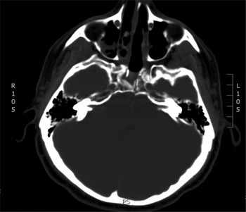
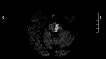
Nasal packing was removed and imaging examinations conducted. Cranial computed tomography showed a breakthrough fracture of the left sphenoid sinus anterior wall and clivus, with posterior dislocation of the fractured clivus wall (Figure 1). The linear course of the clivus breakthrough fracture seemed compatible with osteotome-induced damage. Magnetic resonance imaging showed a haemorrhagic area in the left paramedian pons (Figure 2).
Axial cranial computed tomography image showing the breakthrough fracture of the left sphenoid sinus anterior wall and clivus, with posterior dislocation of the fractured clivus wall. Note the linear course of the clivus breakthrough fracture, compatible with chisel or linear osteotome induced damage. R = right; L = left

Axial diffusion magnetic resonance image (B-1000) showing the haemorrhagic (hyperintense) area in the left paramedian pons. R = right; L = left

Neurosurgical consultation confirmed a cause–effect relationship between the patient's pons haemorrhage and right hemiplegia, and excluded any indication for surgical treatment because of his neurological stability.
Clinical and radiological monitoring was conducted over the next 20 days. Physiotherapeutic rehabilitation was commenced.
One year later, the patient had gained partial recovery of his right leg mobility, but no significant improvement in right arm function was noticed.
Discussion
Nasal obstruction from a deviated septum is one of the most frequent complaints bringing patients to otolaryngologists. Despite the significant number of septoplasties performed each year, severe complications of this procedure are relatively uncommon. The most frequent ones are nasal deformities, infections and septal perforations. Other possible complications include smell disturbance, anterior palate sensory impairment, dental pulp necrosis, cerebrospinal fluid leakage and blindness.Reference Ketcham and Han2–Reference Tawadros and Prahlow7
The case of post-septoplasty unilateral hemiplegia we describe above is, to the best of our knowledge, the only published report of this complication. Based on post-operative imaging, it is likely that the linear clivus breakthrough fracture and subsequent brainstem haemorrhage were caused by the physician's misuse (or loss of control) of the osteotome or chisel, during bony septum surgery.
Satisfactory surgical planning, good intra-operative visualisation and adequate surgical technique are the key factors in preventing complications and achieving a functional nasal airway. In particular, we would emphasise the following ‘learning points’ about osteotome or chisel use during septoplasty: (1) the osteotome or chisel should be used only in the anterior aspect of the nose (maxillary crest), where it can be adequately visualised and manipulated; (2) such instruments do not need to be used to manage the thin vomer and ethmoid in the posterior septum; (3) extreme care must be taken when initially engaging the instrument in the maxillary crest before bone removal; and (4) in general, larger instruments tend to be safer and less likely to dislodge and inadvertently move posteriorly when the mallet is applied.
• A unique case is presented of unilateral hemiplegia as a complication of septoplasty
• This risk should be considered intra-operatively and during the informed consent process
In addition, the septoplasty surgeon must be aware of all the possible complications that may arise during septal surgery, and should discuss all these risks with the patient as part of the informed consent process.
Acknowledgement
The support of Drs Marco Manzini and Piergiorgio Volpini is greatly acknowledged.