Introduction
Major depressive disorder (MDD) is a prevalent psychiatric disorder associated with high morbidity and mortality, frequently characterized by a chronic or recurrent course (Kessler et al., Reference Kessler, Berglund, Demler, Jin, Merikangas and Walters2005). Biased emotional memory has been proposed as a key factor for the development and maintenance of MDD (Leppänen, Reference Leppänen2006; Disner et al., Reference Disner, Beevers, Haigh and Beck2011; Ai et al., Reference Ai, Opmeer, Veltman, van der, Nic, van Buchem, Aleman and van Tol2015; Everaert et al., Reference Everaert, Duyck and Koster2015) and may even underlie the vulnerability for depressive psychopathology (Chan et al., Reference Chan, Goodwin and Harmer2007). Cross-sectional studies suggested that emotional memory biases are state-independent phenomena: better memory for negative information and worse memory for positive information have been reported during both the acute depressive state and during remission [reviewed elsewhere (Bradley and Mathews, Reference Bradley and Mathews1988; Elliott et al., Reference Elliott, Zahn, Deakin and Anderson2010)], mirroring functional brain abnormalities observed in areas critical for memory formation of emotional material, i.e. the amygdala and hippocampus (Ramel et al., Reference Ramel, Goldin, Eyler, Brown, Gotlib and McQuaid2007; Arnold et al., Reference Arnold, Fitzgerald, Fernández, Rijpkema, Rinck, Eling, Becker, Speckens and Tendolkar2011; van Tol et al., Reference van Tol, Demenescu, van der Wee, Kortekaas, Marjan, Boer, Renken, van Buchem, Zitman and Aleman2012). Previously, we however observed hyperactivation of the anterior hippocampus/amygdala during encoding of negative information in acutely depressed patients but not in remitted patients in a cross-sectional comparison (van Tol et al., Reference van Tol, Demenescu, van der Wee, Kortekaas, Marjan, Boer, Renken, van Buchem, Zitman and Aleman2012), suggesting state-dependency instead. However, cross-sectional studies do not allow strong inferences on state-dependency. Importantly, identifying state-dependent neurocognitive markers of MDD may constitute a first step in understanding mechanisms of recovery v. maintenance of depression (Mayberg, Reference Mayberg1997; Maalouf et al., Reference Maalouf, Clark, Tavitian, Sahakian, Brent and Phillips2012; Dohm et al., Reference Dohm, Redlich, Zwitserlood and Dannlowski2017).
While longitudinal neuropsychological studies have found that memory biases resolve upon recovery after treatment (Calev et al., Reference Calev, Korin, Shapira, Kugelmass and Lerer1986; Peselow et al., Reference Peselow, Corwin, Fieve, Rotrosen and Cooper1991) (though not consistently in Sternberg and Jarvik, Reference Sternberg and Jarvik1976), functional neuroimaging studies reported mostly changes in activation of the amygdala and hippocampus following symptomatic improvement during affective processing (i.e. not in the context of memory processing) or rest. Findings have been inconclusive with reports of decreased (Sheline et al., Reference Sheline, Barch, Donnelly, Ollinger, Snyder and Mintun2001; Fu et al., Reference Fu, Williams, Cleare, Brammer, Walsh, Kim, Andrew, Pich, Williams and Reed2004; Redlich et al., Reference Redlich, Bürger, Dohm, Grotegerd, Opel, Zaremba and Dannlowski2017), increased (Goldapple et al., Reference Goldapple, Segal, Garson, Lau, Bieling, Kennedy and Mayberg2004; Neumeister et al., Reference Neumeister, Drevets, Belfer, Luckenbaugh, Henry, Bonne, Herscovitch, Goldman and Charney2006; Victor et al., Reference Victor, Furey, Fromm, Öhman and Drevets2010; Ritchey et al., Reference Ritchey, Dolcos, Eddington, Strauman and Cabeza2011), or unchanged (Fu et al., Reference Fu, Costafreda, Sankar, Adams, Rasenick, Liu, Donati, Maglanoc, Horton and Marangell2015; Opmeer et al., Reference Opmeer, Kortekaas, Tol, Renken, Demenescu, Woudstra, Ter Horst, Buchem, der Wee and Veltman2015) activation following successful short-term pharmacological treatment (Sheline et al., Reference Sheline, Barch, Donnelly, Ollinger, Snyder and Mintun2001; Fu et al., Reference Fu, Williams, Cleare, Brammer, Walsh, Kim, Andrew, Pich, Williams and Reed2004; Victor et al., Reference Victor, Furey, Fromm, Öhman and Drevets2010; Fu et al., Reference Fu, Costafreda, Sankar, Adams, Rasenick, Liu, Donati, Maglanoc, Horton and Marangell2015), electroconvulsive therapy (Redlich et al., Reference Redlich, Bürger, Dohm, Grotegerd, Opel, Zaremba and Dannlowski2017), cognitive behavioral treatment (Fu et al., Reference Fu, Williams, Cleare, Scott, Mitterschiffthaler, Walsh, Donaldson, Suckling, Andrew, Steiner and Murray2008; Goldapple et al., Reference Goldapple, Segal, Garson, Lau, Bieling, Kennedy and Mayberg2004; Ritchey et al., Reference Ritchey, Dolcos, Eddington, Strauman and Cabeza2011), or naturalistic remission (Opmeer et al., Reference Opmeer, Kortekaas, Tol, Renken, Demenescu, Woudstra, Ter Horst, Buchem, der Wee and Veltman2015). Heterogeneity in findings may be partly explained by methodological factors such as small sample size, type of stimuli, effects of the (pharmacological) treatment itself on blood flow, or clinical variation in terms of comorbidity or interval between pre- and post-measurement. Nevertheless, the effects of symptomatic improvement on the neural underpinnings of emotional memory processing have not been studied to date.
Because duration of depression has been associated with more severe structural abnormalities, especially in the hippocampus (MacQueen et al., Reference MacQueen, Campbell, McEwen, Macdonald, Amano, Joffe, Nahmias and Young2003; Frodl et al., Reference Frodl, Koutsouleris, Bottlender, Born, Jäger, Scupin, Reiser, Möller and Meisenzahl2008; Schmaal et al., Reference Schmaal, Veltman, van Erp, Sämann, Frodl, Jahanshad, Loehrer, Tiemeier, Hofman and Niessen2015), persistence of depressive symptoms may be an important additional factor that influences activation of brain areas important for encoding of emotional information. Such ‘persistence’ effects may be related to glucocorticoid-dependent toxic effects of stress (Fossati et al., Reference Fossati, Radtchenko and Boyer2004) and may result in explicit memory deficits (Sapolsky, Reference Sapolsky2000). On a functional level, medial prefrontal involvement during the processing of autobiographical memory was found to be blunted in remitted MDD patients but not in individuals at high risk for developing MDD (Young et al., Reference Young, Bellgowan, Bodurka and Drevets2015), suggesting that memory deficits may be a consequence of having experienced a depressive episode. However, to our knowledge, it has not yet been investigated whether persistence of symptoms may modulate longitudinal functional brain changes related to memory formation.
In the present longitudinal imaging study, we aimed to investigate whether changes in activation of the amygdala and hippocampus during emotional memory encoding are dependent on changes in depressive state and time spent with depressive symptoms. Healthy and depressed participants underwent functional magnetic resonance imaging (fMRI) twice in the context of the naturalistic and observational Netherlands Study of Depression and Anxiety (NESDA) study, with approximately 2 years in between. In this interval, no specific treatment was delivered as part of the study protocol. Given the naturalistic design of our study, participants could receive treatment as usual, which was reconstructed retrospectively based on self-reports at the 2-year follow-up interview. We hypothesized that changes in activation in the hippocampus/amygdala are (1) associated with a change in depressive state, especially during negative word encoding and (2) affected by time spent with depressive symptoms between measurements. Furthermore, we aimed to explore whether activation in regions other than amygdala and hippocampus related to longitudinal treatment responses was associated with severity and time spent with depressive symptoms.
Methods and materials
Participants
Participants were recruited from the ongoing neuroimaging sub-study of the NESDA (Penninx et al., Reference Penninx, Beekman, Smit, Zitman, Nolen, Spinhoven, Cuijpers, De Jong, Van Marwijk and Assendelft2008) and underwent fMRI scanning at the University Medical Center Groningen (UMCG), Academic Medical Center (AMC) of the University of Amsterdam, and the Leiden University Medical Center (LUMC). NESDA has been designed as a longitudinal observational cohort study with measurements at baseline, 1-, 2-, 4-, 6-, and 9-year follow-up, with MRI measurements performed in a subsample at baseline, 2- and 9-year follow-up (9-year follow-up measurement was completed during the preparation of this manuscript). At baseline, patients with MDD (n = 70), MDD and one or more anxiety disorders [i.e. social anxiety disorder (SAD), panic disorder (PD), and/or generalized anxiety disorder (GAD); N = 92], patients with only anxiety disorders (i.e. SAD, PD, and/or GAD; n = 71), and healthy control participants (HC; n = 68) were included. The ethical review board of each participating center approved the study and all participants gave written informed consent.
Exclusion criteria for all participants in the NESDA neuroimaging study at baseline (n = 301) were: age under 18 or over 57 years; current alcohol or substance abuse; presence or history of a neurological or somatic disorder with possible effects on the central nervous system; general 3T MRI contraindications; hypertension. Use of selective serotonin reuptake inhibitors (SSRIs) or infrequent use of benzodiazepines [oxazepam (max 20 mg) or diazepam, maximum of three times a week and not within 48 h before scanning] was allowed. Patients using any other psychopharmacological agent were excluded. Exclusion criteria for the second measurement at 2-year follow-up (S2; N = 199) were identical, with the exception of the age criterion. Also, from a cohort perspective, we were less strict in excluding patients based on the type of medication used at S2 (see Table 1 and online Supplementary Table S1 for details). In line with the observational nature of the NESDA study, no specific treatment was delivered in between measurements, but was monitored retrospectively. Participants were free to consult their general practitioner, psychiatrist or psychologist for the help they wished to receive. Results of the baseline measurement (S1) and their associations with subsequent course related to emotional memory processing have been published elsewhere (van Tol et al., Reference van Tol, Demenescu, van der Wee, Kortekaas, Marjan, Boer, Renken, van Buchem, Zitman and Aleman2012; Ai et al., Reference Ai, Opmeer, Veltman, van der, Nic, van Buchem, Aleman and van Tol2015).
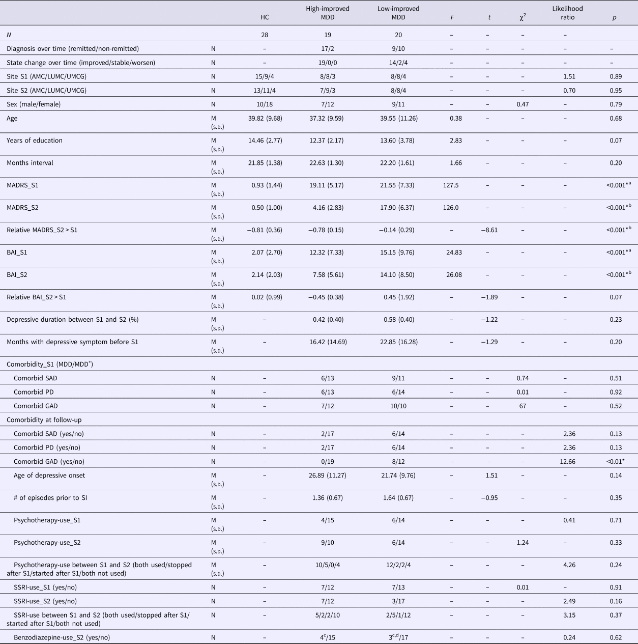
Demographics characteristics

HC, healthy control; S-R, symptom-remitted MDD patients; S-S, symptomatic-symptomatic MDD patients; SAD, social anxiety disorder; PD, panic disorder; GAD, generalized anxiety disorder.
a HC differed from both patient groups, while the two patient groups did not differ.
b All groups differed from each other.
c Infrequent use.
d Two patients used benzodiazepine frequently.
* Significant at p < 0.05.
Complete behavioral data and good quality fMRI data at both S1 and S2 were available for 64 MDD patients and 39 HC. At S1, all patients fulfilled the criteria for a diagnosis of MDD with a half-year recency based on the Composite International Diagnostic Interview (CIDI life time – version 2). An additional diagnosis of SAD, PD, and/or GAD at either S1 or S2 was allowed (see Table 1 for details). Following Opmeer et al. (Reference Opmeer, Kortekaas, Tol, Renken, Demenescu, Woudstra, Ter Horst, Buchem, der Wee and Veltman2015), we included only patients who were in a depressive state at S1 defined as a Montgomery–Åsberg Depression Rating Scale (MADRS) score >10 (Zimmerman et al., Reference Zimmerman, Posternak and Chelminski2004). One participant had a huge increase in MADRS score at S2 and was classified as an outlier (change score >3SD from group mean) and subsequently excluded from the analyses. The final patient sample included 39 individuals. In total, 11 HC were excluded from further analysis based on the presence of possible depressive symptomatology at S2 (i.e. MADRS-score >10; n = 1), too high level of education to be matched to the patient group (n = 1), or unreliable task performance (n = 9; online Supplementary Fig. S1). This resulted in the inclusion of 28 HC without any current or life-time DSM-IV diagnosis and no indication of depressive symptomatology at both S1 and S2 (see online Supplementary Fig. S1 for a flow diagram reflecting data selection).
Task paradigm
All participants performed the event-related, subject-paced, emotional word encoding and recognition task during both fMRI scanning sessions (S1 and S2) (van Tol et al., Reference van Tol, Demenescu, van der Wee, Kortekaas, Marjan, Boer, Renken, van Buchem, Zitman and Aleman2012). During the encoding phase, 20 blocks containing 160 stimuli (positive/neutral/negative words and baseline trials; 40 each) were pseudo-randomly presented. Participants were instructed to evaluate whether the word was positive, negative, or neutral in valence by pressing the right, left, and middle button, respectively. During baseline trials, participants were asked to press the corresponding button to indicate the direction of the arrow. After a retention interval of 10 min (during which the structural T1 scan was acquired), the retrieval phase started and consisted of 120 encoding target words, 120 distracter words, and 40 baseline words that were presented in 20 pseudo-randomized blocks. Participants were instructed to indicate whether they had seen, had not seen, or probably had seen the word. Emotional words in the valence categories were matched based on length, frequency in the Dutch language, and complexity. The same words list was used in both measurements, although the order was changed at the 2-year follow-up measurement. The emotional word encoding task was preceded by an executive planning task (van Tol et al., Reference van Tol, van der Wee, Demenescu, Nielen, Aleman, Renken, van Buchem, Zitman and Veltman2011) and followed by an emotional face viewing task (Demenescu et al., Reference Demenescu, Renken, Kortekaas, van Tol, Marsman, van Buchem, van der Wee, Veltman, den Boer and Aleman2011; Opmeer et al., Reference Opmeer, Kortekaas, Tol, Renken, Demenescu, Woudstra, Ter Horst, Buchem, der Wee and Veltman2015) and a resting state acquisition (Veer et al., Reference Veer, Beckmann, van Tol, Ferrarini, Milles, Veltman, Aleman, van Buchem, van der Wee and Rombouts2010). Based on the hypotheses formulated in our cross-sectional study (van Tol et al., Reference van Tol, Demenescu, van der Wee, Kortekaas, Marjan, Boer, Renken, van Buchem, Zitman and Aleman2012), we only investigated the encoding session.
fMRI data acquisition
Neuroimaging data were collected with 3T Philips MR-scanners located in Amsterdam, Leiden, and Groningen using standard EPI techniques, though with minor differences in acquisition parameters. A detailed description of acquisition specifications can be found in the online Supplementary material.
Data analysis
Independent variables
Firstly, to test for the correlation between symptom change and brain activation change over time, a relative symptom change score representing the difference in depression severity between S1 and S2 while taking into account baseline severity was calculated for each patient [i.e. (MADRS S2–MADRS S1)/MADRS S1]. Furthermore, to be able to compare changes over time in behavior and brain activation following symptomatic change with changes in HC, who were also scanned twice, and to explore, e.g. whether change in the high improved patients represented normalization (i.e. approached activation of HC at S2) or whether change in low improved patients represented further deviations from normal, we divided the patients in two groups based on the median of relative symptom change scores (median = −0.46): a group of high improved (MDD-HI; n = 20, online Supplementary Fig. S1) and a group of low improved patients (MDD-LI; n = 19).
Secondly, to test for the correlation between brain activation change and percentage of time spent with depression (i.e. persistence), presence of depressive symptoms per month for the duration of the interval between S1 and S2 was assessed with the life chart interview (Lyketsos et al., Reference Lyketsos, Nestadt, Cwi and Heithoff1994) at S2. Participants had to rate the severity of depressive symptoms per month and only symptoms with small to severe burden were taken as an indication of the presence of symptoms. Percentage of months experiencing depressive symptoms relative to the overall follow-up period was calculated per patient as the time spent with depression (Ai et al., Reference Ai, Opmeer, Veltman, van der, Nic, van Buchem, Aleman and van Tol2015).
Clinical variables and behavioral data
Effects of symptom change and time spent with depressive symptoms on demographic, psychometric assessment, and memory performance were analyzed in IBM SPSS software (SPSS v.22.0, IBM). We employed analyses of covariance (ANCOVA), χ2 tests and t tests where appropriate for demographic and psychometric data with a significance level of p < 0.05, two-tailed.
For the behavioral data, performance difference scores (S2 − S1) for both reaction times (RT) and accuracy for successfully encoded words (Tulving, Reference Tulving1985) were calculated. We assessed the continuous association between relative symptom change scores and depressive duration, and RT and accuracy difference scores over time in patients. Age and years of education were included as covariates. A sensitivity analysis was performed within patients who showed symptomatic improvement (thus, patients who were equally or more depressed at S2 than at S1 were excluded; n = 6).
Additionally, to investigate whether patients (MDD-HI/LI) performed differently over time as compared to HC, we set up a group (3; HC, HI, LI) × valence (3; positive, negative, neutral) × time (2; S1, S2) repeated-measures ANCOVA, with age and years of education as covariates. Effects were considered significant at p < 0.05. Where appropriate, Bonferroni correction for multiple comparisons was applied.
Imaging data preprocessing
For the fMRI data, preprocessing and task modeling was performed with Statistical Parametric Mapping software (SPM8, Wellcome Trust Center for Neuroimaging, http://www.fil.ion.ucl.ac.uk/spm) implemented in Matlab 7.8 (The Math Works Inc., Natick, MA, USA). A detailed description of the preprocessing steps and first-level modeling can be found in the online Supplementary material.
Effects of change of depressive state
To test for the association between symptom change and change of brain activation during positive and negative encoding over the 2-year interval, scan moments, S2 − S1 contrast maps were entered as dependent variables in a full-factorial model, with valence [successfully encoded positive words>successfully encoded neutral words (S2 − S1), successfully encoded negative words>successfully encoded neutral words (S2 − S1)] as interacting factor with valence. Contrast maps were built for the successful encoding of positive words (v. successful encoding of neutral words) and negative words separately. To control for the possible confounding effects of variations within and between participants in scanning site (which coincided with minor variations in sequence and coil; see online Supplementary material), four dummy variables for site (i.e. both times scanned in AMC; changed from AMC to LUMC; changed from LUMC to AMC; both times scanned in UMCG; both times scanned in LUMC) were defined as covariates of no interest. In addition, age and years of education at S1 were added as covariates.
We repeated our analysis with the following possible confounding factors added separately to the model: percentage of time spent with depression, relative changes in anxiety severity assessed by Beck Anxiety Inventory [(BAI-scores S2 − S1)/BAI-scores S1] (Beck et al., Reference Beck, Epstein, Brown and Steer1988), SSRI-use, and participation in psychotherapy. SSRI-use at/between S1 and S2 was added to the model by means of three dummy variables (used at both S1 and S2, started after S1, stopped after S1, both not used). Psychotherapy-use between S1 and S2 was coded as a dummy variable and added as a covariate to test for the effect of psychotherapy. Use of SSRI and psychotherapy between S1 and S2 are summarized in online Supplementary Table S1.
A sensitivity analysis was planned to test whether associations would hold in the analysis including only patients with symptomatic improvement (n = 33).
Effects of the persistence of depressive symptoms
We built a full factorial model with valence as factor (2; successfully encoded positive words>successfully encoded neutral words and successfully encoded negative words>successfully encoded neutral words) and time spent with depressive symptoms as an interacting covariate with valence. Site (four dummy variables), age, and years of education were added as covariates. We tested for the effects of time spent with depressive symptoms during encoding of positive words and negative words separately. In a subsequent step, relative symptom change of depressive and anxiety symptom severity and treatment at S1 and S2 (medication- and psychotherapy use; yes/no) were added separately as covariates to statistically control for their possible confounding effects.
A sensitivity analysis was planned within patients with symptomatic improvement.
Statistical thresholding
Based on previous studies (see Introduction), we a priori defined the bilateral hippocampus and amygdala as our regions-of-interest (ROI) and built one composite mask encompassing these regions. The regions were defined according to the automated anatomical labels of the Wake Forest University (WFU, Winston Salem, North Carolina) Pick Atlas toolbox. Small volume correction for multiple comparisons was applied within the ROI. F-tests in the main and follow-up analyses were explored separately for positive and negative words at p < 0.001 uncorrected. Post hoc t tests were regarded significant at a threshold of p < 0.05 family wise error (FWE) corrected at voxel-level (with an initial threshold of p < 0.001 uncorrected). We also examined the effects in other brain regions than ROIs, which had to meet p < 0.05, FWE whole-brain corrected to be considered significant.
Results
Demographic characteristics
Demographics and clinical characteristics of all patients and HCs are summarized in Table 1 and online Supplementary material. Clinical characteristics of high-improved and low-improved patient groups that were included in explorative post hoc analyses are listed in online Supplementary Table and results. Thirty-three patients showed symptomatic improvement (S2 < S1), two remained stable (S2 = S1), and four showed more severe symptoms at S2 (S2 > S1).
Behavioral results
No correlations were found between relative depressive symptom change and changes in performance on the memory of positive, neutral, or negative words over time (i.e. RTs and accuracy) (p > 0.05). Sensitivity analyses within symptomatically improved patients only (n = 33) did not change this result. Group × time repeated-measures analysis of variance indicated no changes in the performance and response times in HC nor a difference between HC and HI or LI (ps > 0.05).
There was no association between time spent with depressive symptoms and changes in behavioral performance (p > 0.05).
fMRI results
Correlations with change of depressive state
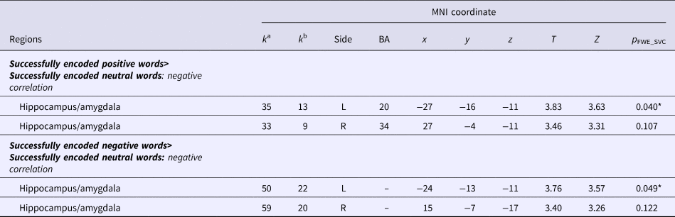
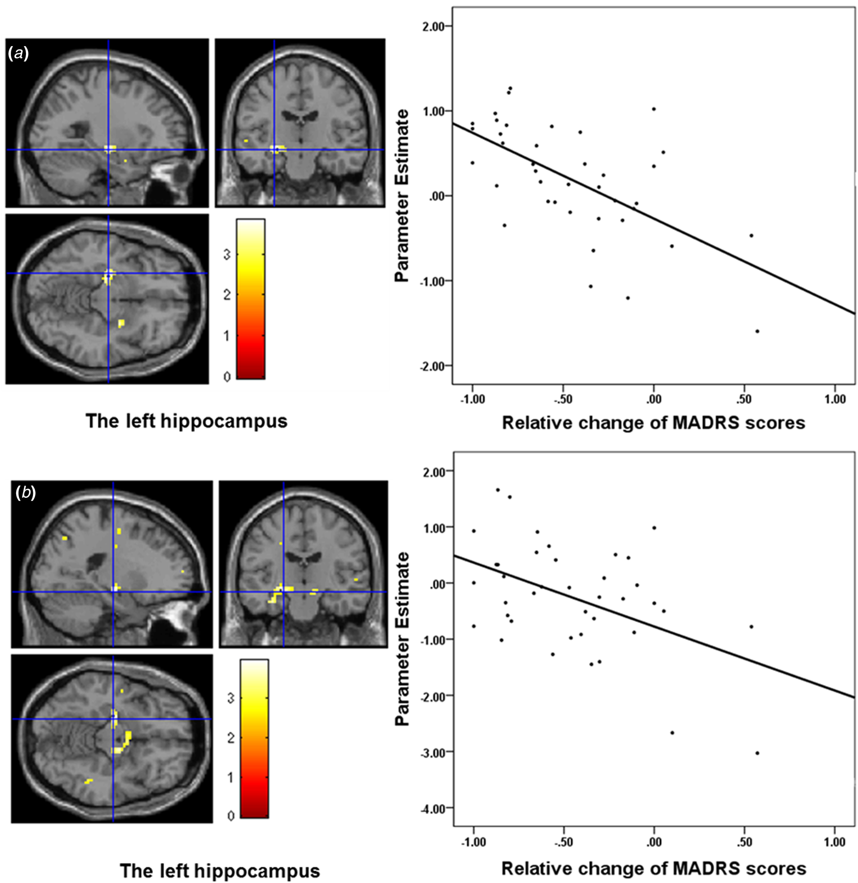
Relative symptom change was negatively correlated with activation change in the bilateral hippocampal/amygdala during both positive and negative word encoding (Table 2; Fig. 1). However, only the effect in the left hippocampus survived multiple comparison correction and indicated that larger symptomatic improvement coincided with a larger increase in left anterior hippocampal activation during encoding of emotional information.
Correlation between state-change scores and brain activation changes across patients

a Cluster size in whole-brain analysis.
b Cluster size after small volume correction.
* Significant at p < 0.05 FWE corrected, voxel-level after small volume correction (SVC).
Brain activation during emotional word encoding. (a) Negative association between symptom change and hippocampal activation change during positive word encoding (peak MNI coordinate: x = −27, y = −16, z = −11); (b) negative association between symptom change and hippocampal activation change during negative word encoding (peak MNI coordinate: x = −24, y = −13, z = −11).

Adding time spent with depressive symptoms in the interval between S1 and S2 as covariate did not change the results [Z = 3.85, p FWE = .019 for successfully encoded positive words>successfully encoded neutral words (pos); Z = 3.95, p FWE = .014 for successfully encoded negative words>successfully encoded neutral words (neg)]. Also, results were not affected by including change in anxiety severity as a covariate to the model (pos: Z = 3.82, p FWE = .021; neg: Z = 3.77, p FWE = .025) or by adding SSRI-use at S1 and S2 as covariates (pos: Z = 3.68, p FWE = 0.034; neg: Z = 3.49, p FWE = 0.06). Results bordered statistical significance after adding psychotherapy as a covariate (pos: Z = 3.59, p FWE = 0.05; neg: Z = 3.54, p FWE = 0.05).
When repeating the analysis in the symptomatically improved patients only, the negative correlation between symptom change and brain activation change in the hippocampus was observed subthreshold [MNI coordinates:(x = −18, y = −13, z = −11), Z = 3.51, p FWE = .09] during positive encoding, and was not significant during negative encoding (p FWE = 0.50).
Furthermore, post hoc group comparison (detailed in the online Supplementary methods and results) showed that activation estimates in our main cluster did not change in HC over time, and plots suggested a trend of normalization during positive but not negative word encoding in the high-improved group (online Supplementary Fig. 3A and 3B).
Correlations with time spent with depressive symptoms and course
No correlation between the percentage of time with depressive symptoms and changes in brain activation was observed across all MDD patients during successful encoding on positive and negative words. Adding change in depressive and anxiety symptoms or medication/therapy use to the model did not change this observation.
Discussion
In this longitudinal study, we examined changes in emotion-related brain activation over time associated with symptomatic improvement and time spent with depressive symptoms in depressed patients. Symptomatic improvement was associated with increased responses in the anterior hippocampus/amygdala during encoding of emotional stimuli over time. Follow-up explorations indicated that increased activation of the hippocampal/amygdala responsiveness occurred in the direction of normalization, especially for the encoding of positive words. The effect was unrelated to changes in anxiety severity, and use of SSRIs, although it became smaller after adding use of psychotherapy as a covariate. No relation was observed between depression duration (i.e. time spent with depressive symptoms) in the 2-year follow-up nor were changes in hippocampal and amygdalar activation observed. These results suggest that hippocampal activation during emotional memory formation changes with symptomatic improvement, but is not subject to functional ‘scarring’ as a result of enduring symptom manifestation. Our results indicate that symptomatic improvement is at least partially associated with normalization of limbic responsiveness to positive material.
Based on previous reports on memory bias-related brain activation abnormalities in depression (Ramel et al., Reference Ramel, Goldin, Eyler, Brown, Gotlib and McQuaid2007; Hamilton and Gotlib, Reference Hamilton and Gotlib2008; Van Wingen et al., Reference Van Wingen, Van Eijndhoven, Cremers, Tendolkar, Verkes, Buitelaar and Fernández2010; Arnold et al., Reference Arnold, Fitzgerald, Fernández, Rijpkema, Rinck, Eling, Becker, Speckens and Tendolkar2011; van Tol et al., Reference van Tol, Demenescu, van der Wee, Kortekaas, Marjan, Boer, Renken, van Buchem, Zitman and Aleman2012) and our previous cross-sectional observations (van Tol et al., Reference van Tol, Demenescu, van der Wee, Kortekaas, Marjan, Boer, Renken, van Buchem, Zitman and Aleman2012), we hypothesized state-dependency of activation of the amygdala and hippocampus specific for negative valence information, and thus changes of activation as a function of symptomatic recovery. In line with this hypothesis, hippocampal reactivity during negative encoding correlated with symptomatic change. Moreover, state-dependency was observed during positive encoding. Although similar linear relations with symptomatic improvement were observed for both positive and negative encoding, changes during positive word encoding showed to be a more specific indicator of symptomatic improvement. This was indicated by the stability of effects when excluding the patients that worsened in terms of symptom severity and by the fact that the post hoc plotting of effects indicated an increase of activation in the improved patients only. This increase followed a pattern of normalization (i.e. approaching activation in the HC). During negative encoding, associations were no longer significant when studied in the symptomatic improved patients only. This suggests that state-dependent changes during positive encoding may be a preferred marker of symptomatic improvement. Notwithstanding, although longitudinal studies did not study emotional encoding for both positive and negative information so far, our study supports findings of altered reactivity to positive information (Fu et al., Reference Fu, Williams, Brammer, Suckling, Kim, Cleare, Psych, Walsh, Mitterschiffthaler and Andrew2007; Victor et al., Reference Victor, Furey, Fromm, Öhman and Drevets2010; Wise et al., Reference Wise, Cleare, Herane, Young and Arnone2014), and suggests normalized reactivity to positive-related effects.
The hippocampus has been proposed as a target for both anti-depressant treatment and cognitive behavioral therapy (CBT) (Goldapple et al., Reference Goldapple, Segal, Garson, Lau, Bieling, Kennedy and Mayberg2004). Treatment studies have confirmed the importance of the hippocampus by consistently reporting normalization of hippocampal activation following pharmacological treatment (Fu et al., Reference Fu, Williams, Cleare, Brammer, Walsh, Kim, Andrew, Pich, Williams and Reed2004; Anand et al., Reference Anand, Li, Wang, Gardner and Lowe2007; Arnone et al., Reference Arnone, McKie, Elliott, Thomas, Downey, Juhasz, Williams, Deakin and Anderson2012b) and CBT (Goldapple et al., Reference Goldapple, Segal, Garson, Lau, Bieling, Kennedy and Mayberg2004; Ritchey et al., Reference Ritchey, Dolcos, Eddington, Strauman and Cabeza2011). In the current study, we studied the neural characteristics related to naturalistic changes in a depressive state, which was not attributable to treatment with antidepressant medication. However, most of our sample received at least one type of psychological care. Therefore, we cannot fully rule out of the effect of psychotherapy and indeed our effects were slightly attenuated when treatment with psychotherapy was added to the model. Together, our observations suggest that increased hippocampal responsiveness to emotional material may not only reflect treatment effects of or symptomatic improvement following anti-depressants or psychological treatment (Fu et al., Reference Fu, Williams, Brammer, Suckling, Kim, Cleare, Psych, Walsh, Mitterschiffthaler and Andrew2007; Victor et al., Reference Victor, Furey, Fromm, Öhman and Drevets2010; Wise et al., Reference Wise, Cleare, Herane, Young and Arnone2014) but also naturalistic improvement.
No other regions were found to change as a function of symptomatic improvement. Although changes in regions such as the ventromedial prefrontal cortex (Ritchey et al., Reference Ritchey, Dolcos, Eddington, Strauman and Cabeza2011), anterior cingulate cortex (Fu et al., Reference Fu, Williams, Cleare, Scott, Mitterschiffthaler, Walsh, Donaldson, Suckling, Andrew, Steiner and Murray2008), frontal pole (Usami et al., Reference Usami, Iwadare, Kodaira, Watanabe and Saito2014), and the extrastriate cortex (Fu et al., Reference Fu, Williams, Brammer, Suckling, Kim, Cleare, Psych, Walsh, Mitterschiffthaler and Andrew2007) have been reported by previous longitudinal treatment studies. They have been reported in the context of emotional processing, but not in the context of memory formation or using verbal stimuli. Additionally, other studies have reported that prefrontal alterations might be a trait marker rather than a state marker of vulnerability to depression (Elliott et al., Reference Elliott, Lythe, Lee, McKie, Juhasz, Thomas, Downey, Deakin and Anderson2012; Tomioka et al., Reference Tomioka, Yamagata, Kawasaki, Pu, Iwanami, Hirano, Nakagome and Mimura2015), which was not the focus of our study.
The second aim of this study was to investigate whether time spent with depressive symptoms was associated with greater functional brain alterations during emotional memory encoding. We found that depression duration was not correlated with changes of activation in the hippocampus, which indicates that the neurotoxic or scarring hypothesis might not be relevant to functional changes over time. Previous cross-sectional and longitudinal studies suggested that hippocampal volume is negatively related to the duration of illness in MDD, represented by the history of psychiatric hospitalization (Zaremba et al., Reference Zaremba, Dohm and Redlich2018), number of episodes (MacQueen et al., Reference MacQueen, Campbell, McEwen, Macdonald, Amano, Joffe, Nahmias and Young2003; Treadway et al., Reference Treadway, Waskom, Dillon, Holmes, Park, Chakravarty, Dutra, Polli, Iosifescu, Fava, Gabrieli and Pizzagalli2015), and duration of untreated illness (Sheline et al., Reference Sheline, Sanghavi, Mintun and Gado1999), though not consistently (Bremner et al., Reference Bremner, Narayan, Anderson, Staib, Miller and Charney2000; McKinnon et al., Reference McKinnon, Yucel, Nazarov and MacQueen2009). At the same time, volumetric changes in the hippocampus have been linked to symptomatic improvement following treatment (Arnone et al., Reference Arnone, McKie, Elliott, Juhasz, Thomas, Downey, Williams, Deakin and Anderson2012a), suggesting state-dependency of hippocampal volume. In the present study, though patients differed in course trajectory of depression, changes of brain activation were not related to the depressive course, indicating that functional longitudinal changes observed in the hippocampus are load-independent. However, the variety in selected clinical variables of current and previous studies might explain some heterogeneity in reported results. Together, our results indicate that functional responsiveness of limbic brain regions may be more related to a depressive state, without exacerbation of abnormalities as a function of unfavorable course of the depression.
Some limitations of our study should be noted. First, although clear strengths of our study are its longitudinal naturalistic design and that we could control for activation changes over the same interval in a healthy sample, the associations we found between changes of brain activation and symptom change over time are correlational in nature and do not imply causation of remission in depression. This effect was not found in a formal group × time × valence interaction. However, testing this was not the aim of our paper because we focused on changes over time within depressed patients. Second, we investigated symptom severity change of depression rather than symptom remission. Although most of our high-improved patients were recovered at the time of the follow-up measurement, our conclusions cannot be generalized to changes associated with stable remission. Third, although adding SSRI-use and psychotherapy use as covariates to the model did not change the observed relations, this does not fully rule out specific medication/treatment effects. Fourth, caution should be taken in interpreting our result as a true memory effect (i.e. hits–misses), because the number of error trials was too low to investigate this. More sensitive measures on behavioral changes in primary emotional and memory processing are necessary in future studies. Fifth, although the site effect was controlled by adding it as a covariate, it might still have a confounding effect on our results. Quality assurance analysis and exploration by excluding patients that switched scanners between measurements (online Supplementary results) revealed similar results. These indicate that our observed effects, especially those observed during positive encoding, were not primarily driven by site-specific changes in signal over time. Next, the retrospective life chart method used to measure the persistence of depressive symptoms might have been subject to patients' mood state, though the reliability and validity have been estimated to be relatively high (Warshaw, et al., Reference Warshaw, Dyck, Allsworth, Stout and Keller2001). Furthermore, although comorbidity of SAD and PD was similar in low- and high-improved MDD groups, GAD was more frequent in low-improved MDD patients, which may have affected our results. Finally, it is possible that the encoding processing was more explicit at S2 than at S1, because people at S2 could have remembered that a recognition phase followed the encoding phase. However, implicit and explicit memory processing have been suggested to be subject to the same encoding factors and rely on similar perceptual processes and representations (Turk-Browne et al., Reference Turk-Browne, Yi and Chun2006), which is corroborated by the lack of differences over time in the HC group in our study.
Conclusion
By characterizing longitudinal changes of activation in the anterior hippocampus/amygdala during emotional memory encoding, our study showed that the neural correlates of emotional memory formation change with the improvement of the depressive state. Furthermore, our findings suggest a normalization of activation, especially for positive information. On the other hand, enduring depressive symptom manifestation was not related to longitudinal changes in hippocampal–amygdalar activation. Taken together, our results suggest that hippocampal activation is a state-dependent characteristic that is not related to the persistence of depression. This may indicate that functional activation patterns in depression are not subject to functional ‘scarring’, a hypothesis that deserves further investigation.
Supplementary material
The supplementary material for this article can be found at https://doi.org/10.1017/S0033291719001259.
Acknowledgments
This study is supported by the participating universities and mental health care organizations: VU University Medical Center Amsterdam, University Medical Center Groningen, Leiden University Medical Center, GGZ inGeest, Arkin, GGZ Rivierduinen, Lentis, GGZ Friesland, GGZ Drenthe, Scientific Institute for Quality of Healthcare (IQ healthcare), The Netherlands Institute for Health Services Research (NIVEL), and The Netherlands Institute of Mental Health and Addiction (Trimbos Institute).
Financial support
Hui Ai is jointly supported by the National Natural Science Foundation of China (grant no. 31700959 and 31871137), Young Elite Scientists Sponsorship Program by CAST, YESS, Shenzhen Peacock Program (827-000235 and KQTD2015033016104926) and Guangdong Pearl River Talents Plan for Postdoctoral researchers. Marie-José van Tol was supported by a VENI grant from the Netherlands Organization for Scientific Research (N.W.O.) (grant no. 016.156.077). Esther Opmeer is partly supported by a grant from N.W.O. ZonMw (grant no. 537001005). The infrastructure for the NESDA study (http://www.nesda.nl) is funded through the Geestkracht program of the Netherlands Organisation for Health Research and Development (ZonMw, grant no. 10-000-1002) and financial contributions by participating universities and mental health care organizations (VU University Medical Center, GGZ inGeest, Leiden University Medical Center, Leiden University, GGZ Rivierduinen, University Medical Center Groningen, University of Groningen, Lentis, GGZ Friesland, GGZ Drenthe, Rob Giel Onderzoekscentrum).
Conflict of interest
Hui Ai, Jan-Bernard C. Marsman, Dick J. Veltman, and Esther M. Opmeer declare no conflict of interest. Nic van der Wee received speaking fees from Eli Lilly and Wyeth; and served on advisory panels of Eli Lilly, Pfizer, Wyeth, and Servier. Marie-José van Tol received speakers fees from Lundbeck n.v. André Aleman received an investigator-initiated unrestricted research grant from Bristol-Myers Squibb and speakers bureau honoraria from Lundbeck n.v. All of these activities are not directly related to the present study and, therefore, do not form a conflict of interest.
Ethical standard
The authors assert that all procedures contributing to this work comply with the ethical standards of the relevant national and institutional committees on human experimentation and with the Helsinki Declaration of 1975, as revised in 2008.