CHDs are among the most common birth defects in humans, with an estimated incidence of 2–10/1000 live births. Non-syndromic CHDs account for ~80% of all CHDs.Reference Hoffman and Kaplan 1 Although the aetiology of non-syndromic CHDs is often poorly understood, most of them are considered to arise from multifactorial influences, including genetic and environmental factors.Reference Srivastava and Olson 2 – Reference Brand 4 The majority of genetic factors predisposing to non-syndromic CHDs remain to be identified. We therefore postulated that mutations in genes that regulate cardiac morphogenesis and development may predispose individuals to CHDs.
Embryonic patterning, especially the left–right asymmetry patterning, is crucial for the proper morphogenesis and development of the heart.Reference Beddington and Robertson 5 – Reference Hamada, Meno, Watanabe and Saijoh 8 Loops and turns, which break the symmetry of the linear heart tube, require accurate expression of associated genes. Nodal signalling pathway has been shown to be essential for the establishment and maintenance of the left–right asymmetry patterning affecting organ morphogenesis, including the heart, lungs, and brain.Reference Srivastava and Olson 2 , Reference Shen 9 , Reference Schier and Shen 10 GDF3 is a member of the transforming growth factor β superfamily of ligands, which are the important components of the Nodal signalling pathway. GDF3 gene was first isolated from a mouse embryonic day 6.5 cDNA embryonic library by homology to Xenopus VG1 gene. Human GDF3 gene is located on chromosome 12p13.1, comprises two exons, and was identified through subtractive hybridisation in activin-treated embryonic carcinoma cell lines.Reference Caricasole, van Schaik and Zeinstra 11 – Reference Chen, Ware and Sato 14 As a ligand of the Nodal signalling pathway, the mutations of GDF3 may disturb the embryonic patterning. It has been shown that GDF3 is a key developmental regulator and that mutations of GDF3 result in a range of ocular and skeletal disorders in humans.Reference Ye, Berry-Wynne and Asai-Coakwell 15 Furthermore, GDF3 has been found to contribute to the formation of anterior visceral endoderm, as well as the migration of the anterior visceral endoderm from the distal tip to the prospective anterior side of the epiblast, which are both crucial for the embryonic patterning and the terminal differentiation of cardiac progenitor cells into beating cardiomyocytes.Reference Andersson, Bertolino and Ibanez 16 , Reference Arai, Yamamoto and Toyama 17
In addition, recent study has shown that GDF1 genetic variations may be associated with elevated risk of CHDs in the Chinese Han population. As GDF3 shows significant homology to GDF1, and they are both transforming growth factor β proteins and share a similar molecular structure and an array of receptors and downstream effectors, besides cooperating during early embryonic development,Reference Shen 9 , Reference Schier and Shen 10 , Reference Andersson, Bertolino and Ibanez 16 , Reference Wharton and Derynck 18 we hypothesised that the mutations of GDF3 may also play potential roles in the pathogenesis of CHDs.
On the basis of the above-mentioned data, we hypothesised that GDF3 plays a previously unknown role in the development of the cardiovascular system. Genetic variations of GDF3 could predispose individuals to increased risk for CHDs. We therefore screened patients with a broad spectrum of non-syndromic CHDs phenotypes for mutations in the coding region of GDF3 gene, aiming to understand the pathogenesis of non-syndromic CHDs.
Materials and methods
Study population
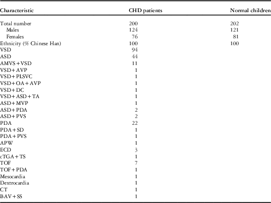
This study was approved by the Ethics Committees of Taiping People’s Hospital of Dongguan. After obtaining their informed consent, 200 non-syndromic CHDs children – 124 male and 76 female – were recruited from the Taiping Renmin Hospital of Dongguan between 2011 and 2013. All cases were confirmed by regular diagnostic tests, such as anthropometric measurement, physical examination for dysmorphism and malformation, electrocardiogram, and ultrasonic echocardiograms, or surgical reports. The individuals of our study have a broad phenotypic spectrum of CHDs (see Table 1).
Characteristics between CHD patients and normal children.

AMVS+VSD=aneurysm of the membranous ventricular septum associated with ventricular septal defect; APW=aortopulmonary window; ASD=isolated atrial septal defect; ASD+MVP=atrial septal defect associated with mitral valve prolapsed; ASD+PDA=atrial septal defect associated with patent ductus arteriosus; ASD+PVS=atrial septal defect associated with pulmonary valve stenosis; BAV+SS=bicuspid aortic valve with supravalvular stenosis; CT=cor triatriatum; cTGA+TS=corrected transposition of the great arteries associated with tricuspid stenosis; ECD=endocardial cushion defect; PDA=patent ductus arteriosus; PDA+PVS=patent ductus arteriosus associated with pulmonary valve stenosis; PDA+SD=patent ductus arteriosus associated with subaortic diaphragm; TOF=tetralogy of Fallot; TOF+PDA=tetralogy of Fallot associated with patent ductus arteriosus; VSD=isolated ventricular septal defect; VSD+ASD+TA=ventricular septal defect associated with atrial septal defect and tricuspid atresia; VSD+AVP=ventricular septal defect associated with aortic valve prolapsed; VSD+DC=ventricular septal defect associated with dextrocardia; VSD+OA+AVP=ventricular septal defect associated with overriding of aorta and aortic valve prolapsed; VSD+PLSVC=ventricular septal defect associated with persistent left superior vena cava
Those cases with a recognised genetic syndrome or chromosome abnormalities, for example, 22q11 deletion or trisomy 21, were excluded from this study. Blood samples were collected from the study cases before surgery and blood transfusion. In addition, 202 unrelated healthy controls – 121 male and 81 female – matched with ethnicity, gender, and no family history of CHDs were analysed. As this was a study on birth defects, we did not match the ages between cases and controls.
Genomic DNA extraction and polymerase chain reaction amplification
Ethylene diamine tetra-acetic acid tubes were used for venous blood collection. Genomic DNA was extracted from peripheral leukocytes using standard methods for genetic analyses. DNA extraction was performed according to standard methodology (QIAamp DNA Mini Kit; Qiagen, Hilden, Germany). Exon 1 of GDF3 gene was amplified using an initial denaturation at 95°C for 3 minute, followed by 35 cycles of 95°C for 30 seconds, 61.3°C for 30 seconds, and 72°C for 45 seconds with a single final extension of 72°C for 8 minutes. Exon 2 of GDF3 gene was amplified using an initial denaturation at 95°C for 7 minutes, followed by 35 cycles of 95°C for 30 seconds, 54.4°C for 50 seconds, and 72°C for 60 seconds with a single final extension of 72°C for 10 minutes. The polymerase chain reaction and sequencing primers are listed in Table 2. Primer sequences of candidate genes were designed on the basis of sequence data obtained from the National Centre for Biotechnology Information database (http://www.ncbi.nlm.nih/org).
PCR primer sequences for GDF3 gene.

DNA analysis
Polymerase chain reaction products were sequenced using the appropriate polymerase chain reaction primers and the BigDye Terminator Cycle Sequencing kit (Applied Biosystems, Foster City, Canada) and run on automated sequencer ABI 3730XL (Applied Biosystems) to perform mutational analyses.
The novelty of all variants found in sequencing was determined from the National Centre for Biotechnology Information human Single Nucleotide Polymorphisms database and the 1000 Genome Project database (http://browser.1000genomes.org/).
Results
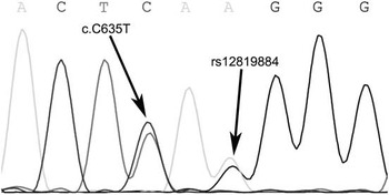
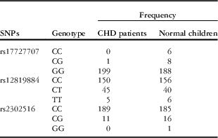
One missense mutation (c.C635T, p.Ser212 Leu, phenotype: isolated muscular ventricular septal defect) was found in the patient with CHD that has not been reported previously and was not detected in 202 normal control DNA samples (Fig 1). The patient is an 8-year-old girl who has no other congenital abnormalities, such as ocular and skeletal abnormalities. We also investigated the family members of this patient and no CHDs were found in her parents and sisters. GDF3 gene of her family members were also amplified and directly sequenced, and no mutation was found. Of the previously reported polymorphisms within the GDF3 coding sequence, three, that is, rs17727707, rs12819884, and rs2302516, were identified in both CHD and control patients (see Table 3).
Electropherogram of the novel identified variation at the position 635 (C>T) of the GDF3 gene as a C/T double peak is shown by the left arrow. One previously reported polymorphism (rs12819884) within the coding sequence of GDF3 gene is shown by the right arrow.

Genotype frequencies of three GDF3 polymorphisms in CHD patients and normal children.

SNP=single nucleotide polymorphism
Discussion
The human GDF3 gene is expressed as a homodimeric preproprotein and can be cleaved extracellularly by the proprotein convertases Furin and Pace4 into mature form, encoding a 364 amino-acid protein. The central portion of the GDF3 protein is structurally similar to the other members of the transforming growth factor β superfamily.Reference Shen 9 , Reference Shi and Massagué 19
Recent studies on mice suggest that it is best to view the total Nodal signalling pathway strength and not just the contribution of a single factor for a particular patterning event. With respect to axis formation in the mouse, a synergistic interaction was observed between GDF3 −/− and GDF1 −/− genotypes, which was not seen in either genotype alone. It has been shown that GDF1 and GDF3 cooperate during formation of the anterior visceral endoderm.Reference Beddington and Robertson 5 , Reference Hyatt and Yost 6 , Reference Andersson, Bertolino and Ibanez 16
The anterior visceral endoderm is an extra-embryonic cell population that is induced at the distal region of the epiblast and subsequently migrates upwards to the prospective anterior side of the pre-gastrulation embryo. At the onset of gastrulation, the anterior visceral endoderm promotes expression of markers in the anterior region of the epiblast that are both essential for the embryonic patterning and the terminal differentiation of cardiac progenitor cells into beating cardiomyocytes. Anterior visceral endoderm play a crucial role in the morphogenesis and development of the heart, especially in the axis asymmetry patterning. In addition, studies indicated that the cardiac progenitor cells require cell interaction with anterior visceral endoderm for their differentiation into cardiomyocytes. The interaction between them may be either contact dependent or contact independent.Reference Andersson, Bertolino and Ibanez 16 , Reference Arai, Yamamoto and Toyama 20 – Reference Perea-Gomez, Rhinn and Ang 22 Besides, it has been showed that the expression of GDF3 gene is significantly upregulated in the right ventricle of patients with hypoplastic left heart syndrome, which is characterised by underdevelopment of the left ventricle and remodelling of the right ventricle, compared with control patients.Reference Ricci, Mohapatra and Urbiztondo 23
We therefore speculated that the GDF3 gene is potentially related to the development of the heart and that GDF3 mutations might play a role in causing CHDs. In this study, we screened potential pathogenic mutations in the exons of GDF3 gene in Chinese patients with CHDs. We found one missense mutation that has not been reported previously and was not detected in 202 normal control patients.
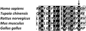
The multiple alignment of amino-acid sequence of the GDF3 gene was taken from different species (Fig 2). It shows that the p.Ser212 Leu mutation lies in the evolutionarily conserved residues of GDF3 among Homo sapiens, Mus musculus, Rattus norvegicus, Tupaia chinensis, and Gallus gallus. It suggested that this position of serine might be critical for the protein’s interaction with other members of the Nodal signalling pathway.Reference Chen, Ware and Sato 14 Functional effect of this mutation, however, remains unclear. The functional studies would be considered in the further research.
Multiple-sequence alignment of partial amino-acid sequence of GDF3 protein in different species is shown. The alignment data indicate that serine at position 212 (indicated by an arrow) is conserved in different species in the GDF3 protein.

It is a challenging work to dissect the full range of genetic and environmental contributors to the non-syndromic CHDs. We hypothesise that mutations must always be considered in the broader context of these interacting factors.Reference Ming and Muenke 24 It will be important to search for evidence of additional genetic and environmental factors. Our studies suggest that mutation in GDF3 may contribute to the CHDs. However, there is a chance that this is a purely incidental finding.
In conclusion, our results expand the spectrum of mutations associated with non-syndromic CHDs and first suggest the potentially disease-related GDF3 gene variant in the pathogenesis of non-syndromic CHDs.
Acknowledgements
The authors thank the patients who participated in this research and also the Social Security Bureau of Dongguan and the Education Bureau of Dongguan for supporting this work.
Financial Support
This work was supported by grant from Science and Technology bureau of Dongguan (grant number 2011105102001), National Basic Research Program of China (grant number 2010CB529504), National Natural Science Foundation of China (grant number 81300131), and National Natural Science Foundation of China (grant number 81470447).
Conflicts of Interest
None.
Ethical Standards
The authors assert that all procedures contributing to this work comply with the Helsinki Declaration of 1975, as revised in 2008, and has been approved by the institutional committees (the Ethics Committees of Taiping People’s Hospital of Dongguan).