Introduction
Tumours of the parapharyngeal space are uncommon, representing about 0.5 per cent of head and neck neoplasms. They include primary neoplasms, extensions from tumours in adjacent regions and metastatic cancer from other sites. Salivary gland tumours are the most common, followed by neurogenic tumours, with most (70–80 per cent) being benign. The diagnosis is usually delayed due to the anatomy of the region.
Several surgical approaches have been described for parapharyngeal space tumours. However, in recent years the transcervical approach has been considered the most useful, especially for extensive neoplasms, because it allows wide intra-operative exposure with minimal side effects both functionally and aesthetically.
The transcervical approach may involve a mandibular osteotomy.
In this report, we discuss our experience with a new approach using a single, subcondylar osteotomy for access to benign parapharyngeal space masses.
Case report
A 43-year-old woman came to our attention in December 2006 due to a swelling of the right oropharyngeal wall which had been present since November 2006. She was otherwise completely asymptomatic.
During clinical examination, a needle aspiration biopsy of the swelling was performed. Cytological examination led to the diagnosis of pleomorphic adenoma.
The patient underwent magnetic resonance imaging (MRI), which revealed a large neoplasm expanding inside the right parapharyngeal space (Figure 1).
Pre-operative (a) coronal and (b) axial magnetic resonance imaging scans, showing the size and position of the neoplasm.

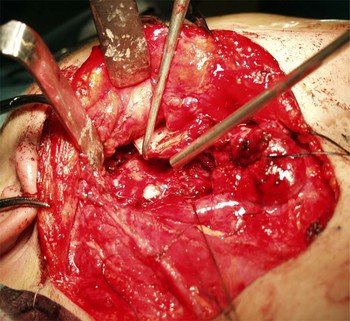
In January 2007, the patient underwent surgery. Following a cervicotomy incision, the submandibular gland was isolated and distracted anterolaterally. A standard dissection of the parapharyngeal space was commenced, and the tumour was exposed and partially isolated. It was not possible to dissect the whole tumour, especially on the most upper and medial side, so a subcondylar mandibular osteotomy was performed (Figure 2). After pre-plating with a titanium mini-plate, the mandibular body and ramus were distracted anteriorly, enabling wider exposure of the tumour (Figure 3). The neoplasm was isolated from the nearby structures and removed. Finally, two titanium mini-plates were positioned to restore mandibular continuity, and the submandibular gland was repositioned.
Intra-operative photograph showing the line of the subcondylar mandibular osteotomy.

Intra-operative photograph showing the wide exposure obtained after tumour removal. Following subcondylar osteotomy, the mandible is distracted anteriorly, while the condyle is elevated laterally. The submandibular gland is distracted anterolaterally.

Histological examination of the surgical specimen confirmed the diagnosis of pleomorphic adenoma.
At the time of writing, the patient was well. Her occlusion was preserved (Figures 4 and 5), and there were no areas of hypoaesthesia or anaesthesia. Yearly follow up with both clinical examination and MRI showed her to be disease-free.
Post-operative views showing the patient's (a) aesthetic result and (b) occlusion.

Post-operative orthopantomography showing the mandibular fixation.

Discussion
Tumours arising in the parapharyngeal space are usually asymptomatic until they become larger than approximately 3 cm, at which point they can begin to cause dysphagia, dysarthria, trismus and pain. When such tumours are large, the intraoral and transparotid approaches are contraindicated because the surgical exposure is poor and the control of the muscular and neurovascular structures is limited. In such cases, most reports describe the use of the transcervical approach, especially when mandibular osteotomies are performed.
In 1954, Ariel et al. Reference Ariel, Jerome and Pack1 first proposed ostectomy of the mandible involving resection of the posterior aspect of the ascending ramus.
Subsequently, various other types of osteotomy have been proposed, involving the angle of the mandible (Berdal and Hall in 1969),Reference Berdal and Hall2 a stepped, vertical osteotomy of the body of the mandible (Stell and Maran in 1978 and Morfit in 1955)Reference Morfit3 or an L osteotomy of the angle of the mandible (Baker and Conley in 1979).Reference Baker and Conley4 Most of these approaches required section of the inferior alveolar nerve, resulting in hypoaesthesia of the inferior lip and mandible.
Another surgical technique, suggested by Flood and HislopReference Flood and Hislop5 in 1991, consisted of an inverted-L osteotomy which preserved the alveolar nerve and allowed the use of a single mandibulotomy.
In 1985Reference GR and Moore6 and 1989,Reference GR7 Seward described a parasymphyseal mandibulotomy without lip-splitting; however, in order to achieve the best surgical exposure, another osteotomy at the condylar neck was often necessary. In our department, we usually follow the Seward technique described by Bozzetti et al.,Reference Bozzetti, Biglioli, Gianni and Brusati8 which involves two mandibular osteotomies: one anterior to the mental foramen, and a second one, for large neoplasms, on the condylar neck.
Osteotomies performed medial to the mental foramen preserve the inferior alveolar nerve but may require lip-splitting, leading to poor aesthetic results.
Kolokythas et al. Reference Kolokythas, Eisele, El-Sayed and Schmidt9 have recently reviewed their experience of gaining parapharyngeal space access via single (paramedian) or double (paramedian and horizontal) mandibular osteotomies, and have concluded that both techniques provide good access to the parapharyngeal space. However, they needed to use a lingual dental splint and rigid fixation in their patients, in order to preserve occlusion and stabilisation, and this involved increased patient discomfort and morbidity.
In 2003, Teng et al. Reference Teng, Genden, Buchbinder and Urken10 proposed a surgical approach using a single subcutaneous midline mandibulotomy for access to the parapharyngeal space. Although the aesthetic results were good, this technique increased tooth morbidity, and, in some cases, the application of a tension band or segmental arch bar was required to prevent rotation of the alveolus.
The use of a single mandibulotomy at the condyle base is a good way to gain access to benign parapharyngeal masses (Figure 6). Using this technique, the inferior alveolar and lingual nerves are preserved. No osteotomy is performed in teeth-bearing areas, so there is no risk to the dental elements (Teng et al.). Distraction of the two parts of the split mandible, with section of the stylomandibular ligament, results in wide exposure without the need for a second mandibulotomy.
Schematic drawing showing the surgical technique: subcondylar mandibular osteotomy is performed and the mandibular body and ramus are distracted anteriorly, enabling wider exposure of the parapharyngeal space.

This technique has other advantages. The use of a single osteotomy reduces mandibular devascularisation and halves the possible post-operative complications, such as bone repair defects and plate infections. The functional results are optimal, without the need for intermaxillary fixation. In addition, lip-splitting is avoided, enabling optimal aesthetic results.
As reported previously, we prefer not to perform a sialoadenectomy of the submandibular gland; rather, the gland is simply distracted anterolaterally (Figure 3).