Anatomical characterisation
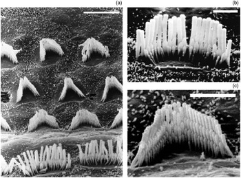
Stereocilia are finger-like processes arranged in rows of graded heights. They form a linear shape on the apical surface of vestibular hair cells, a crescent shape on inner hair cells and a ‘W’ shape on outer hair cells (Figure 11).Reference Wright2
Electron micrographs showing (a) the basal turn of the rat cochlea, with higher power magnifications showing (b) the linear arrangement of the inner hair cell stereocilia and (c) the W pattern of the outer hair cell stereocilia. Scale bar a) 10 µm, b) 3 µm, c) 3 µm. Published with the permission of R Pujol et al. 1

The term stereocilia does not reflect their origin, as these microvilli-derived structures should be clearly distinguished from microtubule-based, true cilia.
Stereocilia are constructed of cross-linked actin filaments in a parallel, paracrystalin array, giving stereocilia their stiffness. The actin cytoskeleton undergoes continuous replenishment via a ‘treadmilling’ process involving actin polymerisation at the tip and actin depolymerisation at the base, which determines the stereocilia length.Reference Rzadzinska, Schneider, Davies, Riordan and Kachar3 Actin filament arrays are continuously remodelled by the addition of actin monomers to the stereocilium tip; the entire core of each stereocilium is renewed every 48 hours.Reference Schneider, Beyantseva, Azevedo and Kachar4 The actin filaments insert with an electron-dense rootlet into a fibrous anchoring structure, the cuticular plate. The cuticular plate is a network of actin filaments which also contains myosin, α-actinin, fimbrin, tropomyosin, fodrin and calcium-binding proteins.Reference Pack and Slepecky5, Reference DeRosier and Tilney6 Stereocilia are rich in actin and fimbrin, and the stereociliary rootlets contain actin and tropomyosin.Reference Slepecky and Chamberlain7 Filamentous actin can be found in pillar cells, Deiter's cells, inner and outer hair cells, and stereocilia. A study investigating F-actin labelling of the cuticular plate and stereocilia found increased labelling after noise and decreased labelling following silence or preconditioning.Reference Hu and Henderson8
Vestibular stereocilia
There are generally approximately 50–80 stereocilia and one kinocilium per vestibular hair cell, arranged in a distinct geometrical alignment. The wider and longer type I vestibular hair cells possess more, taller and thicker stereocilia and a thicker cuticular plate, compared with type II cells.Reference Lapeyre, Guilhaume and Cazals9, Reference Morita, Komatsuzaki and Tatsuoka10
In the vestibular organ, there exists a continuum from loose to tight hair bundles. Interciliary spacing is usually widest along the three hexagon axes closest to the bundle's axis of bilateral symmetry, the presumptive excitatory axis. The spacing between stereocilia alters with distance from the kinocilium.Reference Rowe and Peterson11, Reference Bagger-Sjoback and Takumida12
The kinocilium has no cuticular plate at its origin. Stereo-kinociliar bonds are a regular finding in both cristae and maculae organs, and connect the kinocilium to the tallest stereocilia. The bond unit consists of two parts: the extracellular interciliar filaments of macromolecular size, and an intercellular structure bound to the plasma membrane within the kinocilia. In the cristae, the stereo-kinociliar bonding usually comprises only one bond unit between a single stereocilium and the kinocilium. In the kinocilium maculae, the units are generally paired, with several stereocilia coupled to each kinocilium.Reference Ernston and Smith13
The suprastructure of the macular end-organ consists of two layers: the supramacular substance and the otoconial membrane. The latter supports the otoconia. Kinocilia and the longest stereocilia, as well as the otoconia, are tethered to the otoconial membrane by strands of its material.Reference Ross, Komorowski, Rogers, Pote and Donovan14
The cupula consists of the cupula itself and the subcupular meshwork. The latter consists of long, blanching filaments cross-bridged to one another. The subcupular meshwork may play a role in the transmission of the shear strain force of the cupula to the ciliary bundle and may also exert an additional damping effect in order to prevent unwanted vibrations.Reference Takumida15
Cochlear stereocilia
In contrast to the vestibular system, the kinocilium is lost in the mammalian cochlea during development. However, the basal body usually remains within the hair cell centrosome, which is located in the fonticulus, an area free of actin in the region of the cuticular plate.Reference Raphael, Athey, Wang, Lee and Altschuler16, Reference Steyger, Furness, Hackney and Richardson17 Investigation of kinocilium reformation in post-traumatic hair cells (in a mouse model) indicated a possible role of the kinocilium basal body in the morphogenesis and differentiation of cuticular plates and stereocilia.Reference Sobkowicz, Slapnick and August18
Cochlear stereocilia are usually about 0.2 µm in diameter, and no more than 2–8 µm long in the mammalian cochlea, although much longer stereocilia can be found in other animals (e.g. 50 µm long bundles occur in frog semicircular canals).Reference Flock, Flock and Murray19 Extreme dimensions have been found in mammals that specialise in high frequency hearing, such as bats (which have very short stereocilia) and mole rats (which are low-frequency adapted mammals with very long stereocilia).Reference Vater, Lenoir and Pujol20, Reference Raphael, Lenoir, Wroblewski and Pujol21
The number of stereocilia on each hair cell decreases in a linear fashion with distance from the base of the cochlea. However, stereociliary length increases as a hyperbolic function of distance along the cochlear duct.Reference Wright2, Reference Wright22 Differences in stereociliary gradation also exist between the three rows of stereocilia on the outer hair cells and the two rows on the inner hair cells, with the tallest row positioned laterally.Reference Lim23 Some species have an additional, shorter row of inner hair cell stereocilia on the medial aspect of the tuft, or additional rows of outer hair cell stereocilia.Reference Raphael, Lenoir, Wroblewski and Pujol21 The longest layer of stereocilia leaves an imprint on the undersurface of the tectorial membrane, in the region known as Hardesty's or Kimura's membrane.Reference Gu and Goodwen24 The inner hair cell cilia do not have the same firm attachment to the tectorial membrane as outer hair cell cilia, suggesting different modes of mechanical coupling with the tectorial membrane.Reference Lim23 It has been suggested that the glycoconjugates of the hair cell surface coat attach the tectorial membrane to the stereocilia, and these glycoconjugates are involved in stereociliary fusion following sound exposure and ototoxic drug administration.Reference Santi and Anderson25
Glycocalyx
The glycocalyx resembles a shell-like structure which separately surrounds each stereocilium along its entire length. Stereociliary links (see below) are covered with a layer which is a continuation of the glycocalyx covering the stereocilia.Reference Valk, Oei, Segenhout, Dijk, Stokroos and Albers26 The glycocalyx serves as an anchoring structure for mechanical coupling, not only between the ciliary structures but also between the sensory hair cells and the otolithic membrane of the cupula.Reference Takumida, Wersall and Bagger-Sjoback27 The position and bundle arrangement of the stereocilia are determined by their glycocalyces' strong negative charge.Reference Takumida, Wersäll, Bagger-Sjöbäck and Harada28 This ensures group movement of the stereocilia, maintains distance between them,Reference Takumida, Harada, Wersäll and Bagger-Sjöbäck29 and concentrates cations close to this portion of the hair cell membrane.Reference Prieto and Merchan30 The glycocalyx on sensory and supporting cells forms a filamentous layer that is six times thicker on the endolymphatic side than on the perilymphatic side. This may constitute a barrier mechanism against potassium-induced cell damage.Reference Prieto and Merchan31
Hypothyroidism may alter the carbohydrate composition of the glycocalyx.Reference Gil-Loyzaga, Bueno, Broto and Pérez32
Links
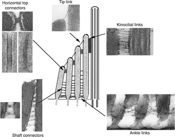
Several distinct cell–surface link types are recognised in hair bundles: tip links, lateral links and tectorial membrane attachment crowns (also known as kinocilial links) (Figure 2Reference Nayak, Ratnayaka, Goodyear and Richardson33–Reference Goodyear, Legan, Wright, Marcotti, Oganesian and Coats36). These appear and fade at different developmental time points in different species.
Central diagram showing types of inner ear hair bundle link: tip links, lateral links (subdivided into horizontal top connectors, shaft connectors and ankle links) and kinocilial links. Peripheral electron micrographs illustrate the ultrastructural appearance of each link type. The horizontal top connector electron micrograph section planes are parallel (top image) and orthogonal (lower two images) to the hair cell's apical surface; the lower right image shows a grazing en face section of a stereocilium. All other electron micrographs show section planes orthogonal to the hair cell's apical surface. Published with permissionReference Nayak, Ratnayaka, Goodyear and Richardson33–Reference Goodyear, Legan, Wright, Marcotti, Oganesian and Coats36 (© 2007 International Journal of Developmental Biology, © 1992 John Wiley and Sons, and © 2003 Society for Neuroscience).

Lateral links can be subdivided into horizontal top connector links, shaft connector links and ankle links.Reference Goodyear, Marcotti, Kros and Richardson37 Lateral links connect stereocilia with each other, either within the same row or across adjacent rows.Reference Osborne and Comis38
Tip links are composed of at least two proteins, procadherin 15 from the lower end and cadherin 23 from the upper end. They are extracellular, cell surface associated, fine filaments which are thought to gate the mechanotransducer channel of the sensory hair cell by deflecting the hair cell bundle towards the taller row.Reference Ahmed, Goodyear, Riazuddin, Lagziel, Legan and Behra39, Reference Siemens, Lillo, Dumont, Reynolds, Williams and Gillespie40 A deflection in the opposite direction leads to hyperpolarisation.Reference Watson and Mire41 Tip links connect the tip of one stereocilium to a longer, adjacent one, and disappear from the hair bundle in response to calcium chelation and lanthane or elastase treatment.Reference Goodyear and Richardson35, Reference Osborne and Comis42 Tip links contain keratin sulphate proteoglycans, which contribute to structural integrity and fibrillar spacing, and which influence the microenvironment of the cell surface.Reference Katori, Hackney and Furness43
The number of glycocalyx tip and lateral links is decreased by noise and aminoglycosides, resulting in hair cell fusion.Reference Lim23, Reference Takumida, Harada, Wersäll and Bagger-Sjöbäck29, Reference Pickles, Comis and Osborne44, Reference Tsuprun, Schachern, Cureoglu and Paparella45 The tip links of outer hair cell stereocilia seem to be equally vulnerable regardless of length, whereas the tip links of inner hair cells are more vulnerable in longer stereocilia compared with shorter stereocilia.Reference Pickles, Osborne and Comis46
Tectorial membrane attachment crown links form a crown-like array around the tips of only the tallest stereocilia, and attach these stereocilia to the tectorial membrane.Reference Tsuprun and Santi47
Molecular characterisation
Several specific molecular compounds are responsible for maintaining the complex, fragile mechanisms of dynamic stereocilia regulation. Dysfunction of any of these compounds leads to various types of inner ear impairment.
Myosins
The myosin motor protein family is subdivided based on the degree of sequence divergence of the motor domain and the different tail domains. Each myosin type (encoded by a separate gene) binds actin and hydrolyses adenosine triphosphate (ATP) to produce force and movement.Reference Mermall, Post and Mooseker48
Several unconventional myosin isoenzymes (i.e. nonfilament-forming myosins) are involved in stereociliary function and integrity, and also in inner ear hearing loss. Unconventional myosins are involved in various hair cell functions, including endocytosis, ion channel regulation, calmodulin localisation, stereociliary anchoring, and movement of vesicles and particles in the cytoplasm.
Myosin Ic acts as an adaptation motor within stereocilia. It facilitates slow adaptation, provides tension to sensitise transduction channels, and may participate in assembly of the transduction apparatus.Reference Gillespie and Cyr49 Myosin Ic interacts with hair cell receptors through its calmodulin-binding IQ domain, and mediates adaptation of the transduction current by amplifying displacements caused by mechanical force into maximal channel opening or closing.Reference Cyr, Dumont and Gillespie50, Reference Gillespie51
Together with cadherin 23 (which corresponds to the stereociliary tip link), myosin 1c is suggested to regulate the activity of mechanically gated ion channels within hair cells.Reference Siemens, Lillo, Dumont, Reynolds, Williams and Gillespie40
Myosin Ib is found along the sides of the stereociliary actin core.Reference Metcalf52
Myosin IIa stabilises the contacts between adjacent inner hair cells.
Myosin VI is found in the cuticular plate associated with the stereociliary rootlets. At this site, it may be involved in anchoring the apical hair cell membrane to the underlying, actin-rich cuticular plate.Reference Self, Sobe, Copeland, Jenkins, Avraham and Steel53 In the absence of normal myosin VI, this apical membrane tends to contract between stereocilia, leading to stereociliary fusion.Reference Self, Sobe, Copeland, Jenkins, Avraham and Steel53
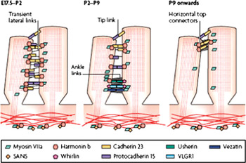
Myosin VIIa is anchored by vezatin to the cadherin–catenins complex, and creates a tension force between adherens junctions and the actin cytoskeleton which is thought to strengthen cell–cell adhesion (Figure 3Reference Goodyear, Marcotti, Kros and Richardson37, Reference Brown, Hardisty-Hughes and Mburu54). The transmembrane protein vezatin is also located at the fibrillar links interconnecting the bases of adjacent stereocilia.Reference Kussel-Andermann, El-Amraoui, Safieddine, Nouaille, Perfettini and Lecuit55 Myosin VIIa helps anchor and hold membrane-bound elements to the actin core of the stereocilium. Mutations mean that hair bundles must be displaced beyond their physiological operating range in order for the mechanotransducer channels to open. In addition, transducer currents also adapt more strongly.Reference Kros, Marcotti, Van, Self, Libby and Brown56
Diagram of development of the the Usher protein network, showing components of the Usher interactome and their involvement in interstereociliary links, based on mouse genetics and protein interaction studies. Transient lateral links appear in the embryo at approximately stage E17.5, then fade after stage P2, the time point at which ankle links begin to appear. Ankle links fade after stage P9, at which point horizontal top connectors are formed. Analysis of the localisation of Usher proteins during mouse development shows that different components are involved with the different links. Tip links and their protein components are also shown. In the mouse, tip links are thought to appear from approximately stage E17.5 onwards.Reference Brown, Hardisty-Hughes and Mburu54 Published with permissionReference Kussel-Andermann, El-Amraoui, Safieddine, Nouaille, Perfettini and Lecuit55 (© 2008 Macmillan Publishers Ltd).

Myosin XIIa appears to be involved in transporting protein and/or lipid complexes, and acts as a channel-gating motor.
Myosin IIIa transports espin to the plus ends of actin filaments, thus facilitating the hearing process.Reference Redowicz57, Reference Salles, Merritt, Manor, Dougherty, Sousa and Moore58
Myosin XV is another compound which is crucial to the normal structure and function of auditory and vestibular hair cells.Reference Anderson, Probst, Belyantseva, Fridell, Beyer and Martin59 Myosin XVa is present at the site of actin polymerisation, in concentrations proportional to the length of the actin filament bundles, and plays a combined role within the treadmill machinery, regulating the steady-state length of these actin protrusions, together with other myosins localised alongside the actin bundles.Reference Lin, Schneider and Kachar60 Myosin XVa is found at the very tips of cochlear and vestibular hair cell stereocilia; it overlaps with the barbed ends of actin filaments and extends to the apical plasma membrane.Reference Belyantseva, Boger and Friedman61 It interacts with the third PDZ domain (for postsynaptic density protein, Drosophila disclarge tumor suppressor, and zona occludens-1 protein). As a result, the scaffolding protein whirlin is delivered to the stereociliary tips; this is probably a key event in hair bundle morphogenesis.Reference Belyantseva, Boger, Naz, Frolenkov, Sellers and Ahmed62
Other molecules
The scaffolding proteins vezatin, harmonin, Sans and whirlin, along with the transmembrane proteins cadherin 23, protocadherin 15, G protein-coupled receptor 98 (formerly termed VLGR1 - very large G protein-coupled receptor-1) and usherin, all form complexes that bind to myosin VIIa and tether the actin cytoskeleton to interstereociliary links.Reference Brown, Hardisty-Hughes and Mburu54 Defects within this complex lead to stereocilia disruption.
The first PDZ domain of harmonin, an F-actin-bundling protein, interacts with myosin VIIa, while the second PDZ domain binds the cytoplasmic region of cadherin 23 (otocadherin). The third PDZ domain, along with the PST (proline-serine-threonine-rich) domain, may interact with F-actin. The interaction of all three proteins forms a transmembrane complex near the tip of the stereocilia, which is essential for the organisation and stabilisation of the stereocilia.Reference Siemens, Kazmierczak, Reynolds, Sticker, Littlewood-Evans and Muller63, Reference Boeda, El-Amraoui, Bahloul, Goodyear, Daviet and Blanchard64
Sans proteins that are underneath the cuticula located aputative scaffolding proteins, are assumed to possess a class-I PDZ-interacting consensus sequence and co-localize with harmonin. Consequently, they are probably part of the cuticular plate molecular complex.Reference Weil, El-Amraoui, Masmoudi, Mustapha, Kikkawa and Laine65
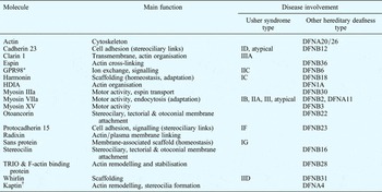
Whirlin is a putative PDZ domain scaffold protein. It is a critical and dynamic organiser of stereocilia elongation and actin polymerisation, and is also involved in DFNB31-type autosomal recessive hearing loss (Table I).Reference Kikkawa, Mburu, Morse, Kominami, Townsend and Brown66, Reference Belyantseva, Labay, Boger, Griffith and Friedman67 Whirlin is present at the base of the stereocilia, and interacts with myosin VIIa which is present along the entire length. The transmembrane netrin G1 ligand binds to PDZ domains 1 and 2 of whirlin, and has an extracellular region that homophilically self-interacts in a calcium-dependent manner. This interaction may be involved in the stabilisation of interstereociliary links.Reference Delprat, Michel, Goodyear, Yamasaki, Michalski and El-Amraoui68
Stereociliary molecules involved in usher syndrome and other hereditary deafness types

Usher syndrome types IE and IIIB are unknown. *Formerly termed VLGR1 (very large G protein-coupled receptor-1). †Also termed 2E4. DFNA = nonsyndromic deafness, autosomal dominant; DFNB = nonsyndromic deafness, autosomal recessive; GPR98 = G protein-coupled receptor 98; HDIA = human homolog of diaphanous; TRIO = TRIO and F-actin-binding proteins
Ezrin-radixin-moesin proteins cross-link actin filaments to plasma membranes, and in so doing integrate the function of cortical layers. Radixin deficiency may cause hearing impairment.Reference Kitajiri, Fukumoto, Hata, Sasaki, Katsuno and Nakagawa69
The TRIO and F-actin-binding proteins (TRIOBP) and F-actin-binding proteins are critical for rootlet formation and function. Mutations of human TRIOBP, a gene encoding multiple isoforms, are associated with profound prelingual deafness DFNB28.Reference Riazuddin, Khan, Ahmed, Ghosh, Caution and Nazli70–Reference Kitajiri, Sakamato, Belyantseva, Goodyear, Stepanyan and Fujiwara72
Physiological characterisation
As stereocilia represent the gate for stimulus detection, they possess a key role in mechanoelectrical transduction, cochlear amplification, frequency selectivity, adaptation and tuning.
Mechanoelectrical transduction
The mechanical strategy for hair cell transduction possesses the advantages of sensitivity and rapidity. The threshold of hearing occurs at a hair bundle deflection of close to 0.3 nm, and channel opening and closing occur thousands of times per second.Reference Hudspeth73
The stereociliary tip links contain the gate cation channels which open during deflection of the hair bundle and allow potassium and calcium to enter via mechanosensitive channels.Reference Beurg, Fettiplace, Nam and Ricci74 Recent findings suggest that mechanoelectrical transduction channels are localised at the bottom of the second and third stereocilia row, rather than near the top of the larger first row.Reference Lumpkin and Hudspeth75 Each tip link has one corresponding transduction channel, with up to 80 channels per cochlear hair cell.
It may be possible that transducer channel gating is determined by rotation rather than translation.Reference Geleoc, Lennan, Richardson and Kros76 Transduction channels flicker between open and closed states; however, when tension is placed on the gating spring, due to deflection of the stereociliary bundle in the excitatory direction, the probability of the channels being open at any given time is increased.Reference Howard, Roberts and Hudspeth77 The hair bundle stiffness has been reported to be directly linked to the state of the mechanoelectrical transduction channel.Reference Martin, Mehta and Hudspeth78, Reference Ricci, Crawford and Fettiplace79 Mechanotransduction in vertebrate hair cells involves a biophysically defined elastic element, the gating spring, which pulls on the transduction channels. Stretching of the gating spring applies force to the gate and increases the probability of it opening. However, the structure of the tip link, composed of cadherin 23 and protocadherin 23, suggests that it is relatively stiff and inextensible. Such behaviour is compatible with the measured elasticity of the gating spring, suggesting that the gating spring lies in series with the helical segment of the tip link,Reference Kachar, Parakkal, Kurc, Zhao and Gillepsie80 or that ankyrin repeats of transient receptor potential ion channels TRPA and TRPN1 serve as the gating spring for mechanotransduction.Reference Sotomayor, Corey and Schulten81 The effectiveness of stereociliary mechanoelectrical transduction is dependent on calcium, which induces active stereociliary bundle movements, producing force feedback in lower vertebrates.Reference Fettiplace, Ricci and Hackney82, Reference Sotomayor, Weihofen, Gaudet and Corey83
Cochlear amplification and frequency selectivity
Together with the structures of the hair cell body, the stereocilia probably contribute actively and/or passively to cochlear amplification. The stereocilia also influence the amplification properties of the outer hair cell body, enabled by bending, which leads to a membrane potential change in outer hair cells. This results in length changes,Reference Brownell, Bader, Bertrand and deRibaupierre84 which feed force back to the basilar membrane on a cycle-by-cycle basis and so tune its otherwise shallow vibrations to the characteristic frequency.Reference Bekesy85 At low frequencies, stereociliary sensitivity is proportional to the cube of the heights of their hair bundles, whereas at high frequencies the sensitivity is proportional to the inverse of their heights.Reference Shatz86 Frequency and stiffness are proportional to each other,Reference Ehret87 and correlate with the height of the stereocilia.Reference Flock and Strelioff88 The cochlear amplification gain is the difference between the peaks in the sensitivity functions for low- and high-intensity tones, or between in vivo and post-mortem responses.Reference Robles and Ruggero89 According to this definition, the cochlear amplification gain, measured in terms of displacement, is 35–58 dB at the 9 to 10 kHz site in the chinchilla,Reference Ruggero, Rich, Recio, Narayan and Robles90 and 35 dB at the 17 to 18 kHz region in the guinea pig.Reference Cooper and Rhode91, Reference Johnstone, Patuzzi and Yates92
It has been suggested that auditory sensitivity is provided by self-tuned critical oscillations of hair cells. Motor proteins generate oscillations at a frequency dependent on the elastic properties of the bundle.Reference Camalet, Duke, Julicher and Prost93 Therefore, these oscillations may represent tuning of the transduction channel through negative feedback from the adaptation process.Reference Ricci and Fettiplace94 The combination of calcium binding to both the transduction channels and the myosin motors may produce self-tuned critical oscillations, which maintain the hair bundle at the threshold of oscillatory instability.
The hair bundle represents an active amplifier, as the characteristic frequency depends on the geometry of the bundle and the calcium dynamics, but is independent of kinetics. If the hair cells are sufficiently perturbed, other movement patterns can occur. These include slow relaxation oscillations, which resemble the hair bundle motion observed in some experimental preparations.Reference Vilfan and Duke95
Blocking transduction with amiloride abolishes nonlinear amplification, whereas eliminating all but the calcium component of the transduction current does not. This supports the thesis that the calcium current drives the active cochlear process.
The hypothesis that active hair bundle motility underlies cochlear amplification is supported by the fact that hair bundles respond to stimulation with a characteristic resonance and a compressive nonlinearity.Reference Chan and Hudspeth96 A slow mechanism limits the mechanical stimulus, through the action of one or more unconventional myosins. A fast mechanism is dependent on Ca2+ regulation of the mechanoelectrical transducer channel, which can generate resonance and confer tuning on transduction. Force generation on a submillisecond timescale, linked to adaptation of the mechanotransducer channels by mammalian hair bundles, support a role in cochlear amplification. As the bundle motor may ultimately be limited by the deactivation rate of the channels, it could theoretically operate at high frequencies.Reference Kennedy, Crawford and Fettiplace97 Change in the conductance or kinetics of mechanoelectrical transduction channels can vary their resonant frequency. The tuning information conveyed in transduction may combine with the somatic motility of outer hair cells to produce an active process that supplies amplification and augments frequency selectivity in the mammalian cochlea.Reference Fettiplace and Ricci98
A bundle is most sensitive to stimulation at its frequency of spontaneous oscillation. Far from that frequency, the sensitivity of an active hair bundle resembles that of a passive bundle. Over most of its range, an active hair bundle's response grows as the one-third power of the stimulus amplitude. The bundle's sensitivity declines in proportion to the negative two-thirds power of the excitation. This scaling behaviour, also found in the response of the mammalian basilar membrane to sound, signals the operation of an amplificatory process at the brink of oscillatory instability, a phenomenon termed a Hopf bifurcation.Reference Martin and Hudspeth99
Adaptation
Adaptation works together with gating compliance to establish mechanical tuning. Different adaptation mechanisms contribute to inner ear function, namely voltage-dependent hair cell properties, afferent transmitter release and hair bundle characteristics. Adaptation warrant tension of the tip links resetting of the neutral set point for the transduction machinery without any significant reduction of its dynamic range. An adaptation model is based on the idea that a calcium dependent negative feedback is responsible for the open probability of the mechanosensitive transduction channels.
Another motor model posits that adaptation relieves the input to the transduction channels (i.e. force applied by the elastic tip links between stereocilia) by repositioning the insertions of the links in the stereocilia. The tip link insertion and the transduction channel are dragged by myosins moving on each stereocilium's actin core.
Adaptation mechanisms attenuate the transduction current at low frequencies, and may be tuned to different corner frequencies according to the stimulus demands of the inner ear.Reference Howard and Hudspeth100, Reference Eatock101 The calcium gradient across the stereocilia appears to provide the energy required for this process.Reference Wu, Ricci and Fettiplace102 The motor force is regulated by calcium via calmodulin binding to myosin, which may promote slippage of myosin relative to the actin core (i.e. it may decrease the amount of time that myosin spends bound to actin in its adenosine triphosphatase (ATPase) cycle).Reference Assad and Corey103 Alternatively, calcium/calmodulin may reduce the stiffness of myosin's force-generating arm.Reference Gillespie and Corey104
In turtle cochleae, calcium regulation has a latency of 0.1–5 milliseconds, whereas in the frog saccule the process is slower, with a myosin-based model indicating a latency of 10–100 milliseconds.Reference Assad, Hacohen and Corey105, Reference Eatock, Corey and Hudspeth106 However, both models may occur together, and the time course of adaptation varies tonotopically.Reference Fettiplace, Ricci and Hackney82 Fast adaptation operates around the most sensitive portion of the hair cell's activation, while the slower form of adaptation requires larger displacements of the hair bundle.Reference Ricci, Crawford and Fettiplace79 Fast adaptation has been identified in both auditory and vestibular hair cells, but is the predominant form in auditory cells. Slow adaptation has been identified in all but mammalian cochlear hair cells, and is the predominant form of adaptation found in vestibular hair cells.Reference Kros, Lennan, Richardson, Flock and Ulfendahl107, Reference Geleoc, Lennan, Richardson and Kros76, Reference Holt, Gillespie, Provance, Shah, Shokat and Corey108 The adaptive shift mechanism preserves sensitivity even in the presence of large stimuli that would otherwise saturate transduction.Reference Holt, Corey and Eatock109
The rate and extent of adaptation to positive steps vary with external Ca2+ concentration, membrane potential and internal Ca2+ buffering, all of which affect the Ca2+ level inside the stereocilium. For example, most, if not all, of the strong apparent voltage dependence of adaptation is an indirect effect of voltage-dependent changes in Ca2+ flux into the stereocilium.Reference Howard and Hudspeth100, Reference Crawford, Evans and Fettiplace110 The stereociliary endogenous calcium buffer, which must be a fast, high-affinity buffer, has a higher effective concentration, and the adaptation time constant is faster in cells at the high frequency end than the low frequency end. The gradient in adaptation time constant and the tuned transducer current at pathophysiological levels of calcium buffer and external Ca2+ suggest that transducer adaptation may contribute to hair cell frequency selectivity.Reference Ricci, Wu and Fettiplace111
Calcium
In the inner ear, calcium is responsible for distinct metabolic processes, cytoskeletal integrity, cell shape and cell excitability. Several transport mechanisms are responsible for calcium homeostasis in stereocilia, namely diffusion, active pumps, and fixed and mobile buffers. Calcium concentrations fall toward resting levels as adaptation proceeds.Reference Lumpkin and Hudspeth112
Different calcium-binding proteins are involved in the above-mentioned functions. Calmodulin is proposed to be the calcium sensor of transducer adaptation. In the guinea pig, calmodulin is found in the stereocilia, cuticular plates and cytoplasm of the inner and outer hair cells. In contrast, calbindin is not expressed in stereocilia, and calsequestrin is only found in the cytoplasm.Reference Slepecky and Ulfendahl113 All of the myosins known to be present in stereocilia possess calmodulin-binding IQ domains, suggesting a role for calcium in regulating their activity.Reference Jurado, Chockalingam and Jarrett114
Plasma membrane Ca2+-ATPases are the principal calmodulin binding proteins. They are predominantly located at the distal ends of stereocilia, and are stronger in the outer than the inner hair cells. Plasma membrane Ca2+-ATPase 2a is the only isoform of this enzyme in stereocilia, and seems to be the main method for calcium export.Reference Caride, Filoteo, Penheiter, Paszty, Enyedi and Penniston115 By this mechanism, it also contributes to endolymph calcium maintenance.Reference Wood, Muchinsky, Filoteo, Penniston and Tempel116
Other important mechanisms for calcium extrusion appear to be free diffusion, buffered diffusion and binding to fixed buffers.Reference Ricci, Wu and Fettiplace111, Reference Lumpkin and Hudspeth112 The stereociliary membrane contains N-type calcium channels (i.e. voltage-gated α1B channels).Reference Lopez, Ishiyama, Ishiyama, Jen, Liu and Balow117 Calcium also controls the probability of the transduction channel resting in its open state; this probability increases as the calcium concentration decreases.Reference Ricci, Wu and Fettiplace111, Reference Ricci and Fettiplace94
Pathophysiological relationships
Inner ear stressors
As described above, molecular irregularities within the stereocilia lead to increased inner ear vulnerability. For example, reduction or omission of otocadherin (also known as CDH23, and encoded by the Ahl gene) weakens the cell and may make stereocilia more vulnerable to physical damage from noise and ageing.Reference Davis, Kozel and Erway118
In addition, stereocilia are the first structures to be damaged by inner ear stressors such as noise or aminoglycosides. Noise affects the stereociliary carbohydrate metabolism, resulting in degeneration of ciliary interconnections together with disarrangement and detachment of cilia.Reference Takumida, Fredelius, Bagger-Sjoback, Harada and Wersall119
Conversely, stereocilia are most vulnerable and tip links are lost when disarray occurs.Reference Clark and Pickles120 Buckling of the supporting cells leads to uncoupling of the stereocilia from the tectorial membrane, which results in a temporary threshold shift. The lower part of the stereocilia and the continuity of the cuticular plate must remain intact to ensure threshold reversibility.Reference Nordmann, Bohne and Harding121, Reference Gao, Ding, Zheng, Ruan and Liu122 Impulse noise trauma predominantly results in the first row of outer hair cell stereocilia disappearing and/or fusing.Reference Slepecky, Hamernik, Henderson and Coling123 The greatest disruption occurs when the hair bundle is deflected away from the basal body of the cell.Reference Patuzzi124 Following overstimulation, the reduction in stereociliary stiffness correlates with the duration, applied noise level and the hair cell row. After several minutes of overstimulation, the threshold shift reaches a plateau the magnitude of which is dependent upon noise exposure intensity. Preconditioning with a low level stimulus can reduce this threshold shift.Reference Canlon125, Reference Saunders, Canlon and Flock126
Adenosine triphosphate gated ion channels with P2X2 receptor subunits are strongly expressed in the stereocilia and in the inner and outer hair cell innervating neurons.Reference Wang, Raybould, Luo, Ryan, Cannell and Thorne127 Their up-regulation could provide a pathway for an ATP-induced reduction in endocochlear potential and inner ear protection, by decoupling the cochlear amplifier in response to stressors.Reference Housley, Kanjhan, Raybould, Greenwood, Salih and Järlebark128
Aminoglycosides alter the hair cell carbohydrate metabolism, leading to deterioration of the glycocalyx and weakening of the ciliary interconnections and tip links, resulting in stereociliary fusion.Reference Takumida, Urquiza, Bagger-Sjoback and Wersall129, Reference Takumida, Bagger-Sjoback, Wersall and Harada130 The presence of myosin VIIa is required in order for gentamicin to accumulate within hair cells.Reference Richardson, Forge, Kros, Fleming, Brown and Steel131
In endolymphatic hydrops, an uncoupling of the outer hair cell stereocilia from the tectorial membrane, an atrophy of short and middle stereocilia first, and a relationship with the CAP (compound action potential) loss can be found.Reference Rydmarker and Horner132 Here, another early pathological feature is loss of stereociliary cross-links, with concomitant disarray of the outer hair cell stereociliary bundles.Reference Ruding, Veldman, Berendsen and Huizing133, Reference Van Benthem, De Groot, Albers, Veldman and Huizing134 A change in glycocalyx characteristics has also been noted.Reference Van Benthem, Albers, De Groot, Veldman and Huizing135 In addition, an alteration in stereociliary stiffness leads to an increase in the hair cell discharge rate, and this has been linked to tinnitus generation.Reference Schwaber136
Ciliary neurotrophic factor can reduce hair cell loss due to noise or ototoxicity, thus protecting the cochlear hair cells and auditory nerves.Reference Zhou, Zhai and Yang137, Reference Kang, He and Shi138
Epithelial growth factor receptor is predominantly found in stereociliary hair bundles and supporting cells. Following exposure to ototoxic drugs, up-regulation of this receptor has been detected.Reference Otterstede, Spandau, Blankenagel, Kimberling and Reisser139
Usher syndrome and other forms of hereditary deafness
Usher syndrome is estimated to account for 3–6 per cent of congenital profound deafness cases in children, and for 50 per cent of cases of deafness and blindness.Reference Kimberling and Moller140–Reference Vernon142
Three types of Usher syndrome have been distinguished, with several subclasses based on the loci of mutation. The first type is characterised by hearing impairment, vestibular dysfunction and retinal degeneration beginning in childhood. In contrast, the second type involves normal vestibular function, less severe hearing loss and a later onset of retinal degeneration. The third type is defined by progressive hearing loss, occasional vestibular dysfunction and a variable onset of retinal degeneration.
Some patients affected with Usher syndrome cannot be easily categorised into one of these three subtypes: they show an atypical clinical designation.Reference Otterstede, Spandau, Blankenagel, Kimberling and Reisser143
In each of these types, retinitis pigmentosa may be accompanied by cataract.Reference Auffarth, Tetz, Krastel, Blanckenagel and Volcker144
In contrast to other deaf children, children with Usher syndrome may show delayed walking development (occurring at a mean of 20 months) as the first sign.Reference Loundon, Marlin, Busquet, Denoyelle, Roger and Renaud145 Affected children cannot sit without support at six months of age, and often do not walk before 18 months.Reference Smith, Berlin, Hejtmack, Keats, Kimberling and Lewis146, Reference Moller, Kimberling, Davenport, Priluck, White and Biscone-Halterman147
The Usher gene products found in the stereocilia are described below.
As mentioned above, myosin VIIa, harmonin, cadherin 23, procadherin 15 and Sans proteins form a complex. These proteins are involved in Usher syndrome type one, in which disorganisation of the hair bundle leads to hearing loss.Reference Adato, Michel, Kikkawa, Reiners, Alagramam and Weil148
Myosin VIIa is a stereocilia-specific molecule. Changes in the myosin VIIa gene are responsible for most Usher syndrome type one forms, one Usher syndrome type three form, and, together with the USH2A gene, for Usher syndrome type two and atypical Usher syndrome. Changes in the myosin VIIa gene are also responsible for the DFNB2 type of nonsyndromic hearing impairment.Reference Keats and Corey149
Myosin VIIa and calmodulin are required for inner hair cell transduction and adaptation. Impaired calmodulin binding of myosin VIIa causes the DFNA11 type of autosomal dominant hearing loss.Reference Bolz, Bolz, Schade, Kothe, Mohrmann and Hess150
Cadherin 23 is expressed in the utriculo-saccular foramen, the ductus reuniens and Reissner's membrane, suggesting that it may be involved in maintaining the ionic composition of the endolymph. Mutations of cadherin 23 have been described in DFNB12-type nonsyndromic hearing loss and Usher syndrome type one D.Reference Wilson, Householder, Coppola, Tessarollo, Fritzsch and Lee151, Reference Bolz, Von Brederlow, Ramirez, Bryda, Kutsche and Nothwang152
Procadherin 15 is involved in deafness caused by Usher syndrome type one F and DFNB23-type nonsyndromic hearing loss.Reference Ahmed, Riazuddin, Ahmad, Bernstein, Guo and Sabar153
Mutations in the gene encoding Sans proteins, which associate with harmonin, have been found to be involved in Usher syndrome type one G.Reference Weil, El-Amraoui, Masmoudi, Mustapha, Kikkawa and Laine65
Myosin XVa is located in the cuticular plate and stereocilia. Mutations of this protein are involved in DFNB3-type nonsyndromic hearing loss.Reference Liang, Wang, Belyantseva, Anderson, Probst and Barber154
Mutations in various actin binding domains are thought to mildly interfere with bundling, gelation, polymerisation or myosin movement, and may cause hearing loss by hindering the repair or stability of cochlear cell structures damaged by noise and ageing. Alterations in the γ-actin gene are associated with the DFNA20/26 type of dominant progressive deafness.Reference Zhu, Yang, Wei, DeWan, Morell and Elfenbein155
Stereocilin and otoancorin are suggested to be responsible for the attachment of the tectorial and otoconial membranes to sensory and nonsensory cells.Reference Jovine, Park and Wassarman156 Stereocilin is involved in DFNB16-type nonsyndromic deafness.Reference Verpy, Masmoudi, Zwaenepoel, Leibovici, Hutchin and Del Castillo157 Otoancorin is defective in DFNB22-type autosomal recessive deafness.Reference Zwaenepoel, Mustapha, Leibovici, Verpy, Goodyear and Liu158
The gene encoding actin-binding protein kaptin (also known as 2E4) is a candidate gene for DFNA4-type autosomal dominant hearing loss. This protein extends beyond the barb ends of actin filaments, at the tips of the stereocilia.Reference Bearer, Chen, Chen, Li, Mark and Smith159 It is thought to play a unique role in actin rearrangement during stereocilia formation.Reference Bearer and Abraham160
Espin actin-bundling proteins, which are located at the stereociliary tips, cause actin depolymerisation in vitro (to a less than twofold degree) and are positively correlated with stereocilium length.Reference Loomis, Zheng, Sekerkova, Changyaleket, Mugnaini and Bartles161 They seem to be associated with autosomal dominant and recessive hearing loss.Reference Donaudy, Zheng, Ficarcella, Ballana, Carella and Melchionda162, Reference Naz, Griffith, Riazuddin, Hampton, Battey and Khan163
Conclusion
Numerous different proteins create the distinct geometrical alignment and biochemical composition of stereocilia, and thus provide the basis for hearing. Stereocilia provide a gating function for key inner ear activities, namely mechanoelectrical transduction, amplification, adaptation, frequency selectivity and tuning. Thus, stereociliary dysfunction leads to various types of sensorineural hearing loss and syndromic deafness.
Further research is needed to elucidate more fully the complex, fragile mechanisms of dynamic stereociliary regulation.