Patients with myopericarditis are typically young adults or adolescents admitted to the hospital with anginal chest pain. Electrocardiographic changes and/or elevated biochemical cardiac markers may be indicative of myocardial involvement. The aetiological factors that cause inflammation in the pericardium and myocardium are similar, so clinical features are used to determine which cardiac structure is predominantly affected. Although the terms myocarditis or perimyocarditis are sometimes used instead of the description of myopericarditis, these are not accurate since there is no wall motion defect in myopericarditis.Reference Imazio and Cooper1 If there is a wall motion abnormality with decreased ventricular function, the term perimyocarditis is appropriate. In the setting of chest pain, myopericarditis and acute coronary syndrome are two leading causes of elevated cardiac enzymes in the emergency room. Differentiating myopericarditis from acute coronary syndrome may not be easy because both present with chest pain, ST segment changes, and elevated cardiac enzymes.
Myopericarditis has been diagnosed more frequently in recent years, resulting in widespread use of biochemical markers. This study is aimed to retrospectively evaluate the clinical, laboratory, and MRI findings of patients with myopericarditis.
Materials and methods
A retrospective, observational single-centre study was performed, which included patients diagnosed with myopericarditis between June 2014 and February 2019. Myopericarditis was diagnosed based on the presence of at least two of the following four criteria (characteristic chest pain, pericardial friction rub, suggestive electrocardiographic findings, and pericardial effusion) suggesting acute pericarditis, concurrent with myocardial inflammation without myocardial dysfunction. Myocardial inflammation was demonstrated by elevated cardiac markers, electrocardiographic changes, or cardiac MRI. Patients with left ventricular systolic dysfunction and coronary artery anomaly were excluded from the study. This resulted in a total of 39 patients diagnosed with myopericarditis being included in the study. Demographic and clinical features, haemogram, C-reactive protein high-sensitivity cardiac troponin I, high-sensitivity cardiac troponin T, and viral serology results were recorded. In our centre, the normal range of C-reactive protein, troponin I, and troponin T is 0–5 mg/L, 0.00–0.04 ng/mL, and 0–14 ng/mL, respectively. A standard 12-lead electrocardiography was performed daily in all patients at the time of diagnosis and during hospitalisation. ST-T segment changes were classified as localised or diffuse. ST segment elevation of >1 mm in two contiguous limb leads, or >2 mm in two contiguous chest leads, was considered as a pathological finding.
Echocardiographic evaluation was performed on all patients. The left ventricular end-diastolic diameter, left ventricular ejection fraction, and shortening fraction were measured with M-mode. Ejection fraction >50% and shortening fraction >25% were considered normal. The presence of pericardial effusion was evaluated by two-dimensional echocardiography. Diagnostic catheter angiography or coronary CT angiography was performed in patients with elevated cardiac biomarkers and persistent chest pain or in those with electrocardiographic changes, to exclude acute coronary syndrome. Cardiac MRI was not performed in one patient with a Kohler implant and 10 patients who refused the test.
Statistical analysis
All statistical analyses were performed using IBM SPSS for Windows version 20.0 (SPSS, Chicago, IL, USA). Continuous variables are presented, depending on distribution, as either mean ± standard deviation for parametric data or for non-parametric data median and interquartile range (25th–75th percentile). Categorical variables were summarised as counts (percentages).
Results
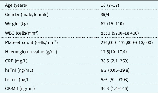
A total of 54 patients with chest pain and elevated troponin levels were admitted to the outpatient clinic between June 2014 and February 2019. Thirteen patients with left ventricular systolic dysfunction and two patients with coronary artery anomaly were excluded from the study. Finally, 39 patients diagnosed with myopericarditis were analysed in the study. The mean ± standard deviation age was 14.69 ± 2.59 (range 7–17) years and 89.7% were male (male/female 35/4). Patient demographic characteristics and laboratory findings are depicted in Table 1. All patients fulfilled at least two criteria for pericarditis and additionally had troponin and creatinine kinase myocardial band elevation, electrocardiographic findings, or MRI findings indicative of myocardial inflammation. Twenty-six patients (66.6%) had a history of upper respiratory tract infection or viral gastroenteritis within the preceding two weeks. In addition, five (12.8%) had palpitations, eight (20.5%) had fever and weakness, and one (2.6%) had dyspnoea.
Demographic characteristic and laboratory findings of the patients (n = 39)

CK-MB = creatine kinase-myocardial band; CRP = C-reactive protein; hsTnI = high sensitivity troponin I; hsTnT = high sensitivity troponin T; WBC = white blood cell.
The median white blood cell count was 8350 (5700–18,400) per mm3 and platelets were 276,000 (172,000–610,000) per mm3. The median C-reactive protein was 38.5 (2.1–269) mg/L. Serum electrolyte and kidney function assays were normal in all patients, while 11 (28.2%) patients had slightly elevated liver aminotransferases. Serum was assayed for troponin I in 11 (28.2%) patients, for Troponin T in 28 (71.8%), and for both biomarkers in 10 (25.6%). The median levels were 6.3 (0.05–29.8) ng/mL for high-sensitivity cardiac troponin I and 586 (51–9398) g/L for high-sensitivity cardiac troponin T. Viral serology was investigated in 24 of 39 (61.5%) patients. Twenty-one patients were negative for available viral tests. Two patients were positive for parvovirus B19 and one was positive for cytomegalovirus.
Electrocardiography was normal in four (10.3%) patients. Ventricular premature beats were present in two (5.1%) patients, non-specific repolarisation changes in four (10.3%), sinus tachycardia in one (2.6%), and supraventricular tachycardia in one (2.6%). In 11 of 23 (47.8%) patients with ST-elevation, changes were limited to the anterolateral or inferolateral leads, while the remaining cases had diffuse elevation.
Chest radiography was normal in all patients. On echocardiography, the left ventricular ejection fraction mean value was 65.5 ± 6.2% (range 54–75), and shortening fraction was 35.36 ± 4.79% (range 28–44). All patients’ left ventricular end-diastolic diameter was within normal limits according to their age. Two patients had mild mitral regurgitation, and two had mild pericardial effusion. For the differential diagnosis of coronary anomaly or acute coronary syndrome, 18 (46.1%) patients underwent coronary CT angiography while five (12.8%) underwent catheter angiography, and all were normal.
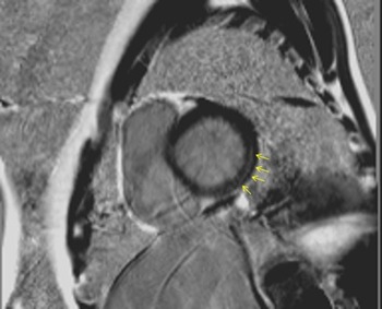
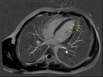
Ten of 28 (35.7%) patients evaluated with cardiac MRI had oedema and late gadolinium enhancement, suggesting myocardial or pericardial involvement. Of these 10 patients, the interventricular septum was affected in one and eight had late period gadolinium enhancement in the left ventricular free wall from the basal or mid zone to the apical region (Figs 1 and 2). Pericardial involvement was evident in the remaining patient.
Late gadolinium enhancement with phase-sensitive inversion recovery reconstruction image. MRI short-axis basal view of the left ventricle demonstrates late gadolinium enhancement in the sub-epicardial area in the free wall (yellow arrows).

Late gadolinium enhancement with phase-sensitive inversion recovery reconstruction image. Yellow arrows show patchy, transmural, late gadolinium enhancement in the left ventricular free wall myocardium on the four-chamber view.

All patients were hospitalised and treated with Ibuprofen at a dose of 30 mg/kg/day. The average length of hospital stay was 3.87 ± 1.56 (range 1–7) days. There was no significant correlation between troponin values and length of hospital stay (for high-sensitivity cardiac troponin T; r = 0.070, p = 0.746 and for high-sensitivity cardiac troponin I; r = −0.035, p = 0.884). The patient with supraventricular tachycardia responded to adenosine treatment, and tachycardia did not recur during subsequent beta-blocker therapy. One patient admitted for limited pericarditis experienced an increase in pericardial effusion so that pericardiocentesis was performed. None of the patients developed left ventricular systolic dysfunction over mid-term follow-up.
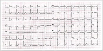
Twelve lead electrocardiograms showing ST elevation in inferolateral leads on admission. Besides, late gadolinium enhancement was observed in the left ventricular free wall by MRI at the same patient.

Discussion
Myopericarditis is a clinical condition in which pericardial tissue is primarily affected, with mild myocardial involvement. Sometimes, the terms myocarditis or perimyocarditis are incorrectly used instead of the term myopericarditis. The term myopericarditis is used when there are mainly pericarditis symptoms with concomitant myocardial involvement, demonstrated by imaging studies or increased levels of troponin I. Patients with myopericarditis do not have ventricular dysfunction. If there is evidence of new-onset, reduced left ventricular function with elevated troponin levels and clinical criteria for acute pericarditis, the diagnosis is interpreted as perimyocarditis.Reference Imazio and Cooper1,Reference Manda and Baradhi2 Unlike perimyocarditis and myocarditis, both of which predominantly cause myocardial involvement and may lead to impaired cardiac function, myopericarditis is a benign self-limiting condition. Myopericarditis, acute coronary syndrome, and coronary artery anomalies may be confused with each other due to chest pain, electrocardiographic changes, and elevation in serum troponin which are common to all.
The aetiology can be idiopathic, or may be related to infectious and immune-mediated conditions. Viruses are the most common infectious agents in developed countries. The primary cardiotropic viruses are Coxsackie virus, adenovirus, herpes viruses, echovirus, Epstein-Barr virus, cytomegalovirus, influenza virus, hepatitis C virus, and parvovirus B19. Bacterial, fungal, and parasitic infections have also been implicated in the aetiology of myopericarditis. Drugs that cause cardiotoxic or hypersensitivity reactions, radiation exposure, systemic inflammatory diseases, malignancy, vaccine administration, and cases of myopericarditis due to some metabolic disorders have also been reported.Reference Imazio and Trinchero3 Due to the retrospective nature of the current study, a standard serological viral panel was not performed in all patients. Three of 24 patients tested had positive viral serology (Parvovirus B19 n = 2; cytomegalovirus n = 1). A male preponderance in myopericarditis has been reported.Reference Imazio and Trinchero3 In our study, 89.7% of the patients were male, which was consistent with the literature.
In many cases, myopericarditis may show subclinical or mild cardiac symptoms, so that systemic signs of viral infection may shadow these cardiac signs. Chest pain is the most common presenting complaint in the patients. Non-specific findings such as decreased exercise capacity, palpitations, fever, or weakness may also be present. All of our patients had chest pain, and 66.6% of the patients had febrile, upper respiratory tract infection, or viral gastroenteritis within two weeks preceding the presentation. Four patients had palpitations, seven patients had fever and abdominal pain, and one patient had dyspnoea. Accompanying fever, weakness, and palpitations increase the possibility of myocardial involvement in cases of pericarditis.
Elevation in serum cardiac biomarkers is an essential indicator of myocardial involvement. In recent years, especially in emergency departments, these assays have become widely used. Therefore, it has been possible to diagnose patients with myopericarditis more frequently. There are studies reporting that a high troponin level has a prognostic predictive value for patients with myocarditis. However, when it comes to myopericarditis, a high troponin level does not appear to be as helpful for prognosis. Kobayashi et al evaluated 12 patients with myopericarditis and found no relation between troponin level and the prognosis of the patients.Reference Kobayashi, Aggarwal, Kheiwa and Shah4 In the current study, troponin levels were high in all patients, but there was no correlation between length of hospital stay and troponin values. Additionally, there was no difference between troponin I and troponin T when evaluated as an indicator of myocardial damage. However, because of the retrospective nature of this study, there were changes in troponin test kit lots that may have affected the reliability of the comparison analysis. Prospective studies are needed to clarify if there is a difference between Troponin I and T in indicating myocardial involvement in young patients with myopericarditis.
The distinctive electrocardiographic pattern for pericarditis consists of diffuse ST-segment elevation and PR interval depression at the first stage, then normalisation of ST-segment and PR interval. At the last stage, diffuse T-wave inversions emerge. Studies conducted in recent years have defined atypical electrocardiographic changes in myopericarditis in comparison to pericarditis.Reference Imazio and Cooper1 ST-segment elevation localised in the anterolateral and inferolateral leads has been detected in almost half of the cases, and it was also observed that some patients presented with T wave inversion before ST segment normalisation. Cardiac arrhythmias were detected five times more frequently than pericarditis. ST-elevation was detected in 23 (59%) of our patients. In two patients, ST elevation was localised in the anterolateral leads while in nine it was localised in the inferior and inferolateral leads, while other patients had diffuse ST elevation that is common to pericarditis (Fig 3). Arrhythmias, such as ventricular and supraventricular premature beats, can be seen and even non-sustained ventricular tachycardia has been reported in more severe cases. In our study, arrhythmia was detected in four (10%) patients, premature ventricular contraction in two, supraventricular tachycardia in one, and sinus tachycardia in the remaining patients. Özyurt et al Reference Özyurt, Baykan and Pamukçu5 observed arrhythmia in 9 of 28 (32%) patients. The rate of arrhythmia in this study was higher than in ours, likely due to the inclusion of patients with left ventricular systolic dysfunction. The prognosis of patients with ventricular dysfunction is usually worse. It has been suggested that patients with ventricular dysfunction should be described as having perimyocarditis.Reference Manda and Baradhi2
Acute coronary syndrome and early repolarisation pattern may occasionally present with similar electrocardiographic findings to myopericarditis. J point/ST segment elevation seen in pericarditis should be differentiated from the early repolarisation pattern which is common in young adolescents.Reference Sharma, Fermandes, Alvarez and Khanna6 Early repolarisation pattern is characterised by prominent j point, which manifests as notching or slurring of the terminal part of the QRS with concave upward ST-segment elevation. This pattern is often considered benign. Isoproterenol and exercise may normalise ST segment.Reference Sharma, Fermandes, Alvarez and Khanna6
Myopericarditis and acute coronary syndrome are two leading causes of elevated cardiac enzymes in the emergency room.Reference Yoldaş and Örün7 Focal or diffuse ST segment elevation can be seen with PR segment depression, especially in lead II and precordial leads, and is an important electrocardiographic finding in myopericarditis. Porela et al showed that PR segment depression is the useful sign to differentiate myopericarditis from myocardial infarction in adults.Reference Porela, Kayto and Nikus8 However, this feature may not be present in all cases. It is not always possible to make a differential diagnosis using only electrocardiographic findings between acute coronary syndrome and myocarditis. In the differential diagnosis with acute coronary syndrome, findings such as a previous history of infection, young age, and characteristic chest pain suggesting pericarditis can eliminate the possibility of acute coronary syndrome in most cases.Reference Sharma, Fermandes, Alvarez and Khanna6,Reference Yoldaş and Örün7 Coronary artery imaging may be required for definitive differential diagnosis in challenging cases who have family history and additional risk factors.Reference Kobayashi, Aggarwal, Kheiwa and Shah4
Echocardiography is a valuable and easily accessible diagnostic method for demonstrating pericardial effusion and detecting wall motion defects and dysfunctions. Echocardiography is an important diagnostic tool for documenting pericardial and myocardial involvement and recommended as a Class 1 indication in the evaluation of acute pericarditis. Pericardial effusion is seen more commonly in acute pericarditis than in myopericarditis. Increased brightness of the pericardium, resulting from deposition of fibrin within inflamed pericardial layers, is more specific for myopericarditis.Reference Imazio, Cecchi and Demichelis9 However, it should be noted that echocardiographic evaluation may be normal in myopericarditis. Pericardial effusion or increased pericardial brightness can be observed. In addition, echocardiography allows the evaluation of additional cardiac defects. In the echocardiographic evaluation of our patients, the left ventricular end-diastolic diameter was within normal limits according to age. Two patients had mild mitral regurgitation, and two had mild (4–5 mm) pericardial effusion. Echocardiography is also important in follow-up. During hospitalisation, all patients underwent control echocardiography, and none developed systolic dysfunction, though pericardiocentesis was performed in one patients due to increased pericardial effusion during follow-up.
Coronary CT can provide useful information in terms of pericardial and myocardial inflammation and is also an important diagnostic tool in the exclusion of acute coronary syndrome, coronary artery anomaly, pulmonary embolism, and aortic dissection.Reference Ferreira, Bettencourt and Rocha10,Reference Takakuwa, Ku and Halpern11 Coronary abnormalities may be detected during echocardiography in infants and younger patients. However, this may not be that easy in adolescents or adults due to decreasing echogenicity. We found coronary artery anomaly (right coronary artery originating from left coronary sinus) in two of our patients who presented with persistent chest pain and high troponin levels by CT angiography. Although it is not necessary to recommend CT or conventional angiography with typical presentation, since these tests are mostly normal and give a low yield. Coronary artery imaging may be reserved for patients with recurrent, persistent chest pain, unclear history of viral infection, and who have concomitant risk factors such as family history, familial hyperlipidemia, or obesity.Reference Kobayashi, Aggarwal, Kheiwa and Shah4
We may have used CT angiography more than needed because of these alarming patients with coronary artery abnormalities. Although the gold standard for demonstrating myocardial involvement is an endomyocardial biopsy, recently MRI has been shown to diagnose myocarditis with an accuracy of 78%.Reference Lurz, Eitel and Adam12,Reference Hundley, Bluemke and Finn13 MRI is superior to echocardiography in imaging the characteristics of the pericardial effusion, it allows for measurement of pericardial thickness and also shows areas of inflammation through contrast enhancement. MRI is also used to assess myocardial involvement by observing myocardial oedema or early and late contrast enhancement, as outlined in the Lake Louise criteria.Reference Friedrich, Sechtem and Schulz-Menger14 Cardiac MRI is useful in detecting coronary artery diseases as well as showing pericardial and myocardial involvement. However, high cost, the limited number of experienced physicians, and low patient compliance restrict its use. It should also be noted that MRI during the acute period increases the possibility of making a correct diagnosis. In our study, MRI was performed in 28 of the patients, and myocardial involvement was detected in 35.7%. We think that positive MRI findings in only one-third of the cases is related to the presence of subclinical cases in our cohort without marked fibrosis in the myocardium, due to the widespread use of troponin for diagnosis in emergency clinics.
Patients suspected of myopericarditis should be hospitalised for differential diagnosis and clinical follow-up. Aside from a single patient requiring pericardiocentesis during follow-up, none developed complications. The fact that this one patient suffered from effusion requiring pericardiocentesis highlights the importance of close monitoring. Patients should have six weeks of absolute movement restriction followed by a total of six months of limitation of heavy exercise.Reference Pelliccia, Corrado and Bjørnstad15 There are insufficient data on the long-term prognosis of myopericarditis, though recent studies show us that prognosis is good for myopericarditis caused by viral infections or idiopathic causes. In the study by Imazio et al in which 40 cases of myopericarditis were analysed, cardiac functions were evaluated up to 12 months after diagnosis and found to be normal in 39 of them.Reference Imazio and Trinchero3 All of our patients are under regular annual follow-up, and none of them has developed heart failure.
The degree of myocardial involvement is likely to be the most important prognostic factor. We advise observing the patient until cardiac biomarkers become normal and to be sure that cardiac contractility is preserved during follow-up.
Limitation
Due to the retrospective nature of our study, it was not possible to use the same troponin test kits and viral serology panel for all patients. This made it unreliable to perform a correlation analysis of the duration of hospitalisation, MRI, and electrocardiographic findings and troponin levels. It was also not possible to make an aetiological evaluation, since a standard viral serology panel was not used in all patients.
Conclusion
The possibility of myopericarditis should be considered in younger patients who are admitted to the hospital complaining of chest pain and have high troponin levels and ST segment-T wave changes in the inferior or inferolateral leads. Male adolescents are more likely to be affected. Even though myopericarditis almost always has a good prognosis, clinicians should be alert for possible complications and differential diagnoses. Echocardiography is the primary and most reliable diagnostic tool to determine cardiac function and pericardial effusion. The diagnostic approach to myopericarditis must include a complete blood count, troponin level, acute phase reactants, electrocardiography, echocardiography, and, if possible, contrast-enhanced cardiac MRI. Routine coronary CT angiography use is not recommended unless there are additional risk factors. MRI is valuable to determine myocardial involvement and is helpful in assessing coronary artery morphology and function and may help to resolve challenging cases.
Acknowledgements
None.
Financial support
This research received no specific grant from any funding agency, commercial, or not-for-profit sectors.
Conflict of interest
None.
Ethics standards
09.2020.685.