Introduction
The present paper describes the mineralogical and crystal chemical description of the new species novograblenovite, (NH4,K)MgCl3·6H2O, based on its chemical composition, crystal structure and physical properties. It also contains a discussion on topological and genetic interconnections of the new mineral and related natural and synthetic phases.
Novograblenovite is named after Prokopiy Trifonovich Novograblenov (August 14, 1892–January 1, 1934), a researcher of Kamchatka Peninsula, a teacher, naturalist, geographer and geologist. He published papers on botany, geology, zoology and archaeology. The most important of Novograblenov's works are “Catalogue of Kamchatka Volcanoes” (Novograblenov Reference Novograblenov1932) and “Hot Springs of Kamchatka” (Novograblenov Reference Novograblenov1931). P.T. Novograblenov perished due to political repressions in 1934.
The new mineral and its name have been approved by the IMA Commission on New Minerals, Nomenclature and Classification (IMA2017-060, Okrugin et al., Reference Okrugin, Kudaeva, Karimova, Yakubovich, Belakovskiy, Chukanov, Zolotarev, Gurzhiy, Zinovieva, Shiryaev and Kartashov2017). The cotype specimen is deposited in the systematic collection of the Fersman Mineralogical Museum, Russian Academy of Sciences, Moscow, Russia, with the registration number 5003/1.
Occurrence, morphology and physical properties
The mineral novograblenovite was discovered on the basaltic lava of the 2012–2013 Tolbachik fissure eruption at the southern slope of the Plosky Tolbachik volcano, Kamchatka Peninsula, Far-Eastern Region, Russia (55°45′28.8″N, 160°18′39.3″E). Here, novograblenovite occurs in association with gypsum and halite.
Plosky Tolbachik is spatially and genetically connected with two radial linear zones of slag cones (Tolbchinsky Dol – a broad lava plain with an area of ~875 km2), extending in the submeridional direction on either side of it at distances up to 20 km to the northeast and ~50 km to the south. Plosky (= flat) Tolbachik volcano has the form of a truncated cone, its top is cut by a Hawaiian-type caldera of 3 km diameter (Okrugin, Reference Okrugin2013). It is one of two volcanoes in the Tolbachik volcano complex.
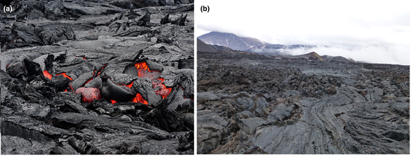
The mineral samples were collected from black basaltic lava that had a thick (5–10 mm) hardened vitreous crust and columnar parting (Fig. 1). The surface of these lava flows is characterised by a peculiar needle-like glassy texture. The basaltic lava under the crust has a porous texture with cavities up to 10 mm across. Long-prismatic, needle-like transparent crystals of the new mineral were found within these cavities together with gypsum and halite. Novograblenovite was formed due to exposure of the host rock to eruptive gas exhalations enriched in HCl and NH3. Basalt was the source of potassium and magnesium for the mineral formation.
Plosky Tolbachik volcano eruption: the black basaltic lava flows in June 2013 (a) and in October 2013 (b).

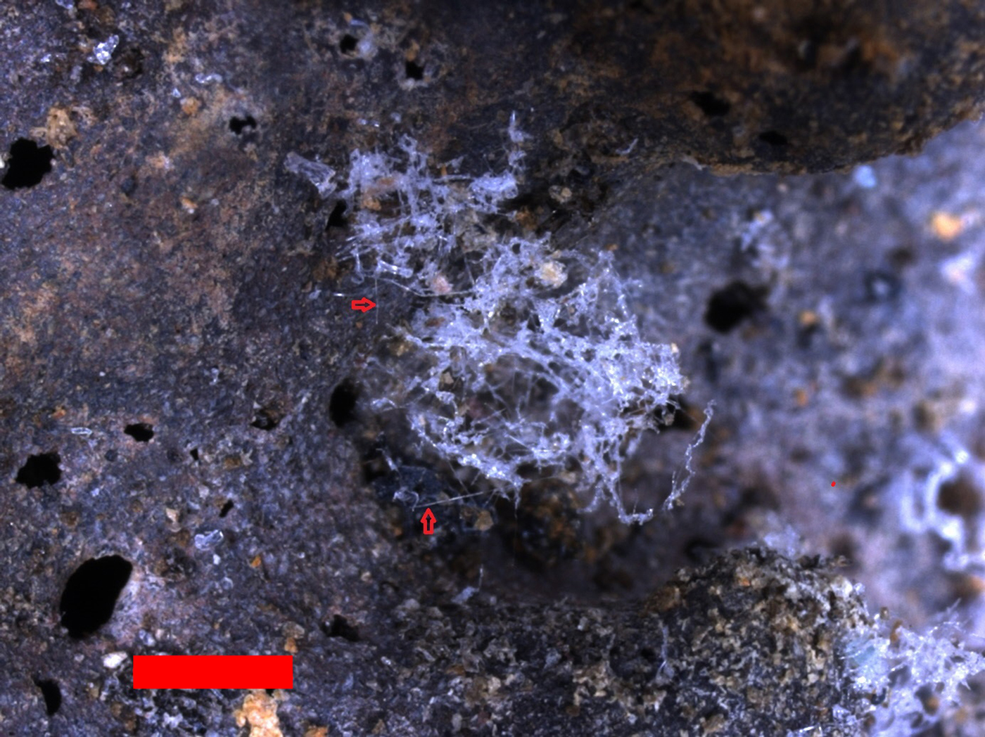
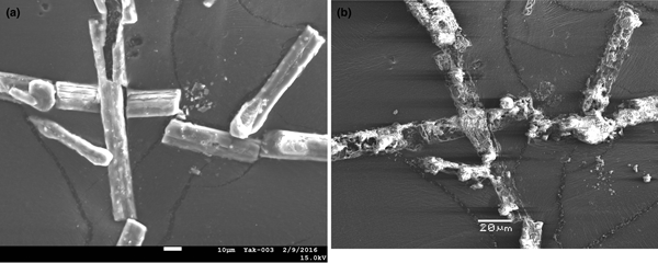
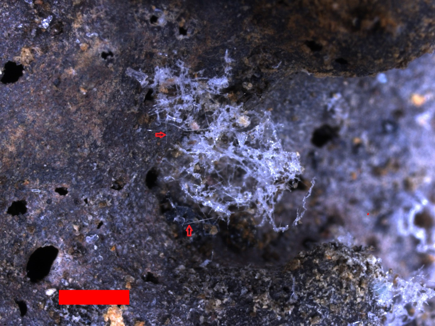
The mineral forms isolated transparent, colourless, imperfect acicular crystals up to 1 mm (typically, 0.2–0.5 mm) long and open-work fibrous aggregates up to 2 mm across (Figs 2, 3). It is characterised by a white streak and vitreous lustre, and is non-fluorescent. Novograblenovite is brittle; thin fibres are flexible. Its Mohs hardness is 1–2. Cleavage or parting is not observed and the fracture is even. The density could not be measured directly because of the small size of individual crystals and the open-work character of aggregates. Calculations from the empirical formula and the X-ray data gives 1.504 g cm–3. Novograblenovite is soluble in water, alcohol and acetone; it dissolves in hydrochloric acid without gas evolution.
Fibrous aggregate of novograblenovite crystals on basalt. Scale bar = 1000 µm.
Novograblenovite crystals: (a) in February 2016, scale bar = 10 µm; (b) in June 2016, scale bar = 20 µm. Scanning electron microscopy (secondary electron) images; specimen number 5003/1.

The mineral is biaxial (+) with α = 1.469(2), β = 1.479(2) and γ = 1.496(2) (λ = 589 nm); 2Vmeas. = 80(10)° and 2Vcalc. = 75.7°.
Dispersion of the optical axes r > v is very weak. In the sections parallel to the optical axes plane c ∧ Z = 40°. In transmitted light the mineral is colourless and non-pleochroic.
Infrared spectroscopy
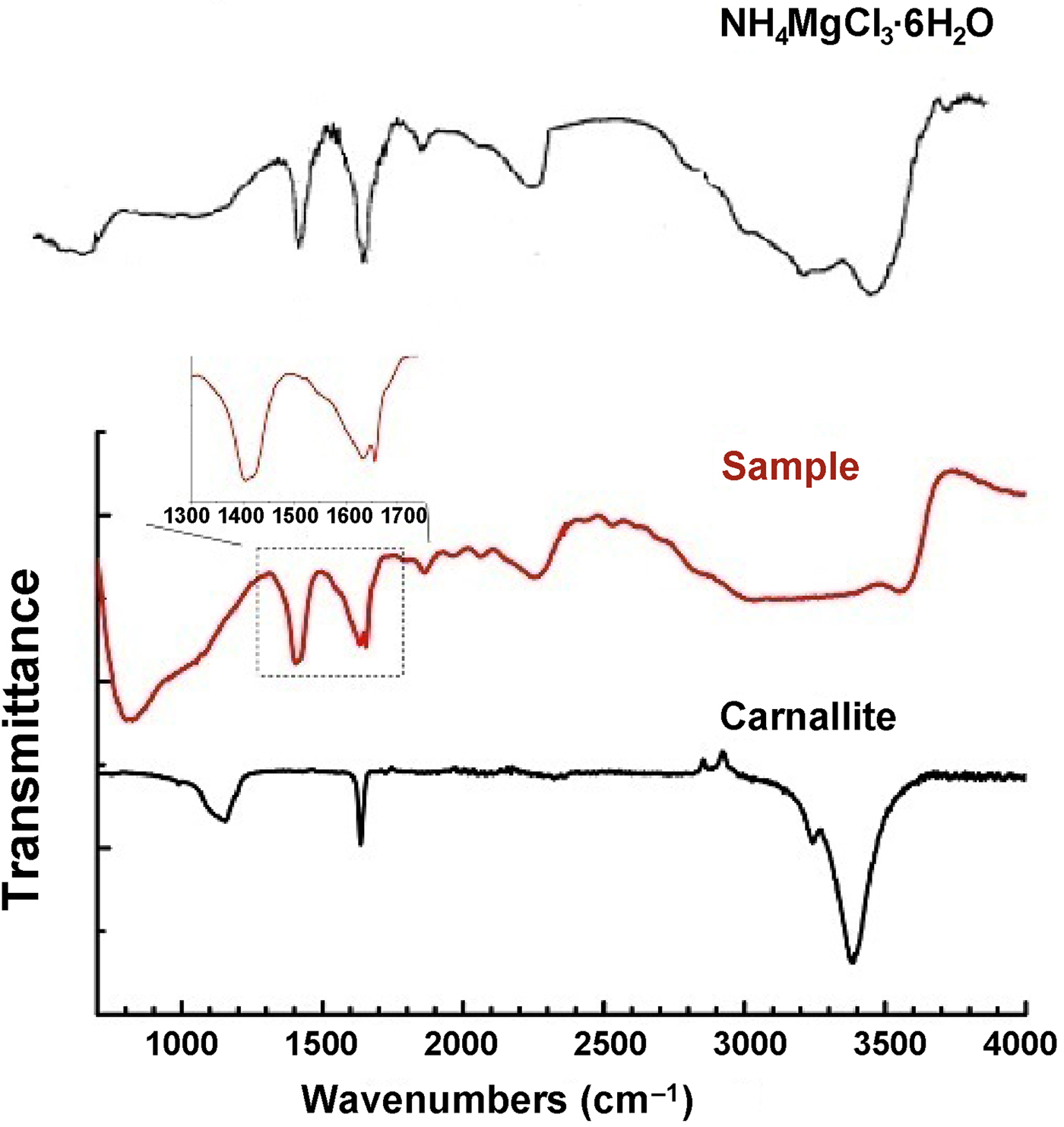
An infrared (IR) spectrum of novograblenovite (Fig. 4) was obtained using a SpectrumOne FTIR spectrometer coupled with an AutoImage microscope with a spectral resolution of 1 or 2 cm–1; 256 scans were collected. The sample was placed on a gold mirror, and the spectrum obtained is a superposition of transmission and reflectance signals with strong predominance of the former. Rectangular apertures matching crystal dimensions (i.e. 50 × 150 or 30 × 150 µm) were employed; several different spots were analysed from every crystal to assure consistency of the results.
Infrared spectra of novograblenovite (‘sample’), natural carnallite (RRUFF R050353) and synthetic compound NH4MgCl3·6H2O (from Wheeler and Wypyski, Reference Wheeler and Wypyski1993).

For comparison, an ATR spectrum of natural carnallite from the RRUFF database (entry R050353 http://rruff.info/)/R050353) and an IR spectrum of synthetic NH4MgCl3·6H2O (Wheeler and Wypyski, Reference Wheeler and Wypyski1993) are also shown (Fig. 4). The spectrum of novograblenovite is close to that of the synthetic NH4-carnallite analogue (Wheeler and Wypyski, Reference Wheeler and Wypyski1993). In the spectrum of the novograblenovite specimen studied a strong feature at 1409 with a shoulder at 1420 cm–1 characterises bending modes of ammonium ions (ν4); superposition of broad bands in the range from 2200 to 3200 cm–1 is due to stretching modes of NH4+ ions. A sharp peak at 1655 cm–1 probably corresponds to the ν2 mode of NH4Cl. The broad band in the interval 2800–3800 cm–1 matches O–H and N–H valence vibrations. The relatively narrow peak at 3553 cm–1 might be related to a bridging hydroxyl. The band at 1630 cm–1 and the absorption above 3200 cm–1 are due to O–H vibrations. A peak at 2256 cm–1 and weak bands in the range 1800–2200 cm–1 most probably characterise combination bands. An absence of the 2256 cm–1 peak in the pure carnallite spectrum suggests ammonia ion librations (broad band at ~800 cm–1) contributed to this band in our case.
Chemical data
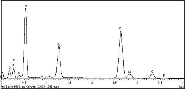
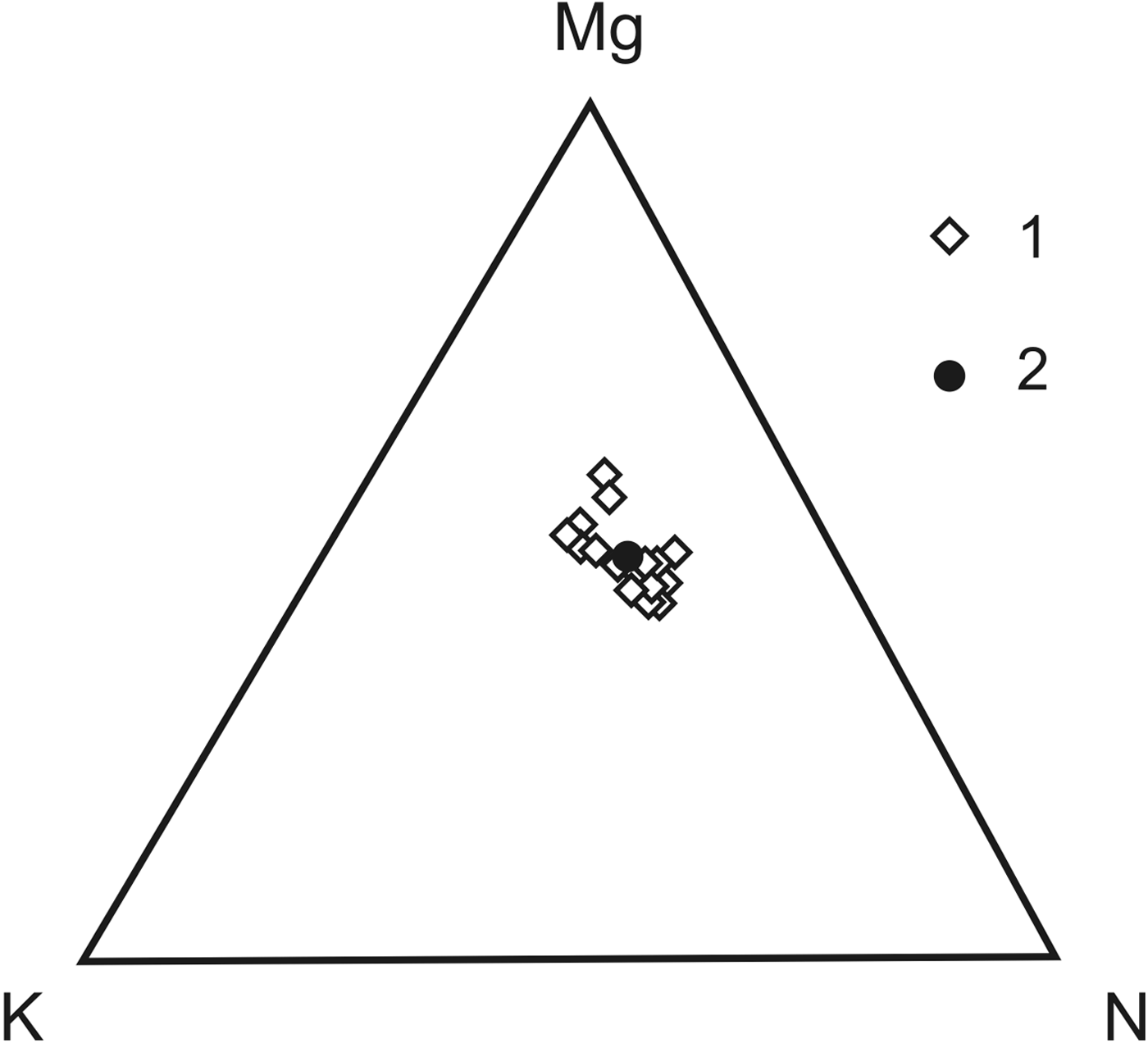
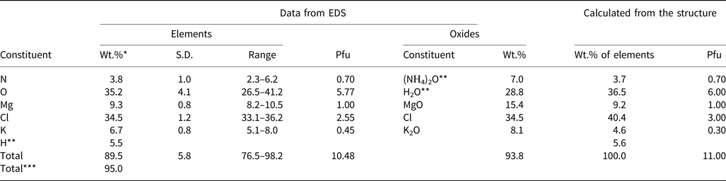
The chemical composition of cotype material was acquired using a Jeol JSM-6480LV scanning electron microscope equipped with an Oxford X-MaxN energy-dispersive spectrometer (EDS) (Laboratory of Analytical Techniques of High Spatial Resolution, Department of Petrology, Geological Faculty, Moscow State University). Samples of novograblenovite were carbon coated and analysed using the following conditions: 10 kV acceleration voltage; 1.4 nA beam current; and analysed area 14 μm2. The data acquisition rate was 10,000–12,000 counts/s, with 20–25% dead time and 25 s live time. Under these conditions the dispersion of measured concentrations of main elements did not exceeded 0.5 relative%, and detection limits were 0.05–0.1 wt.% for all elements analysed. Element concentrations were measured using the Kα lines for N, O, K, Mg and Cl. The standards employed were: microcline (K), Al2O3 (O), MgO (Mg), NaCl (Na and Cl), and internal standard BN for N. The presence of N was also evident in the EDS spectrum (Fig. 5). X-ray intensities were converted to wt.% by the XPP quantitative analysis software of Oxford Instruments (UK). This analytical method was chosen because the sample would be severely damaged by using the wavelength dispersive spectroscopy technique, even with a low voltage and a large diameter beam. The mineral is destroyed by polishing techniques and damaged under the electron beam (Fig. 3). Therefore, the analysis of the chemical composition of novograblenovite was performed on an unpolished surface; 17 areas (14 μm2), near to planar, were chosen for the scanning electron analyses. The mean analytical results are reported in Table 1 and Fig. 6. The chemical formulae obtained from the chemical analysis and the crystal structure are: [(NH4)0.70K0.45]1.15Mg1.00Cl2.55·6H1.92O0.96 and [(NH4)0.70K0.30]1.00MgCl3·6H2O, respectively. The difference in the formulae and the scatter of the sums of the novograblenovite analyses are related to the fact that the EDS experiment was carried out on an unpolished surface; and also that the mineral is destroyed under the electron beam. The lowered O content, as well as the lowered content of calculated H may be due to partial dehydration of the mineral (see Fig. 3b). The H2O content was calculated based on the structure refinement (see below). The presence of H2O is also confirmed by the IR spectrum.
Energy-dispersive spectrum for novograblenovite.

Diagram of EDS-analyses (at.%) made on 17 relatively planar areas (14 μm2) of unpolished crystals of novograblenovite (1) and their mean composition (2).

Composition (in wt.%) of novograblenovite.

*The mean analytical results of 17 analyses; **calculated from the structure; ***includes additional H calculated from the structure.
S.D. – standard deviation. Pfu – per formula unit.
The simplified formula is (NH4,K)MgCl3·6H2O. The end-member formula is (NH4)MgCl3·6H2O, which requires N 5.46, Mg 9.47, Cl 41.41, O 37.38, H 6.28, total 100.00 wt.%.
The Gladstone-Dale compatibility index (Mandarino, Reference Mandarino1981) calculated from the empirical formula and unit-cell parameters determined from powder X-ray diffraction data is 1 – (K P/K C) = –0.058 (good).
X-ray crystallography
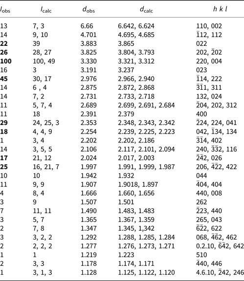
Powder X-ray diffraction data (Table 2) were obtained using a Rigaku R-AXIS Rapid II diffractometer equipped with a cylindrical image plate detector, with Debye-Scherrer geometry (diameter = 127.4 mm; CoKα radiation). The data were integrated using the software package OSC2XRD (Britvin et al., Reference Britvin, Dolivo-Dobrovolsky and Krzhizhanovskaya2017). The indexing of the powder-diffraction pattern was made by comparison with the pattern calculated after the crystal-structure determination. The powder data show good agreement with those calculated from the structure refinement. Parameters of the monoclinic unit cell refined from the powder data are: a = 9.2734(3) Å, b = 9.5176(3) Å, c = 13.2439(4) Å, β = 90.187(2)° and V = 1168.91(2) Å3. No twinning of the crystals is apparent. The most common forms are: {100} and {010}.
Observed and calculated powder X-ray diffraction data (d in Å) for novograblenovite*.

* The strongest lines are given in bold.
X-ray diffraction intensities of reflections were collected for the best tested sample on a Bruker DUO CCD single-crystal diffractometer equipped with a micro-focus X-ray tube (MoKα radiation). Data collection included a total of 10,084 reflections. The intensities were integrated and corrected for Lorenz and polarisation effects, for background, and for beam decay using the Bruker SAINT program. A semi-empirical absorption correction was applied on the basis of intensities of equivalent reflections using the Bruker SADABS program, however, it was found to be negligible.
All calculations were performed with SHELX programs (Sheldrick, Reference Sheldrick2015a,Reference Sheldrickb) in the framework of a WinGX software package (Farrugia, Reference Farrugia2012). The crystal structure was solved via direct methods in the space group C2/c and refined against the F 2 data to the final R factor of 0.0423 (for 1074 unique reflections with I > 2σ(I)) with anisotropic displacement parameters for non-hydrogen atoms. A refinement of the occupancies of the N sites revealed significant substitution of the ammonium ions for K+. The eight independent positions of H atoms were obtained by difference-Fourier techniques and most of them were refined in an isotropic approximation. The coordinates of H7 and H8 atoms forming ammonium groups were fixed in the last run of refinement in order to maintain a reasonable geometry of the NH4+ ion and N–H····Cl hydrogen bonds. The formula resulting from the structure refinement [(NH4)0.70K0.30]MgCl3·6H2O (for Z = 4) reasonably coincides to that obtained by scanning electron microanalysis (taking into account that an unpolished sample was used).
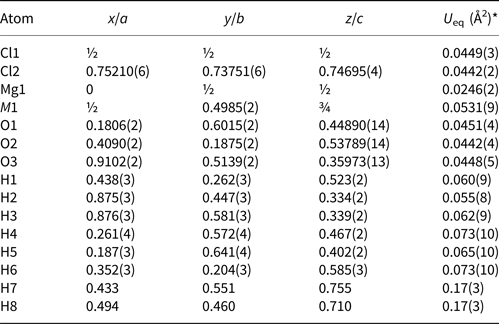
The crystallographic characteristics, the experimental parameters of data collection and structure refinement are summarised in Table 3. The final results for the atom positions are presented in Table 4, while Table 5 reports characteristic distances and angles. The crystallographic information files have been deposited with the Principal Editor of Mineralogical Magazine and are available as Supplementary material (see below).
Crystal information and details of X-ray data collection and refinement.

Atom coordinates and equivalent isotropic displacement parameters.

* Isotropic displacement parameters for hydrogen atoms. Occupancy factors for M1: 0.302(8) K + 0.698(8) N; H7: 0.70; H8: 0.70.
Selected interatomic distances (Å) and angles (°).

Symmetry code for H7′ and H8′: 1 – x, y, 1.5 – z; for Cl2′: x – 0.5, y – 0.5, z.
Crystal structure description
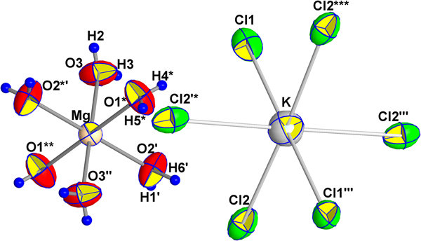
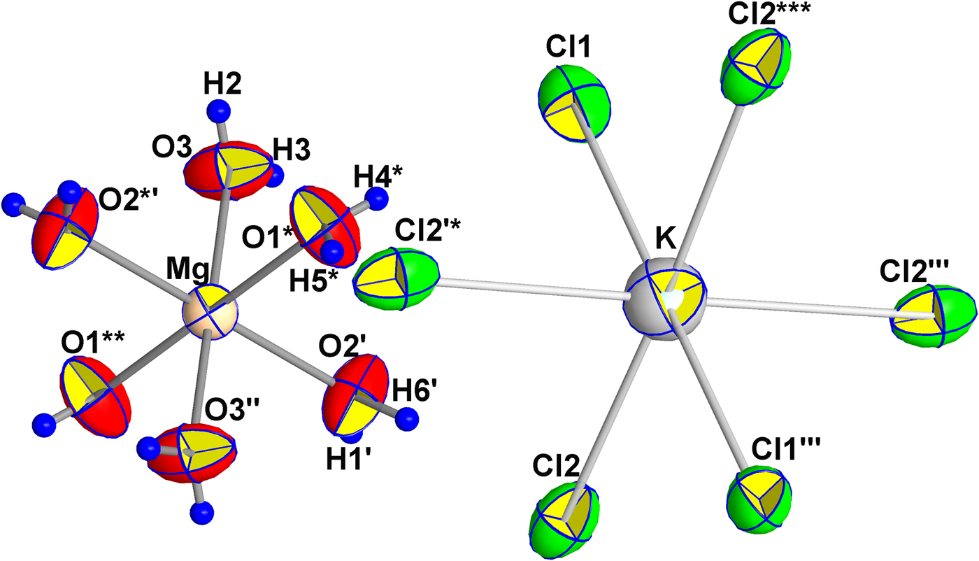
The main units forming the novograblenovite structure are shown in Fig. 7. These are Mg2+ cations in an octahedral environment of H2O molecules and NH4+/K+ ions at the centre of octahedra built by Cl atoms. Note, that (NH4)+ and K+ ions are distributed statistically in one position in a ratio of ~2:1. In nearly regular MgO6 octahedra, the Mg–O distances vary from 2.034(2) to 2.045(2) Å. The (NH4,K)–Cl distances range from 3.257(2) to 3.375(2) Å (Table 5).
Novograblenovite. Basic structural units with atom labelling scheme. Displacement ellipsoids at the 70% probability level. [Symmetry codes (*) 1 − x, 1 − y, 1 − z; (**) 1 + x, y, z; (***) −½ + x, −½ + y, z; (') ½ + x, ½ + y, z; ('') 2 − x, 1 − y, 1 − z; (''') 1 − x, y, 1½ − z; (*') 1½ − x, ½ − y, 1− z; ('*) 1½ − x, −½ + y, ½ − z].
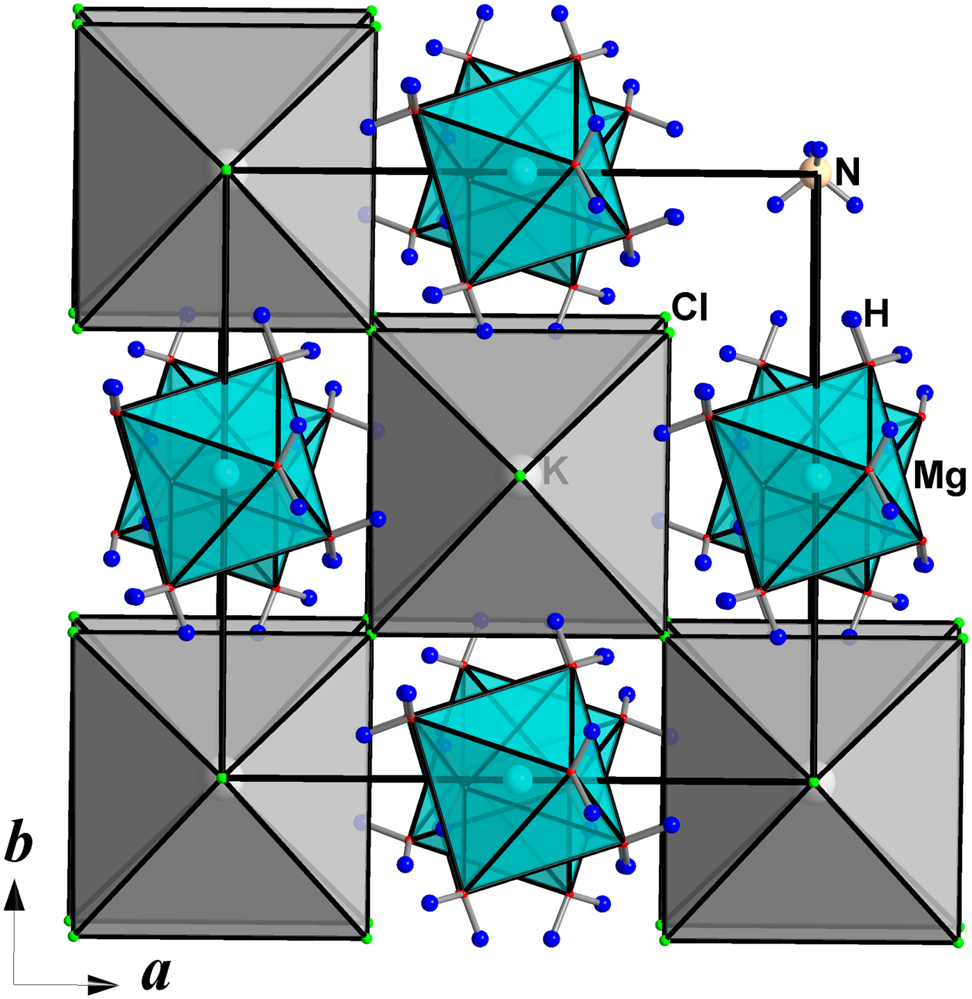
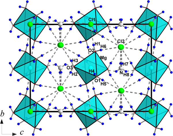
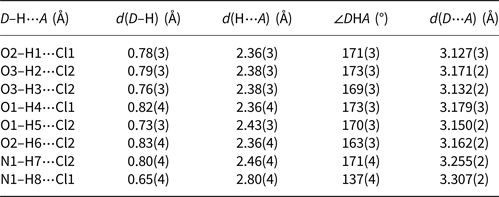
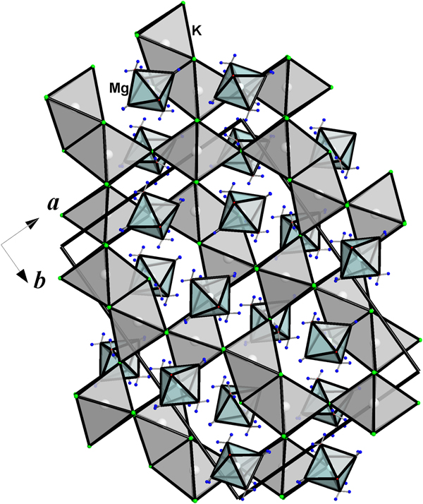
Large (NH4,K)Cl6-octahedra share chlorine vertices to form a three-dimensional framework topologically identical to that built from TiO6 octahedra in perovskite. The perovskite-type network is stabilised by [Mg(H2O)6]2+ cations, which occupy positions in framework channels parallel to [001] and [110] directions (Fig. 8), and are connected with (NH4,K)Cl6-polyhedra through the system of O–H····Cl asymmetric hydrogen bonds formed by water molecules and chlorine atoms (Table 6). Obviously, the N–H····Cl hydrogen bonds act in ~⅔ cases in accordance with the statistical occupancy of the MCl6 (M = NH4 or K) octahedra by the ammonium ions. Each chlorine atom accepts six hydrogen bonds: four from O atoms at the vertices of Mg(H2O)6 octahedra and two from the ammonium ions. The O–H····Cl hydrogen bonds lie in the interval 3.127(3)–3.178(3) Å, while N–H····Cl bonds are normally longer and vary from 3.255(2) to 3.375(2) Å (Table 6). The N–H7····Cl2 distance of 3.255(2) Å is somewhat shorter compared to the expected value of ~3.3 Å. This bond shortening is evidently due to the mixed N/K occupancy of the corresponding site in the structure. An even shorter N/K–Cl distance of 3.220(2) Å has been reported in the crystal structure of adranosite-(Fe) (Mitolo et al., Reference Mitolo, Demartin, Garavelli, Campostrini, Pinto, Gramaccioli, Acquafredda and Kolitsch2013). Thus, all oxygen, nitrogen and chlorine atoms in the crystal structure are involved in the system of hydrogen bonding, acting as donors or acceptors. Hydrogen bonding in this structure serves as an important mechanism providing linkage between the main structural fragments (Fig. 9).
The crystal structure of the novograblenovite in xy projection.

The scheme of hydrogen bonding in the crystal structure of novograblenovite.

Geometric characteristics of hydrogen bonds*.

* D – donor of hydrogen bond, A – hydrogen bond acceptor.
A bond-valence calculation has been performed using the algorithm and parameters given by Pyatenko (Reference Pyatenko1972) (Table 7). Valence contributions of the H atoms in the O–H.⋯Cl hydrogen bonds were estimated from the H–Cl distances using the algorithm of Brown and Altermatt (Reference Brown and Altermatt1985), and bond-valence parameters (R o and b) suggested by Malcherek and Schlüter (Reference Malcherek and Schlüter2007). Data from Table 7 clearly confirm the assignment of H2O ligands.
Bond-valence data for novograblenovite*.

*Multiplicity is indicated by 2↓ and 2→. **M1: K0.30(NH4)0.70.
Crystal chemical and genetic relationships with formula analogues
Novograblenovite has a natural formula analogue, the mineral carnallite, KMgCl3·6H2O. Despite similar chemical compositions these mineral species are characterised by diverse crystal structures (Fischer, Reference Fischer1973; Schlemper et al., Reference Schlemper, Sen Gupta and Zoltai1985). The perovskite, CaTiO3-type, structure of novograblenovite where large Ca atoms are replaced by the Mg(H2O)6 complexes affects the comparison with carnallite significantly. In the novograblenovite structure there is exclusively corner linkage between (NH4,K)Cl6 octahedra, however in carnallite ⅔ of the KCl6 octahedra share faces (Schlemper et al., Reference Schlemper, Sen Gupta and Zoltai1985) (Fig. 10). A similar interconnection of TiO6 octahedra sharing faces occurs in a hexagonal BaTiO3-type structure (Fischer, Reference Fischer1973; Schlemper et al., Reference Schlemper, Sen Gupta and Zoltai1985). Carnallite is thought to crystallise in saline marine deposits by reaction of pre-existing saline minerals with fluids high in potash, while novograblenovite has an effusive origin; it forms under exposure to eruptive gas exhalations enriched in HCl and NH3, and by extracting potassium and magnesium from the basaltic lava. This is similar to the conditions of adranosite formation in the La Fossa crater, Sicily – rather high activities of free ammonia and volatile chlorides in the gas prevent the dissociation of novograblenovite, ensuring its stability (Demartin et al., Reference Demartin, Gramaccioli and Campostrini2010). Thus, the distinct conditions of development of the formula analogues of carnallite and novograblenovite may initiate their structural dissimilarity.
The carnallite, KMg(H2O)6Cl3 crystal structure in xy projection.

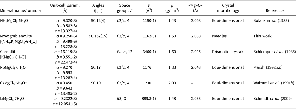
Several synthetic phases in the framework of AMg(H2O)6Cl3 composition (A = NH4, Rb or Cs) obtained in the course of fractional crystallisation and slow evaporation from a solution, have been studied (Solans et al., Reference Solans, Font-Altaba, Aguiló, Solans and Domenech1983; Waizumi et al., Reference Waizumi, Masuda, Ohtaki, Burkov and Scripkin1991a,Reference Waizumi, Masuda, Ohtaki, Scripkin and Burkovb; Marsh, Reference Marsh1992a,Reference Marshb). All three varieties exhibit the same monoclinic perovskite-like crystal structure as established for novograblenovite (Table 8).
Pseudocubic minerals and synthetic compounds related to novograblenovite.

* Converted from the originally reported triclinic unit cell in view of the transformation made by Marsh (Reference Marsh1992a,Reference Marshb) for the Rb analogue.
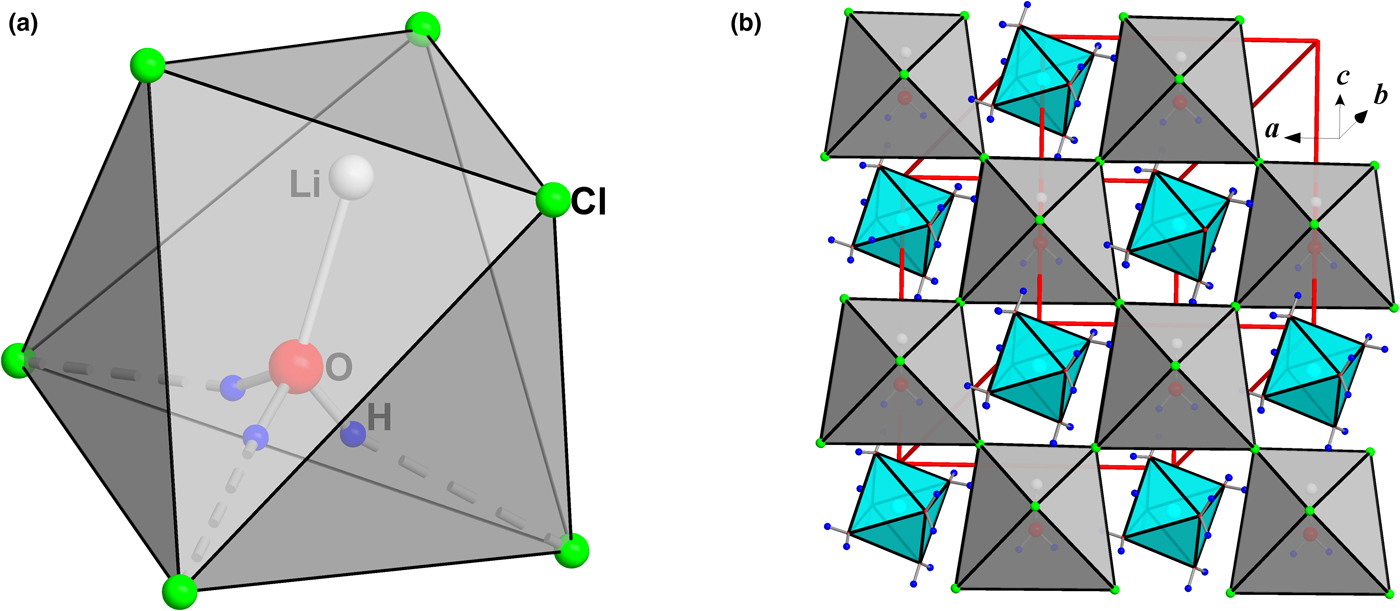
Generally, the mineral carnallite (Schlemper et al., Reference Schlemper, Sen Gupta and Zoltai1985) and its synthetic analogue KMg(H2O)6Cl3 (Fischer, Reference Fischer1973) demonstrate alternate orthorhombic structures as discussed above. In agreement with the observation made in Wizumi et al. (Reference Waizumi, Masuda, Ohtaki, Scripkin and Burkov1991b), the size of ionic radius, being somewhat smaller for K+ compared to NH4+, Rb+ and Cs+, plays a crucial role here, and hence may be responsible for a change of structure type. The synthetic compound LiMg(H2O)7Cl3 is also close in composition and shows a trigonal crystal structure based on a network of Mg(H2O)6 octahedra and Li(H2O)Cl3 tetrahedra connected via O–H····Cl hydrogen bonds (Schmidt et al., Reference Schmidt, Euler, Voigta and Heide2009). In order to simulate a perovskite-like network of MCl6 octahedra, the small alkaline Li adopts the water molecule at 1.890 Å to form [Li(H2O)]+ cation complexes. The [Li(H2O)]+ unit as a coordination centre is surrounded by Cl anions to form five-vertex polyhedra Li(H2O)Cl4 with three Li–Cl bonds of 2.3806(10) Å and two H····Cl bonds of 2.53(5) Å (Fig. 11a). The Li(H2O)Cl4 polyhedra share chlorine vertices with a three-dimensional network as shown in Fig. 11b. As in the previously described alkaline hydrate chlorides, the crystal structure is stabilised by Mg(H2O)6 octahedra, which are connected to Cl atoms via O–H····Cl hydrogen bonds.
‘Li carnallite’: the [Li(H2O)]+ unit in an octahedral surrounding of Cl atoms (only two H atoms of the water molecule are distributed equally over three energetically equivalent positions) (a) and a three-dimensional network stabilised by Mg(H2O)6 octahedra (b).

Acknowledgements
We thank an anonymous member of the IMA-CNMNC who informed us that nograblenovite is likely to be very close to “redikortsevite”, described without IMA approval as fumarole product from a burning mine dump in the Chelyabinsk coal basin, Russia (Chesnokov, et al., Reference Chesnokov, Bazhenova, Shcherbakova, Michal and Deriabina1988), also found later in Germany (Witzke, Reference Witzke1996) and Poland (Stachowicz, et al., Reference Stachowicz, Parafiniuk, Baginski, Macdonald and Wozniak2011). We also acknowledge partial support of M. V. Lomonosov Moscow State University Program of Development. The XRD experiments were carried out using the facilities of XRD Resource Centres of St. Petersburg University. We are much obliged to the Editors, Stuart Mills and Peter Leverett, for handling the manuscript and for useful suggestions.
Supplementary material
To view supplementary material for this article, please visit https://doi.org/10.1180/mgm.2018.88.