Introduction
Dementia is associated with a range of psychiatric symptoms and behavioral disturbances including agitation, aggression, sleep disturbance, wandering, depression, hallucinations and delusions. Agitation and sleep disorders are among the most important of these symptoms, occurring in the majority of people with dementia, causing stress to their carers and promoting institutionalization (Burns et al., Reference Burns, Jacoby and Levy1990; Donaldson et al., Reference Donaldson, Tarrier and Burns1997). Cohen-Mansfield and Billig (Reference Cohen-Mansfield and Billig1986) defined agitation as inappropriate verbal, vocal or motor activity not judged by an outside observer to result directly from the needs or confusion of the agitated individual. An association between agitated behaviors and sleep disturbance has been documented (Cohen-Mansfield and Marx, Reference Cohen-Mansfield and Marx1990).
The mainstay of treatment is with neuroleptic drugs, but there have been recent concerns over the safety of both first- and second-generation antipsychotics (Wang et al., Reference Wang2005) in terms of an increased risk of stroke and mortality (Schneider et al., Reference Schneider, Dagerman and Insel2005) and a detrimental effect on cognition (McShane et al., Reference McShane, Keene, Gedling, Fairburn, Jacoby and Hope1997). The side effects of medication have prompted the search for non-pharmacological treatments and these include cognitive stimulation, music therapy and, specifically for agitation, aromatherapy and bright light therapy. Bright light therapy (BLT) has been used with mixed results in people with Alzheimer's disease (AD) and agitation (Lyketsos et al., Reference Lyketsos, Veiel, Baker and Steele1999; Graf et al., Reference Graf2001; Haffmanns et al., Reference Haffmans2001) but has shown benefit in terms of reducing agitation (Burns et al., Reference Burns, Byrne, Ballard and Holmes2002) The beneficial effect most often reported is improved night-time sleep, reduction in agitation and improvement in cognitive performance. A multifaceted approach can also be helpful (Martin et al., Reference Martin, Marler, Harker, Josephson and Alessi2007)
Control of the circadian rhythm is provided by various phase prompts particularly light, but the environment is also extremely important (Schnelle et al., Reference Schnelle, Alessi, Ouslander and Simmons1993). These zeitgebers exert an influence through the suprachiasmatic nucleus (SCN) (Moore Reference Moore, Swaab, Hofman, Mirmiran, Ravid and van Leeuwen1992), which acts as the pacemaker for circadian functions such as temperature rhythm, sleep/wake cycle and melatonin secretion. People with AD have pathological changes in the SCN and this can lead to disruption of circadian rhythms (Stopa et al., Reference Stopa1999). Patients with dementia have greater night-time motor activity indicative of fragmented sleep, amplitude of the activity cycle, phase delayed core-body temperature circadian rhythm and phase delayed activity rhythm (Satlin et al., Reference Satlin, Volicer, Stopa and Harper1995). Martin et al. (Reference Martin, Marler, Shochat and Ancoli-Israel2000) demonstrated circadian rhythms in agitated behavior in nursing home residents that were not related to seasonality but were related to light exposure, sleep and medication use.
The institutions where people with AD reside often have low ambient light levels and residents are not adequately exposed to daylight (Campbell et al., Reference Campbell, Kripke, Gillin and Hrubovcak1988; Shochat et al., Reference Shochat2000). This leads to an environment weak in phase prompts, which, coupled with neuropathological damage causing poor sensitivity to such prompts, can lead to rhythm disorders. There have been some reports of a seasonal influence on some behavioral abnormalities and a suggested link (Bliwise et al., Reference Bliwise, Carroll and Dement1989; van Someren et al., Reference van Someren1996) between sundowning and the circadian timing system (Vitiello et al., Reference Vitiello, Bliwise and Prinz1992).
The primary objective of this study was to assess the benefit of bright light therapy (BLT) on people with dementia in a nursing care setting, specifically an effect on sleep disturbance. To our knowledge, there have been no reports of the influence of day length on the effectiveness of bright light therapy in dementia.
Method
Study protocols
This was a randomized controlled trial of full spectrum BLT versus normal light for agitation in dementia. The study was approved by the Local Research Ethics Committee. Consent was obtained from the patients where possible, and in all cases from the next of kin. People who had no next of kin were excluded from the study. Throughout the study the investigators were careful to ensure continued consent by the patients, and if any became distressed by the treatment, they were helped to leave the treatment room. Information was provided verbally and in writing to the participants and their next of kin.
The inclusion criteria were (i) a diagnosis of dementia (WHO, 1993); (ii) sleep disruption at least two nights per week; or (iii) the presence of one or more agitated behaviors (Cohen-Mansfield and Billig, Reference Cohen-Mansfield and Billig1986). The exclusion criteria were (i) failure to satisfy the inclusion criteria; and (ii) cataracts.
Outcome measures
The primary outcome measure was the Cohen-Mansfield Agitation Inventory (CMAI; Cohen-Mansfield et al., Reference Cohen-Mansfield and Billig1986), a 29-item scale assessing a range of abnormal behaviors on a scale of 1–7, with particular emphasis on agitation, completed by an independent rater (Cohen-Mansfield and Billig, Reference Cohen-Mansfield and Billig1986). A high score indicates more severe behavior.
The secondary outcome measures were:
• Mini-mental State Examination (MMSE; Folstein et al., Reference Folstein, Folstein and McHugh1975) – a 30-item measure of cognitive function with a maximum score of 30 points. A low score indicates greater cognitive impairment. The standardized version, sMMSE (Molloy et al., Reference Molloy, Alemayehu and Roberts1991), was used.
• Cornell Scale for Depression in Dementia (CSDD; Alexopoulos et al., Reference Alexopoulos, Abrahams, Young and Shamoian1988) – a 19-item scale assessing depressive symptoms in people with dementia, rated on a 3-point scale of “absent,” “mild or intermittent,” and “severe” (a score of 8 or above indicating significant depressive symptoms).
• Manchester and Oxford Universities Scale for the Psychological Assessment of Dementia (MOUSEPAD; Allen et al., Reference Allen, Gordon, Hope and Burns1996) – a rating of psychopathology in people with dementia, scoring 59 symptoms on a 4-point scale of “absent,” “mild,” “moderate,” and “severe.”
• Crichton Royal Behavior Rating Scale (Robinson, Reference Robinson1961) – a scale measuring a number of behaviors such as mobility, communication, cooperation, restlessness, dressing, feeding, incontinence, sleep, mood, and orientation on a 5-point scale of increasing severity.
• Sleep charts: nursing staff were asked to record whether the patient was asleep or not, in 30-minute blocks, during the course of the study.
Patients wore an actiwatch (supplied by Cambridge Neurotechnology Ltd., Cambridge, U.K., www.camntech.com (last accessed 28 March 2008)) on the wrist, which recorded the patients' activity. This wristwatch-size actigraph has been validated as a measure of agitation in people with dementia (Witting et al., Reference Witting, Kwa, Eikelenboom, Mirmiran and Swaab1990). It uses a peizo-electric accelerometer to record the intensity and duration of movement in all directions and is sensitive to movements over 0.05 G. Two principal measures of motor activity were assessed. First, the mean activity counts per hour of the 10 most active consecutive hours in the 24-hour period (most active, M10) and, second, the five least active consecutive hours (least active, L5), computed by averaging the 10 highest and 5 lowest hourly means of activity as recorded by the actigraph. This gives a measure of the amplitude of the diurnal activity. The time of onset of the M10 and L5 gives a measure of the phase of the activity. Measurements of activity using the actigraphs were made in the baseline week and in week 4.
Sample and method of randomization
Patients were recruited from two nursing homes specializing in the care of patients with dementia and behavioral disturbances. A power calculation was based on the method described by Altman (Reference Altman1991) using data from Satlin et al. (Reference Satlin, Volicer, Ross, Herz and Campbell1992). Assuming a 60% response for BLT and 20% response for placebo, at 80% power and a significance level of 5%, the sample size required was 24 in each group. Variables that may influence responsiveness to BLT include mood, severity of dementia, age and gender. Randomization was carried out by the trial statistician using lists drawn up from www.randomization.com prior to the start of the study. Subjects were stratified according to high (10–30)/low (0–9) sMMSE (Molloy et al. Reference Molloy, Alemayehu and Roberts1991) and high (≥8) /low(0–7) CSDD scores (Alexopoulos et al., Reference Alexopoulos, Abrahams, Young and Shamoian1988). Age and gender were not included as criteria, but were analyzed post hoc for between-group differences. Researchers who conducted patient interviews were blind to which treatment the patients had received.
Operation of the study
During the first week, patients were seen for confirmation of diagnosis, examined physically, and were checked for the presence of significant cataracts. The diagnostic ratings were performed by a member of the research team who was not involved in the clinical care of the patients.
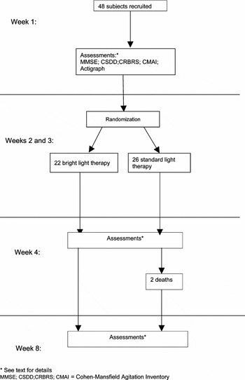
During the second and third weeks, patients were exposed to full spectrum BLT 10000 lux or standard fluorescent tube light at 100 lux for two hours between 10 a.m. and 12 noon. A nurse was present during each bright and standard light condition; the research nurse sat with the resident in front of the light box during the treatment, engaging them in conversation where appropriate and distracting them if they tried to leave. No restraint was used. During the fourth week patients wore the actigraph again, and MMSE, Cornell, CRBRS, MOUSEPAD and CMAI measurements were repeated. The five rating scales were again repeated during the eighth week. On each occasion the independent rater completed the CMAI. Any changes in psychotropic medication were noted. A schematic chart of the study is shown in Figure 1. Assessments were carried out during the baseline week and in weeks 4 and 8.
Description of the study.

A record of the date, time and duration of treatment was made. Standardized MMSE, CRBRS, Cornell scale and MOUSEPAD were completed by the research nurse, and an independent rater (blind to the treatment condition) completed the CMAI. Nursing staff in the homes were also asked to complete categorized sleep charts for all patients (asleep/awake), on which a recording was made in 30-minute blocks of whether or not the patient was asleep. A record was made of prescribed psychotropic medication.
Statistical methods
Baseline measures in the two groups were compared using the χ2 test or Fisher's exact test for categorical measures, and the t-test for continuous measures. Changes from baseline to follow-up on continuous measures in the two groups were compared using analysis of covariance with baseline score as covariate. Associations between changes in questionnaire scores and day length were assessed using Pearson correlations in the separate treatment groups. Changes in sleep duration from baseline to follow-up, as measured by actigraph or the nurses on the two groups, were compared using analysis of covariance with baseline score as covariate, and were repeated for short and long days separately. Associations between changes in sleep duration and day length were assessed using Pearson correlations in the separate treatment groups. Similar analyses were carried out for M10 and L5 levels and onset of M10 and L5.
Results
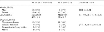
Forty-eight patients were enrolled in the study: 26 were randomized to standard light, 22 to BLT (see Figure 1). One patient was hospitalized after three days of treatment for unrelated causes, and one withdrew from treatment after three days as the BLT caused her to recall memories of being tortured during the Second World War. These two patients completed all follow-up interviews and their results were included, as we have used intention-to-treat analyses. Characteristics of the sample at baseline are shown in Table 1. There were no significant differences between the two groups on any of the questionnaire scores or in age, gender or diagnosis. All subjects had at least one agitated behavior as rated on CMAI at baseline, and 25 (52%) had sleep disturbance according to the MOUSEPAD ratings.
Analysis of variables at baseline

BLT = bright light therapy.
Forty-three of the patients (90%) were able to tolerate light exposure for a minimum of 90 minutes per day. Four patients (8%) tolerated a minimum of 60 minutes per day, and one patient (2%) tolerated 30–60 minutes per day. Only one change to medication was recorded – a person in the treatment group who had her trazodone increased from 50 mg to 100 mg. Two patients died between the week 4 and week 8 assessments, both of whom had received placebo.
Effect on behavioral and psychological symptoms and on cognition
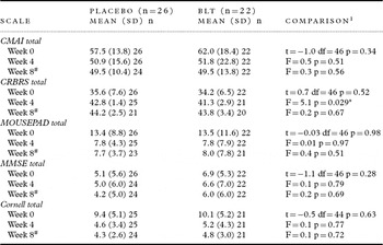
Changes in behavioral and psychological symptoms from baseline to the immediate post-treatment scores (week 4), and baseline to one month after the end of treatment, week 8, were calculated. The comparison between groups is shown in Table 2. Agitation (measured by the CMAI) improved in the immediate post-treatment phase in both the BLT and placebo group, but the between-group comparison was not significant (p = 0.51) at week 4, nor at week 8 (p = 0.56).
Comparison of placebo and active treatment over the study period

1Comparisons made using the t-test at week 0, and using analysis of covariance (with week 0 score as covariate) for the changes from week 0 to weeks 4 and 8.
#Two patients in the placebo died between week 4 and week 8.
*p<0.05.
BLT = bright light therapy; CMAI = Cohen-Mansfield Agitation Inventory; CRBRS = Crichton Royal Behavior Rating Scale; MMSE = Mini-mental State Examination.
The CRBRS change score showed a significantly greater increase (i.e. deterioration) in the placebo than the BLT group at 4 weeks (p = 0.029), but not at week 8 (p = 0.67). There was no significant difference between groups on any of MOUSEPAD, MMSE or Cornell scores at either 4 or 8 weeks. Similar improvements were obtained in both groups. When Bonferroni adjustments are made to allow for the fact that five measures have been compared at two time points, then the only significant effect (for CRBRS at 4 weeks) also becomes non-significant.
Effect of mood
There was a significant correlation between CMAI change score and change in Cornell scores at the immediate post-treatment period (week 4) for the placebo group (Pearson correlation, r = 0.40, p = 0.046), and a trend towards a significant correlation for the BLT group (r = 0.39, p =0.080).
Effect of day length
The study took place between the months of November 2000 and August 2001. Twenty-five subjects (15 placebo, 10 BLT) were entered in the study during the summer months and 23 subjects (11 placebo, 12 BLT) during the winter months – the total group being divided by the date of the spring equinox, 21 March.
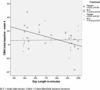
The change in Cornell scores from baseline to week 4 was not significantly correlated with day length for either group (BLT: r = 0.14 p = 0.55: placebo: r = −0.11, p = 0.61). Change in agitation (as measured by the CMAI) from 0 to 4 weeks was significantly negatively associated with day length in the BLT group (Pearson correlation = −0.52, p = 0.013), but not in the placebo group (Pearson correlation = 0.03, p = 0.90) (see Figure 2). Neither age nor diagnosis influenced the changes in CMAI by day length.
Scatterplot of change in CMAI score from baseline to immediate post-treatment by day length in minutes (22 BLT and 26 placebo subjects).

Actigraphic assessments
The mean duration of nocturnal sleep as measured by the actigraph was 8.3 hours and 8.6 hours before and after BLT respectively. The mean duration of sleep as recorded by the nursing staff was 9.4 hours and 9.6 hours before and after BLT respectively. There was no significant difference between active and placebo groups before or after BLT, nor for the change in sleep duration, as measured by either actigraph or the nurses.
Day length correlated with sleep duration as measured by the nurses at baseline. Patients slept longer when the day length was longer (Pearson correlation = 0.42, p = 0.016), although this was not the case when sleep was measured using the actigraph (r = 0.15, p = 0.32). There was no significant difference between groups with respect to sleep duration, as measured by either the nurses or the actigraph, either for short or long days, and no significant difference between groups by day length interaction.
There was no correlation between improvement in sleep duration and change in Cornell scores (Pearson correlation = 0.15, p = 0.37 for the actigraph, and correlation = −0.19, p = 0.35 for the nurses.).
Diurnal phase and amplitude
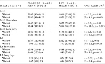
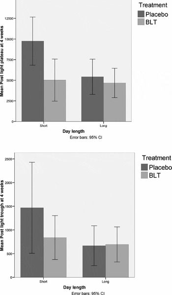
There was no significant pre-treatment difference between the groups in the amplitude of activity (M10), but there was a significant difference between the groups at week 4, with the BLT group showing a significantly greater reduction in mean score (p = 0.004) (Table 3). This was also the case for the patients treated on short days, but not for those treated on long days. There were similar changes found for the trough of activity (L5), but these did not reach statistical significance. Figure 3 shows the M10 and L5 mean values at 4 weeks by treatment group and day length.
Comparison of placebo and active treatment by day length

1Comparisons made using the t-test at week 0, and using analysis of covariance (with week 0 score as covariate) for the changes from week 0 to week 4.
BLT = bright light therapy; M10 = most active consecutive 10 hours in a 24-hour period; L5 = least active consecutive 5 hours in a 24-hour period.
Level of activity rated on actigraph.

Discussion
This study has shown that, in people with dementia, bright light therapy can have some effect in reducing agitation (as measured by an actigraph) and improving sleep and is acceptable to patients and their carers. Non-pharmacological approaches to the treatment of dementia are becoming more popular as an alternative to drugs, whose effectiveness and side-effect profile have raised safety concerns about their use in practice (O'Brien, Reference O'Brien2008). BLT was used because of its effect on the melatonin system, itself implicated in the regulation of abnormal motor behavior (Haffmanns et al., Reference Haffmans2001).
Mechanism of action
Circadian rhythms are controlled by the suprachiasmic nucleus (Moore Reference Moore, Swaab, Hofman, Mirmiran, Ravid and van Leeuwen1992), an area affected by Alzheimer type pathology which may partly explain the observed disruption of sleep. Retinal ganglion cells, directly affected by light, have a role to play in the photoentrainment of circadian rhythms aided by phase prompts (called zeitgebers). Day length is a powerful force in controlling seasonal and daily activity in animals and in humans, although other factors such as social interaction play a part. Perhaps in people with dementia, those higher cortical factors are disabled by illness, with more basic mechanisms taking over. The improvement of behavior mediated by bright light has a demonstrable beneficial effect, which is more marked in winter when the external light cues are compromised. Environmental factors, and in particular the nursing home environment, can have a powerful influence in determining circadian rhythms (Schnelle et al., Reference Schnelle, Alessi, Ouslander and Simmons1993).
For the overall duration of the study both groups improved on the main outcome measure of behavior, the CMAI, and on a secondary measure of behavior, the CRBRS. Depression, as measured by the CSDD, also improved in both groups. This indicates the significantly beneficial effect on agitation and other behaviors, and of the regular presence of a skilled nurse practitioner (Martin et al., Reference Martin, Marler, Harker, Josephson and Alessi2007).
The timing of the morning BLT in our study was between 10 a.m. and 12 noon each day. By choosing this period we probably inadvertently avoided the temperature nadir (of the circadian temperature cycle) in our subjects. Harper et al. (Reference Harper2001) observed that BLT administered on or before the temperature nadir may result in paradoxical changes in circadian phase shifts in male AD patients (no females were included in this study). Avoiding the temperature nadir enhanced our chances of observing real changes in circadian phase due to BLT. Because the study took nearly a full calendar year to perform, the power of our study was effectively halved with roughly equal numbers being treated before and after the spring equinox.
Ours was a diagnostically heterogeneous patient population. Although the figures above are measures of change, they reflect the broad range of baseline values found for both BLT and placebo groups. This is consistent with the highly variable nature of the dementia syndrome, particularly in the moderate to severe stages, and with the previous reports of disruption of rest-activity rhythm in patients with severe illness. As a consequence, there is a large range of responses to BLT, which may be due to the heterogeneous pathology in the sample. The mean CMAI score and mean sMMSE score of 58.9 and 6.0 respectively shows the extent to which this group was disturbed and impaired.
Previous reports have shown that sleep disturbance is proportional to the degree of dementia, and that there is a high degree of intersubject and intrasubject variability in diurnal rhythm (Witting et al. Reference Witting, Kwa, Eikelenboom, Mirmiran and Swaab1990). Benca et al. (Reference Benca, Obermeyer, Thisted and Gillin1992) showed that sleep becomes less efficient, and more fragmented in dementia, and stage 1 sleep increases. Our patients had low sleep efficiency and a high degree of fragmentation, but BLT did not affect these. BLT only marginally altered sleep latency and duration. This is not consistent with reports from Satlin et al. (Reference Satlin, Teicher, Lieberman, Baldessarini, Volicer and Rheaume1991) and van Someren et al. (Reference van Someren, Kessler, Mirmiran and Swaab1997). Lyketsos et al. (Reference Lyketsos, Veiel, Baker and Steele1999) reported that BLT improved sleep but not agitation, whereas we have found the reverse. A consequence of improved night-time sleep might have been a reduction in day-time napping. The reason for these negative findings is not clear. It may be that some of the sleep characteristics are more susceptible to change than others, or that there were more nocturnal disturbances in the environment of our sample than for those previously reported. This finding may also be a consequence of the control of sleep quality being neurologically separate from the control of sleep timing. It may be that as the majority of our patients had agitation rather than sleep disturbance, the overall level of sleep disturbance in the sample was not so high as to be influenced by BLT.
There were consistent but not statistically significant changes in measures of diurnal phase. The pre-treatment gender difference was consistent with the later start for the male temperature acrophase found by Moe et al. (Reference Moe, Prinz, Vitiello, Marks and Larsen1991), i.e. females were phase advanced compared to males. Previous reports (Ancoli-Israel et al., Reference Ancoli-Israel2003; Satlin et al. Reference Satlin, Volicer, Stopa and Harper1995) have shown a lower amplitude for patients with more severe dementia. If BLT was improving day-time wakefulness and promoting night-time sleep, increased amplitude would have been found, but in the BLT group there was a reduction in plateau level, most particularly shown in short days. This suggests that BLT had a direct calming effect on the activity of the patients rather than an indirect effect by improving sleep.
Some patients showed a positive outcome for agitation or sleep or both, but some showed no response for either symptom. This is consistent with our finding that BLT is effective during the winter months. There is a suggestion that BLT may actually be harmful if given during the summer months. The season of earlier studies may explain the divergent conclusions previously reported.
The timing of BLT in this study may explain the positive effect found compared to other studies. Colenda et al. (Reference Colenda, Cohen, McCall and Rosenquist1997) administered light immediately on waking and found a positive effect for only one patient of the five studied. Other studies have started BLT at 9 a.m. and had mixed results. Harper et al. (Reference Harper2001) suggest that light therapy on or before the temperature nadir could shift the diurnal rhythm in the opposite direction to expectation. Our use of light at 10 a.m. for 2 hours may have reduced the chance of a patient being treated with BLT on or before their temperature nadir.
In this study, three different methods of measuring outcomes were used: recording by nursing staff, independent blinded interviewers, and actigraphs. There is a consistent effect of day length found for these different methods of data collection indicating that BLT has a beneficial effect on agitation, sleep and diurnal rhythm during the winter season in severely cognitively impaired patients with sleep disturbance or agitation in nursing homes. In this study, the group size was effectively halved by splitting the subjects into those treated on short or long days, and we suggest that, had we carried out the study only in winter, we would have found statistically significant results.
This study shows that BLT treatment of severely impaired patients with dementia is possible in the setting of nursing homes. Patients were able to tolerate the light treatment, except in the very unusual circumstances relating to previous personal history. We suggest that wall-mounted light boxes that are placed at eye level for the seated patient so that BLT can be administered for a minimum for 60 minutes over breakfast may have a beneficial effect on those patients with agitation and/or sleep disturbance, and may reduce the need for medication. However, the lights must be turned on no earlier than the autumn equinox, and turned off no later than the spring equinox.
Conflict of interest
Professor Burns has received research funding, honoraria and expenses for consultancy work from companies involved in the manufacturing and marketing of drugs for dementia (Eisai, Pfizer, Shire, Baxter, Lundbeck, Janssen, Novartis and Wyeth). Dr Allen and Dr Byrne have received sponsorship and hospitality from several pharmaceutical companies; for Dr Allen these include Novartis, Pfizer, Shire, Astra-Zeneca, Lundbeck, and Janssen-Cilag.
Description of authors' roles
Alistair Burns, Jane Byrne and Harry Allen conceived the study, secured funding and supervised Debbie Duignan in the conduct of the trials. Barbara Tomenson analyzed the results.
Acknowledgments
We are grateful to the homes and their residents who took part in the study, to the Mental Health Foundation for funding, to Cambridge Neurotechnology for the supply of equipment and to Lesley King for providing independent ratings.
Editor's note
As indicated in the Abstract, this randomized controlled trial (RCT) is not listed in any publicly accessible clinical trials register. However, it was accepted for publication after discussion with the authors because the trial was undertaken several years prior to the announcement that International Psychogeriatrics would accept for publication only those RCTs that had been so registered, and because both the anonymous reviewers and the editor felt it would make a significant contribution to the literature. However, no further exceptions will be made in future to the requirements for publication of RCTs set out in the journal's Instructions to Contributors available as a pdf file at www.journals.cambridge.org.jid_IPG and published in the first issue of each annual volume of the journal.
David Ames
Editor-in-Chief, International Psychogeriatrics
Email: ipaj-ed@unimelb.edu.au