Introduction
Tumefactive fibroinflammatory lesions are rare, benign, fibrosclerosing masses which can behave in a malignant fashion in the head and neck.
Similar soft tissue disorders involving fibrosclerosis outside of the head and neck include retroperitoneal fibrosis, sclerosing cholangitis, mediastinal fibrosis, Riedel's thyroiditis and orbital pseudotumour. In fact, it is likely that 23 per cent of patients presenting with a tumefactive fibroinflammatory lesion of the head and neck will have one or more of these associated fibroinflammatory lesions.Reference Laurenzo and Graham1
Most published evidence on tumefactive fibroinflammatory lesion is at least 10 years old; hence, we believed that a current report highlighting the condition would be of definite value. Furthermore, although 41 per cent of these lesions reportedly occur in the sinonasal tract, the described case appears to represent the first report of tumefactive fibroinflammatory lesion involving the frontal sinus.Reference Laurenzo and Graham1
Clinical presentation
A 65-year-old man, previously in good general health, presented to our tertiary referral centre with a nine-month history of a painless swelling of the central forehead. He had previously consulted specialists in dermatology and neurosurgery.
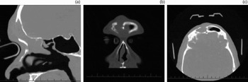
A computed tomography (CT) scan (Figure 1) showed a mass lesion filling the frontal sinus and ethmoid sinuses and with erosion of the anterior table of the frontal sinus.
Computed tomography images of the lesion. (a) Sagittal scan showing the lesion together with erosion of the anterior table of the right frontal sinus; (b) coronal scan showing the lesion opacifying the right frontal sinus; and (c) axial scan showing the eroded anterior table of the right frontal sinus.

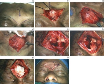
After discussion with the patient, a decision was made to undertake surgery via a combined open and endoscopic approach. The endoscopic approach involved a total uncinectomy, ethmoidectomy and frontal sinusotomy; however, this revealed only inflammatory tissue, with no discernable tumour. An open approach was then utilised via a gull wing brow incision, as depicted in Figures 2a and 2b. A mass was discovered deep to the periosteum. As the lesion was shelled out (Figures 2c and 2d), it was apparent that the mass not only extended superiorly, anterior to the right frontal sinus, but also filled the sinus itself. In order to completely remove the lesion, which extended down to the inferior part of the sinus but not beyond the ostium, it was therefore necessary to drill out the entire right frontal sinus. Once the lesion was removed, there was a significant defect in the anterior table of the right frontal sinus (Figure 2e), as well as a small defect in the left frontal sinus, although a bony fragment was used to close this latter defect (Figure 2f). A decision was made to obliterate the right frontal sinus, as the mucosa had by this stage been removed entirely; demineralised bone matrix was used to fill the sinus (Figure 2g). Finally, the incision was closed in two layers (Figure 2h) and a middle meatal spacer placed lateral to the middle turbinate.
Sequential operative steps following initial endoscopic ethmoidectomy and frontal sinusotomy.

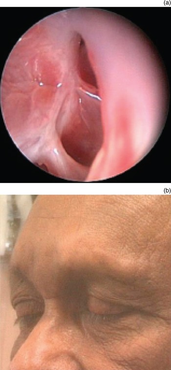
Post-operatively, the patient made a good recovery (Figure 3). Further blood tests (i.e. liver and thyroid function tests) and a CT scan of the chest and abdomen all proved negative for any signs of associated fibrosclerosing disorders.
(a) Post-operative endoscopic view of the right frontal recess. (b) Post-operative appearance of the external gull-wing incision.

Pathology
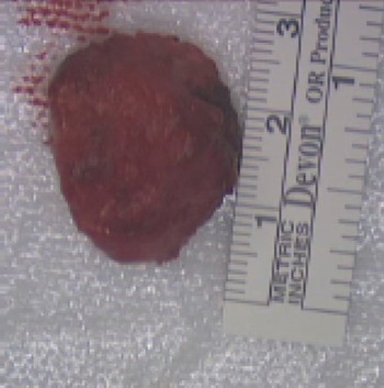
The typical macroscopic features of tumefactive fibroinflammatory lesion include a tan-white to grey colour with a firm, homogeneous texture (Figure 4).
Macroscopic appearance of the lesion (anterior portion).

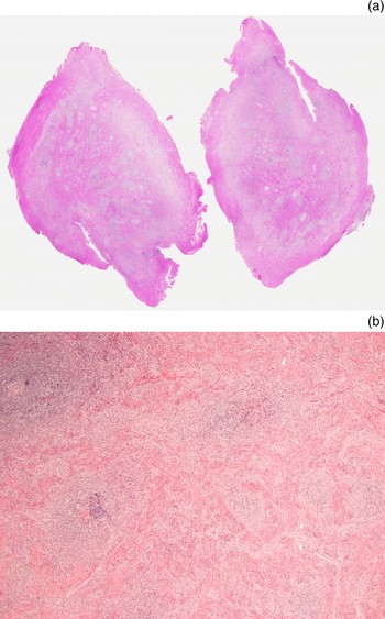
Microscopically, tumefactive fibroinflammatory lesions are usually composed of dense fibroconnective tissue with a marked plasma cell infiltrate and lymphoid follicles. Other cell types seen include apparently normal fibroblasts and lymphocytes, plus a few polymorphonuclear leukocytes (Figure 5).Reference Prichard, Colloby, Barton and Heaton2 Invasion of associated muscle, bone and neurovascular structures is frequently identified, and these lesions may be mistaken for malignant neoplasms clinically.Reference Olsen, DeSanto, Wold and Weiland3 However, tumefactive fibroinflammatory lesions appear to be only locally destructive, and no haematological or lymphatic spread has ever been demonstrated.Reference Laurenzo and Graham1
Photomicrograph showing sections of the lesion. (H&E; ×25)

Discussion
Tumefactive fibroinflammatory lesions of the head and neck were first described in 1983 by Wold and Weiland, with a limited number of subsequent reports; the presented case is the 23rd described.Reference Laurenzo and Graham1, Reference Wold and Weiland4, Reference Holodny, Kirsch, Hameed and Sclar5 The sinonasal tract appears to be the most commonly reported site. We could find no previously reported case involving the frontal sinus.
Lesions commonly present with a mass appearance and pain.Reference Patel, Pellitteri, Vrabec and Szymanski6
Given the current case's size and location, an open approach was an inevitable surgical option.
However, tumefactive fibroinflammatory lesion is not a neoplasm and has no propensity to metastasise, despite its locally aggressive characteristics. Thus, with a confirmed biopsy in hand, published evidence suggests that clinicians treating this condition have the option of administering high dose prednisolone to shrink the lesion.Reference Olsen, DeSanto, Wold and Weiland3, Reference Patel, Pellitteri, Vrabec and Szymanski6, Reference Husband and Knudsen7 However, some reports indicate that cessation of steroids leads to regrowth of the lesion, and that maintenance therapy may be indicated.Reference Laurenzo and Graham1 Steroids may also play a role where recurrences occur after surgical excision, or even for pre-operative shrinkage of the tumour. Radiotherapy has even been utilised in one case series.Reference Olsen, DeSanto, Wold and Weiland3 The key point here is that any surgical or medical plan will be dependent on obtaining an adequate (open) biopsy, and on the cooperation of the pathology department in identifying the lesion.
• Tumefactive fibroinflammatory lesions are rare
• They have previously been reported in the sinonasal tract, but not, as here, in the frontal sinus
• Each case should be managed on its merits, with a biopsy and screening for associated fibroinflammatory lesions
• Isolated lesions are probably best managed surgically, in patients suitable for general anaesthesia
• The literature suggests that these lesions have an association with other fibroinflammatory lesions, and may be amenable to systemic steroid therapy
Conclusion
Tumefactive fibroinflammatory lesions are a rare cause of sinonasal pathology. Key management steps include obtaining a biopsy, establishing a histopathological diagnosis and investigating for the presence of associated lesions elsewhere. The decision of whether to pursue surgical or medical treatment will be determined by these findings, and by the presence of any medical co-morbidity. Once this information is in hand, discussion with the patient will help determine their preference between the two options. Albeit with limited experience, we would certainly recommend a surgical approach in the presence of an isolated lesion.
Acknowledgement
We thank Dr Joanne Wright, Department of Pathology, St Paul's Hospital, Vancouver.