Loeys–Dietz syndrome is a newly recognised, rare autosomal dominantly inherited connective tissue disorder caused by heterogeneous mutations in the genes encoding the transforming growth factor beta receptor one or two.Reference Loeys, Chen and Neptune1 This syndrome is characterised by the triad of arterial tortuosity, aneurysm or dissections, hypertelorism, and bifid uvula or cleft palate.Reference Loeys, Schwarze and Holm2 Here, we present a patient prenatally diagnosed with complex congenital heart disease and confirmed with Loeys–Dietz syndrome after birth.
Case report
A 31-year-old pregnant woman was referred to our paediatric cardiology unit at the 36th week of gestation because of foetal congenital heart disease and dilatation of the pulmonary artery.
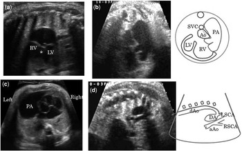
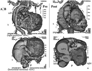
The first foetal echocardiography revealed a huge aneurysm of the main pulmonary artery and complex congenital heart disease – double-outlet right ventricle and interruption of the aortic arch (Fig 1). Detailed multi-planar scanning showed that there was no pulmonary valve stenosis, because of no acceleration in pulmonic flow, and no absent pulmonary valve. Therefore, we suspected a connective tissue disorder, such as Marfan syndrome. The foetus was followed up weekly for foetal decompensation and signs of hydrops until the 39th week of gestation, and an elective caesarean section was then performed. The male infant weighed 2834 grams at birth. After delivery, the infant developed dyspnoea and was intubated for artificial ventilation. Subsequently, a cleft of the soft palate and bifid uvula were noted. To treat the interruption of the aortic arch, we started him on a prostaglandin infusion to maintain patent ductus arteriosus and on nitrogen inhalation to prevent pulmonary blood flow increase. Computed tomography and angiocardiography confirmed the heart disease diagnosed prenatally (Fig 2a). Loeys–Dietz syndrome was strongly suspected because of the presence of cardiovascular lesions, thin skin, and facial appearance. On the 9th day, as the patient had suffered a pulmonary haemorrhage due to pulmonary blood flow increase, emergency bilateral pulmonary artery banding was performed. However, during surgery, it became apparent that application of normal pulmonary artery banding was impossible because of the very thin condition of the pulmonary artery wall. Therefore, the surgeon performed bilateral banding with the clip, not the usual tape, but the banding was insufficient. This may be a reason why his haemodynamics and respiratory status were not subsequently stable. We again performed computed tomography, which showed a further significant expansion of the right pulmonary artery and descending aorta caused by the pressure of the expanded artery (Fig 2b). Therefore, we started internal use of Losartan. On the 21st day, he developed sudden hypotension and massive bleeding from the thoracic cavity, thought to be caused by right pulmonary rupture, and he died the same day. Subsequently, as the genetic analysis showed p.Thr200Pro (c.598A > C) mutation of the transforming growth factor beta receptor one, he was definitively diagnosed with Loeys–Dietz syndrome. The mutation was de novo.
Foetal echocardiography shows a large ventricular septal defect (*) of the double-outlet right ventricle (a), aneurysmal pulmonary artery (b, c), and interruption of the aortic arch (d). aAo = ascending aorta; Ao = aorta; DA = ductus arteriosus; dAo = descending aorta; LV = left ventricle; LSCA = left subclavian artery; PA = pulmonary artery; RSCA = right subclavian artery; RV = right ventricle; SVC = supra caval vein.

Computed tomography (day 0) shows the interruption of the aortic arch and aneurysmal main pulmonary artery before operation (a). Computed tomography (day 18) shows progress of the significant expansion of the right and left pulmonary arteries and descending aorta after operation (b). A = anterior; aAo = ascending aorta; dAo = descending aorta; L = left; ltpa = left pulmonary artery; P = posterior; mPA = main pulmonary artery; rtPA = right pulmonary artery; R = right.

Discussion
Loeys–Dietz syndrome is a recently described connective tissue disorder characterised by aggressive ascending aortic aneurysm and dissection. The clinical features are similar to Marfan syndrome,Reference Mizuguchi, Collod-Beroud and Akiyama3 but this is a more severe syndrome because life-threatening aortic dissection may occur even in early childhood.Reference Yamawaki, Nagaoka and Morishige4, Reference Akutsu, Morisaki and Okajima5 Most patients have the triad of vascular aneurysms, hypertelorism, and bifid or broad uvula/cleft palate associated with variable features. Heterogeneous mutations in the genes encoding for transforming growth factor beta receptors one and two are a consistent finding among affected patients.
In addition, this syndrome shows various cardiovascular manifestations involving not only aortic lesions – such as distortion, aneurysm, and dissections – but also congenital heart diseases.Reference Muramatsu, Kosho and Magota6 The case described in this report was also complicated with congenital heart disease. The patient's pulmonary artery showed an abnormal expansion because of his heart defect. That is, because he had an interruption of the aortic arch, much more blood than normal flowed through the pulmonary artery and the artery was stressed by “volume overload”. Furthermore, the pulmonary artery was stressed by high “pressure overload” because the patient had double-outlet right ventricle and a large ventricular septal defect. It is thought that a pulmonary artery spread for both reasons from the foetal period.
Muramatsu et alReference Muramatsu, Kosho and Magota6 reported a case that was complicated with a ventricular septal defect and showed aortic and pulmonary expansion. It is thought that, in the Muramatsu case, the mechanism producing pulmonary artery dilatation was similar to that in the case reported herein. After birth, the patient's pulmonary blood flow increased due to the ventricular septal defect, which led to acute heart failure. He then underwent pulmonary artery banding on the 12th day. After surgery, however, the root of the main pulmonary artery, which was stressed by pressure, had spread in the shape of an aneurysm and intracardiac surgical repair, that is, closure of ventricular septal defect, was performed on the 42nd day. After the operation, the vascular expansion stopped worsening, and in conclusion they recommended early radical operation. However, because our case was a Fontan candidate, he required gradual surgery and radical operation was impossible in early infancy. Therefore, we performed bilateral pulmonary artery banding as a life-saving procedure, but, owing to mural abnormal thinning, the banding was insufficient, and his vascular expansion and thinning progressed, which finally led to explosion and bleeding to death.
In the case reported herein, significant pulmonary expansion from the foetal period led us to suspect a connective tissue disorder such as Marfan syndrome. Viassolo et alReference Viassolo, Lituania and Marasini7 reported a similar case in a female patient with Loeys–Dietz syndrome, who showed dilated aortic root from the foetal period. Only aortic dilatation was noted in screening foetal echocardiography at 19 gestational weeks and a connective tissue disease was suspected. She underwent genetic analysis and Loeys–Dietz syndrome was confirmed after birth. At present, the Viassolo case and the one we report herein are the only two cases showing a manifestation of Loeys–Dietz syndrome from the foetal period.
Some cases of Loeys–Dietz syndrome are complicated with congenital heart diseases.Reference Loeys, Schwarze and Holm2, Reference Muramatsu, Kosho and Magota6, Reference Watanabe, Sakai and Nishimura8 However, those reported hitherto are associated with “simple” congenital heart diseases such as ventricular septal defect, atrial septal defect, patent ductus arteriosus, and aortic bicuspid valve. There is no previous report of Loeys–Dietz syndrome combined with complex congenital heart disease, such as double-outlet right ventricle and interruption of the aortic arch. In such a case, the cardiovascular lesion as an expansion of the great vessels, that is, the aorta or pulmonary artery, may be aggravated during the foetal period. Consequently, the foetus may die in utero. Even if they can be born, their great vessels are continuously or more strongly stressed after birth. Therefore, their arteries expand and finally explode, leading to an early death without undergoing any surgery.
This may be the reason why this is the first reported case of complex heart disease with Loeys–Dietz syndrome.